. 2014 May;28(5):2260-71.
doi: 10.1096/fj.13-244004. Epub 2014 Jan 27.

Kindlin-3 enhances breast cancer progression and metastasis by activating Twist-mediated angiogenesis

Affiliations

Kindlin-3 enhances breast cancer progression and metastasis by activating Twist-mediated angiogenesis

Khalid Sossey-Alaoui et al. FASEB J. 2014 May.

Abstract

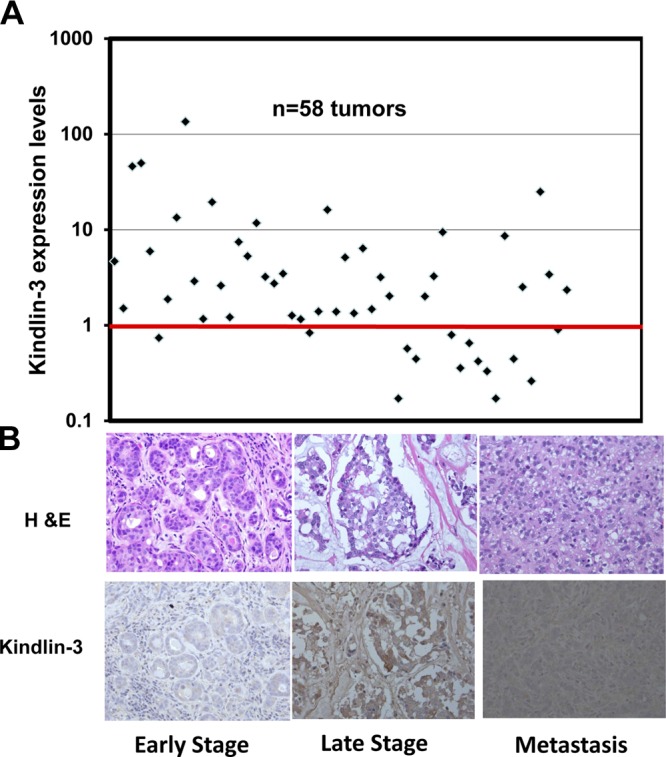
The FERM domain containing protein Kindlin-3 has been recognized as a major regulator of integrin function in hematopoietic cells, but its role in neoplasia is totally unknown. We have examined the relationship between Kindlin-3 and breast cancer in mouse models and human tissues. Human breast tumors showed a ∼7-fold elevation in Kindlin-3 mRNA compared with nonneoplastic tissue by quantitative polymerase chain reaction. Kindlin-3 overexpression in a breast cancer cell line increased primary tumor growth and lung metastasis by 2.5- and 3-fold, respectively, when implanted into mice compared with cells expressing vector alone. Mechanistically, the Kindlin-3-overexpressing cells displayed a 2.2-fold increase in vascular endothelial growth factor (VEGF) secretion and enhanced β1 integrin activation. Increased VEGF secretion resulted from enhanced production of Twist, a transcription factor that promotes tumor angiogenesis. Knockdown of Twist diminished VEGF production, and knockdown of β1 integrins diminished Twist and VEGF production by Kindlin-3-overexpressing cells, while nontargeting small interfering RNA had no effect on expression of these gene products. Thus, Kindlin-3 influences breast cancer progression by influencing the crosstalk between β1 integrins and Twist to increase VEGF production. This signaling cascade enhances breast cancer cell invasion and tumor angiogenesis and metastasis.

Keywords: VEGF; VEGFR2; epithelial-to-mesenchymal-transition; integrins; tumor macrophages.


Figures

Figure 1.
Figure 1.
Kindlin-3 expression levels are increased in human BC tumors. A) Real-time qRT-PCR of Kindlin-3 transcripts in 58 BC tumors. Each data point represents the average fold change of Kindlin-3, from 3 independent PCRs, in 1 BC tumor compared with the adjacent normal mammary tissue. GAPDH was used as an internal control for normalization. Points under the red bar represent tumors in which Kindlin-3 is down-regulated; points above the red bar represent tumors in which Kindlin-3 is up-regulated. B) Immunohistochemical staining of BC human tumor sections with H&E (top panels) and Kindlin-3 antibody (bottom panels). Intensity of brown staining correlates with Kindlin-3 expression levels; low in early stage BC and high in metastatic tumors. Four tumors from early stage, 4 tumors from late stage, and 4 metastatic tumors were stained; 1 representative section from each stage is shown.
Figure 2.
Figure 2.
Kindlin-3 enhances migration and invasion of BC cells in vitro. A) Flow cytometry of untransfected (red histogram), EGFP-expressing (gray histogram), and EGFP-Kindlin-3-overexpressing (solid black histogram) MDA-MB-231 cells. B) Immunoblots of total cell lysates from MDA-MB-231 cells expressing EGFP alone or EGFP-Kindlin-3, probed with anti-Kindlin-3 (top panel), anti-GFP (middle panel), or anti-β-actin as a loading control (bottom panel). C) Representative micrographs of EGFP-expressing and Kindlin-3-overexpressing MDA-MB-231 cells that were induced to migrate into denuded wounds over a 16 h period. D) Size of the remaining open wound after 16 h was measured from 12 different wounds and plotted as the percentage of wound at time 0. E) Representative micrographs of EGFP-expressing and Kindlin-3-overexpressing MDA-MB-231 cells that were induced to invade through a Matrigel-coated membrane. F) Invading cells were counted from 6 different fields and plotted as an average of the number of invading cells per field. All data are representative of 3 independent experiments or are means ± se (n=3; P<0.05, Student's t test).
Figure 3.
Figure 3.
Kindlin-3 enhances primary tumor growth and metastasis of BC in vivo in mouse models. Effect of Kindlin-3 on primary tumor growth (A) and spontaneous lung metastasis (B, C) and in lung colonization model (D–F). A) Tumor growth curves for EGFP-expressing and Kindlin-3-overexpressing MDA-MB-231 cells. Data points represent means ± sd of tumor volume, showing a significant difference (P<0.05) between the EGFP- and Kindlin-3-expressing cells. Mice (SCID) were monitored for 70 d after implantation of cancer cells in their mammary fat pads, after which all were analyzed for lung metastases. B) H&E staining of lung sections of mice implanted with the EGFP-expressing MDA-MB-231 cells (GFP) and mice implanted with Kindlin-3-overexpressing MDA-MB-231 cells. Metastases are delineated by dark lines. C) Quantification of lung surface metastasis nodules from the groups of mice injected with the EGFP-expressing and EGFP-Kindlin-3-overexpressing cells and plotted as an average of the number of metastatic nodules per lung lobe (P<0.001, Student's t test). D) Representative confocal micrographs of lung sections and their corresponding H&E staining from mice injected with EGFP-expressing and Kindlin-3-overexpresing MDA-MB-231 cells. Each green area represents a lung metastasis and is delineated by a dark line in the H&E staining micrographs. E) Quantification of lung metastasis from the 2 groups of mice described in D. F) Genomic PCR analysis of DNA from lungs of mice injected with the EGFP-expressing or Kindlin-3-overexpressing cells. Human-specific ERVK6A gene can be amplified in human DNA (231 and H-Ctrl) but not in mouse DNA (M-Ctrl). Presence of the human-specific ERVK6A DNA sequences in the mouse tissues (GFP and K3) indicates the presence of human cells in these tissues. Intensity of the PCR product of the ERVK6A gene correlates with the level of metastasis. Mouse Wave3 gene is used as a control for integrity of the mouse DNA in each sample. N, number of mice per group.
Figure 4.
Figure 4.
Kindlin-3 enhances tumor angiogenesis by increasing VEGF-A expression. A) Representative immunofluorescence confocal micrographs of sections of mammary fat pad tumors from mice implanted with EGFP-expressing (GFP) and Kindlin-3-overexpressing MDA-MB-231 cells stained with anti CD31 (red) to detect tumor-associated blood vessels. Cell nuclei were counterstained with DAPI (blue). B) Quantification of angiogenesis from the EGFP and Kindlin-3 groups, as determined by the average blood vessel area per tumor section (n=number of mice per group). C) qRT-PCR of the CD31 transcripts from the mammary fat pad tumors described in A. D) Representative confocal micrographs of human BC tumor sections stained with a vWF antibody and their corresponding H&E staining. Intensity of the brown staining correlates with expression of vWF protein; low in early stage and high in late stage BC tumors. E) qRT-PCR of the VEGF-A transcripts from the mammary fat pad tumors described in A. F) Representative immunofluorescence confocal micrographs of sections of mammary fat pad tumors described in A, stained for the phosphorylated form of VEGFR2 (green fluorescence). Cell nuclei were counterstained with DAPI. G) Immunoblots of protein lysates from mammary fat pad tumors from mice implanted with EGFP-expressing (GFP-T1 to -T3) and Kindlin-3-overexpressing (K3-T1 to -T3) MDA-MB-231 cells with anti-phospho-VEGFR2. β-Actin antibody is a loading control.
Figure 5.
Figure 5.
Blockade of secreted VEGF-A from cancer cells inhibits angiogenesis. A) Quantification of secreted VEGF-A in the conditioned media of EGFP-expressing (GFP) and Kindlin-3-overexpressing (K3) MDA-MB-231 cells using ELISA; n = number of repeats. B) Bright-field microscopy of tube-formation structures of HUVECs treated with serum-free medium (SFM), SFM supplemented with VEGF-A (10 ng/ml), or conditioned media from EGFP-expressing and Kindlin-3-overexpressing MDA-MB-231 cells. C) Quantification of tube formation. Number of closed tubes was counted in 12 different fields and plotted as the average ± se number of closed tubes per field. Each assay was repeated ≥3 times. *P < 0.05, **P < 0.05; Student's t test. D) Bright-field micrographs of tube structures of HUVECs treated with SFM supplemented with VEGF-A (10 ng/ml) or conditioned media from EGFP- and Kindlin-3-expressing MDA-MB-231 cells, in the absence (top panels) or the presence (bottom panels) of sFLT, a soluble VEGFR inhibitor.
Figure 6.
Figure 6.
Kindlin-3 modulates angiogenesis through the regulation of Twist expression and macrophage recruitment. A) Immunoblotting with the indicated antibodies of protein lysates from EGFP-expressing (GFP) and Kindlin-3-overexpressing (K3) MDA-MB-231 cells and their respective mammary fat pad-derived tumors (GFP-T1 to -T3 and K3-T1 to -T3, respectively). B) Representative immunofluorescence confocal microscopy of EGFP-expressing (top panels) and Kindlin-3-overexpressing (bottom panels) MDA-MB-231 cells stained with Twist antibody. Cell nuclei were counterstained with DAPI. C) Quantification of Twist nuclear localization in the EGFP-expressing and Kindlin-3-overexpressing MDA-MB-231 cells, plotted as the average percentage of cells with nuclear Twist staining per field. (n=number of fields per experiment from ≥3 repeats). D) Immunoblotting of protein lysates from the Kindlin-3-overexpressing MDA-MB-231 cells with anti-Twist. Cells were first transfected with either an NT-siRNA (NT-si) or a Twist-targeting siRNA (si-Twist). Anti-β-actin serves as a loading control. E) Quantification of secreted VEGF-A into the conditioned medium of Kindlin-3-overexpressing MDA-MB-231 cells using ELISA after transfection with either NT-si or si-Twist; n = number of repeats. F) Representative immunofluorescence confocal micrographs of sections of mammary fat pad tumors derived from EGFP-expressing and Kindlin-3-overexpressing MDA-MB-231 cells, stained with anti-F4/80 macrophage specific antibody (green fluorescence). Cell nuclei were counterstained with DAPI. G) Quantification of macrophage infiltration in the tumors described in F, plotted as the average of the percentage of F4/80-positive area per field; n = number of fields.
Figure 7.
Figure 7.
Kindlin-3-Twist-VEGF-angiogenesis signaling axis is integrin dependent. A) Quantification of cell surface β1-integrin activation in the EGFP-expressing (GFP) and Kindlin-3-overexpressing (K3) MDA-MB-231 cells, as measured by HUTS4 antibody binding, normalized to total β1-integrin expression levels and plotted as a fold change to the EGFP-expressing cells; n = number of repeats. B) Quantification of cell surface β1-integrin expression levels from the cells described in A; n = number of repeats. C) Immunoblotting with the indicated antibodies of protein lysates from the Kindlin-3-overexpressing MDA-MB-231 cells. Cells were first transfected with either an NT-siRNA (NT-si) or with a β1-integrin-targeting siRNA (si-β1). Anti-β-actin is a loading control. D) Quantification of cell surface β1-integrin activation levels in the EGFP-expressing and Kindlin-3-overexpressing MDA-MB-231 cells transfected with either NT-si or β1-si. β1-Integrin activation levels were measured by HUTS4 antibody binding, normalized to total β1-integrin expression levels, and plotted as a fold change in the EGFP-expressing cells transfected with the NT-siRNA. *P < 0.05, Student's t test. E) Quantification of secreted VEGF-A in the conditioned medium of Kindlin-3-overexpressing MDA-MB-231 cells using ELISA after transfection with NT-si or si-β1; n = number of repeats. F) Immunoblotting with the indicated antibodies of protein lysates from the Kindlin-3-overexpressing MDA-MB-231 cells that were transfected with either an NT-siRNA (NT), a Kindlin-3 siRNA (si-K3), si-β1, or a Twist siRNA (si-Twist). G) Quantification of secreted VEGF-A in the conditioned media of cells described in F using ELISA; n = number of repeats. *P < 0.05, **P < 0.05, ***P < 0.05; Student's t test.
Figure 8.
Figure 8.
Kindlin-3-mediated regulation of Twist activates the EMT program in BC. A) Immunoblotting with the indicated antibodies of protein lysates from the EGFP-expressing (GFP) and Kindlin-3-overexpressing (K3) MDA-MB-231 cells and their respective mammary fat pad-derived tumors (GFP-T1 to -T3 and K3-T1 to -T3, respectively). Anti-β-actin antibody serves as a loading control. B, C) Representative immunofluorescence confocal micrographs of sections of mammary fat pad tumors derived from EGFP-expressing and Kindlin-3-overexpressing MDA-MB-231 cells, stained with anti-fibronectin (B) and anti-N-cadherin (C) (green fluorescence). Cell nuclei were counterstained with DAPI. D) Model describing how the β1-integrin-Kindlin-3 interplay modulates the molecular signaling pathways that regulate BC progression and metastasis.

References

    1. Ganguly K. K., Pal S., Moulik S., Chatterjee A. (2013) Integrins and metastasis. Cell Adh. Migr. 7, 251–261 - PMC - PubMed
    1. Cance W. G., Kurenova E., Marlowe T., Golubovskaya V. (2013) Disrupting the scaffold to improve focal adhesion kinase-targeted cancer therapeutics. Sci. Signal. 6, e10 - PMC - PubMed
    1. Desiniotis A., Kyprianou N. (2011) Significance of talin in cancer progression and metastasis. Int. Rev. Cell. Mol. Biol. 289, 117–147 - PMC - PubMed
    1. Shibue T., Brooks M. W., Weinberg R. A. (2013) An integrin-linked machinery of cytoskeletal regulation that enables experimental tumor initiation and metastatic colonization. Cancer Cell 24, 481–498 - PMC - PubMed
    1. Malinin N. L., Plow E. F., Byzova T. V. (2010) Kindlins in FERM adhesion. Blood 115, 4011–4017 - PMC - PubMed

Publication types

MeSH terms