Review
. 2014 Jan-Feb;9(1):37-52.
doi: 10.1002/cmmi.1551.

Nanoparticle contrast agents for computed tomography: a focus on micelles

Affiliations
Review

Nanoparticle contrast agents for computed tomography: a focus on micelles

David P Cormode et al. Contrast Media Mol Imaging. 2014 Jan-Feb.

Abstract

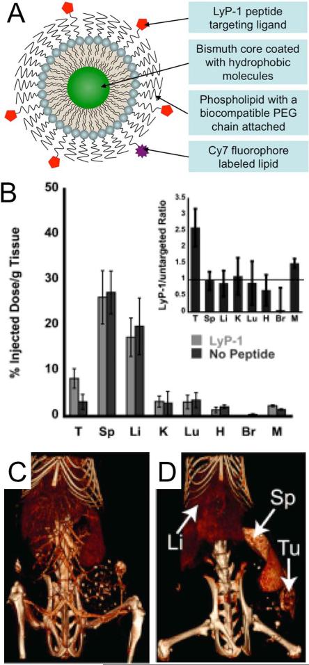
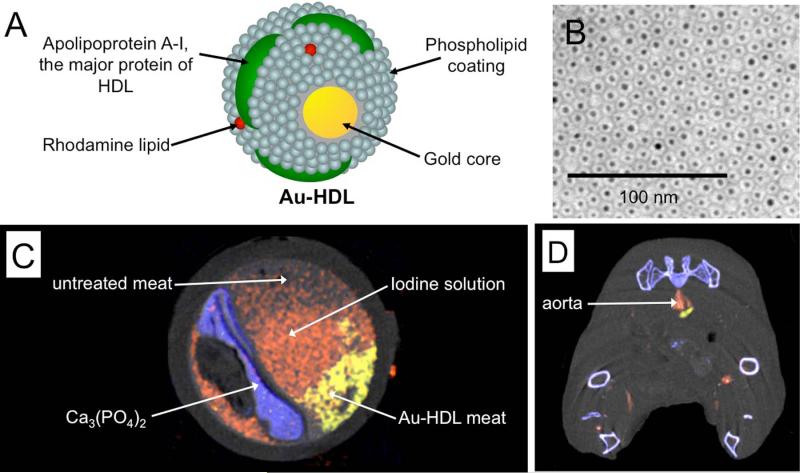
Computed tomography (CT) is an X-ray-based whole-body imaging technique that is widely used in medicine. Clinically approved contrast agents for CT are iodinated small molecules or barium suspensions. Over the past seven years there has been a great increase in the development of nanoparticles as CT contrast agents. Nanoparticles have several advantages over small molecule CT contrast agents, such as long blood-pool residence times and the potential for cell tracking and targeted imaging applications. Furthermore, there is a need for novel CT contrast agents, owing to the growing population of renally impaired patients and patients hypersensitive to iodinated contrast. Micelles and lipoproteins, a micelle-related class of nanoparticle, have notably been adapted as CT contrast agents. In this review we discuss the principles of CT image formation and the generation of CT contrast. We discuss the progress in developing nontargeted, targeted and cell tracking nanoparticle CT contrast agents. We feature agents based on micelles and used in conjunction with spectral CT. The large contrast agent doses needed will necessitate careful toxicology studies prior to clinical translation. However, the field has seen tremendous advances in the past decade and we expect many more advances to come in the next decade.

Keywords: X-ray; bismuth; computed tomography; gold nanoparticle; iodine; lipoprotein; micelle; molecular imaging; nanoparticle; spectral CT.


Figures

Figure 1
Figure 1
A) The chemical structure of iohexol. Graphs of the increase in B) publications and C) citations in the area of computed tomography and nanoparticles over the past 17 years. Data acquired from a search of ‘computed tomography’ refined by ‘nanoparticles’ in the Web of Science database. The data is reproduced with permission from Thomson Reuters. D) Generalized schematic depiction of CT nanoparticle contrast agent.
Figure 2
Figure 2
A) A schematic of a CT scanner. B) A grayscale image of the heart of a patient who had been injected with an iodinated contrast agent. C) A false color, 3D-rendered image of the heart of a patient. Images reproduced with permission from (171,172).
Figure 3
Figure 3
A) Mass attenuation coefficients of a variety of elements. Data downloaded from (64). B) Photon energy distribution generated from the X-ray tube of a CT scanner run at 80 or 140 kV. Adapted with permission from (61).
Figure 4
Figure 4
Schematic depiction of a number of nanoparticle types formed from aggregates of lipids. Reproduced with permission from reference (173).
Figure 5
Figure 5
A) chemical structure of an iodinated polymeric lipid. B) Schematic depiction of a micelle formed from the iodinated polymeric lipid. C) CT images of a rat at the level of the heart, liver and spleen before and after injection with the iodinated micelles. Images reproduced with permission from reference (46).
Figure 6
Figure 6
A) Schematic depiction of a CT/MRI/fluorescence active, micelle-based gold nanoparticle contrast agent. B) Transmission electron microscopy characterization of the nanoparticles. C) CT and D) MR images of a mouse liver pre- and 24 hours post-injection with the gold nanoparticles. E) Fluorescence imaging of the livers of mice injected with either fluorescent (left) or non-fluorescent (right) gold nanoparticles. Figure adapted with permission from (28).
Figure 7
Figure 7
A) Schematic depiction of the structure of a micelle-based, targeted bismuth core nanoparticle. B) Biodistribution of targeted and non-targeted nanoparticle formulations at 24 hours post-injection. T=tumor, Sp=spleen, Li=liver, K=kidney, Lu=lungs, H = heart, Br = brain, M = muscle. CT images acquired C) immediately and D) 24 hours post-injection of targeted nanoparticles. Figure adapted with permission from (48).
Figure 8
Figure 8
A) Structure of Au-HDL, a macrophage targeted CT contrast agent. B) Negative stain transmission electron microscopy characterization of the nanoparticle. C) Spectral CT image of an artery phantom. D) Spectral CT image of an atherosclerotic mouse acquired 24 hours after injection with Au-HDL and directly after injection of an iodine nanoemulsion. In C) and D) gold (yellow), iodine (red) and photoelectric (blue) images are overlaid on a Compton image (greyscale). Images reproduced with permission from (44).

References

    1. Yu S-B, Watson AD. Metal-based X-ray contrast media. Chem Rev. 1999;99:2353–2377. - PubMed
    1. Rumberger JA. Coronary computed tomography angiography. J Am Coll Cardiol. 2008;52(21):1733–1735. - PubMed
    1. Lumbroso P, Dick CE. X-ray attenuation properties of radiographic contrast media. Med Phys. 1987;14:752–758. - PubMed
    1. Yu S-B, Droege M, Segal B, Kim S-H, Sanderson T, Watson AD. Cuboidal W3S4 cluster complexes as new generation X-ray contrast agents. Inorg Chem. 2000;39(6):1325–1328. - PubMed
    1. Engelbrecht V, Koch JA, Rassek M, Modder U. Gadodiamid und Gadolinium-DTPA als intravenöse Kontrastmittel in der Computertomographie [Gadolinium-DTPA and gadodiamide as an alternative contrast medium for CT]. Rofo. 1996;165:24–28. - PubMed

Publication types