. 2014 May;66(5):1272-81.
doi: 10.1002/art.38376.

Interleukin-17+CD8+ T cells are enriched in the joints of patients with psoriatic arthritis and correlate with disease activity and joint damage progression

Affiliations
Free PMC article

Interleukin-17+CD8+ T cells are enriched in the joints of patients with psoriatic arthritis and correlate with disease activity and joint damage progression

Bina Menon et al. Arthritis Rheumatol. 2014 May.
Free PMC article

Abstract

Objective: Psoriatic arthritis (PsA) is associated with HLA class I genes, in contrast to the association with HLA class II in rheumatoid arthritis (RA). Since IL-17+ cells are considered important mediators of synovial inflammation, we sought to determine whether IL-17-producing CD8+ T cells may be found in the joints of patients with PsA and whether these cells might contribute to the disease process.

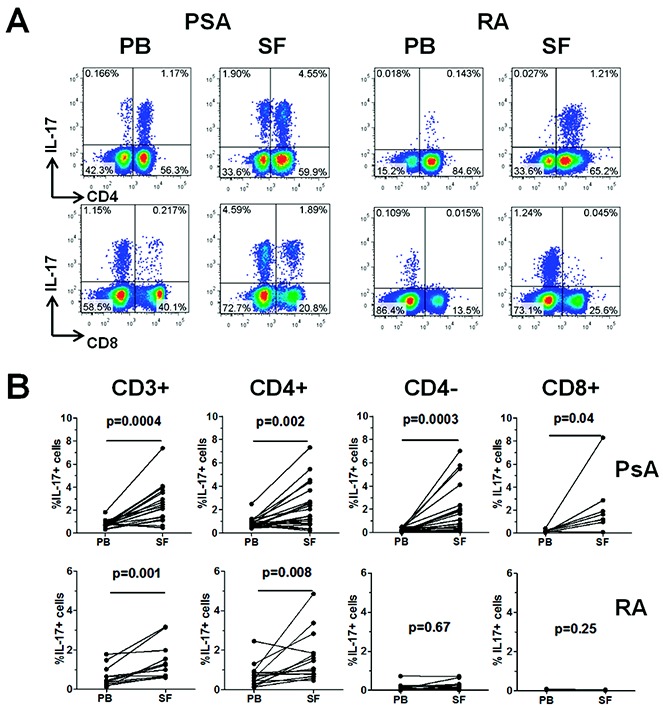
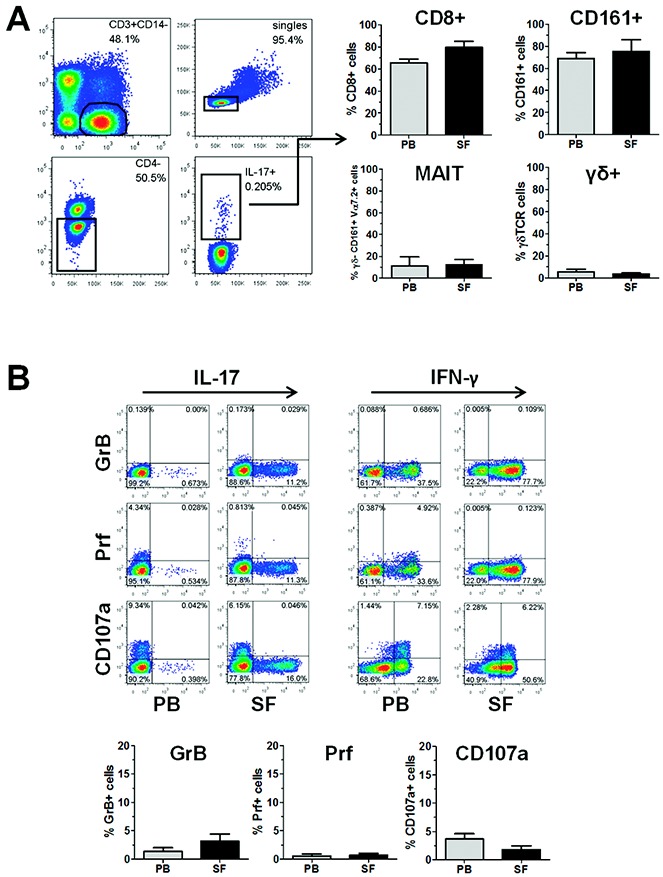
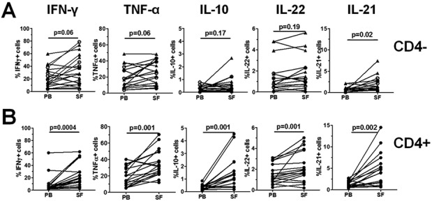
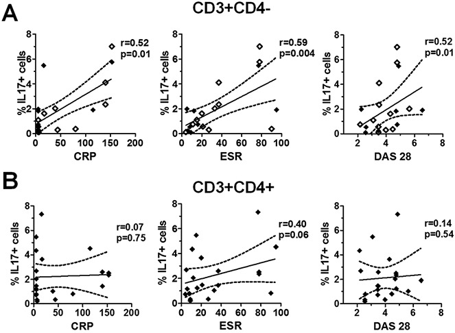
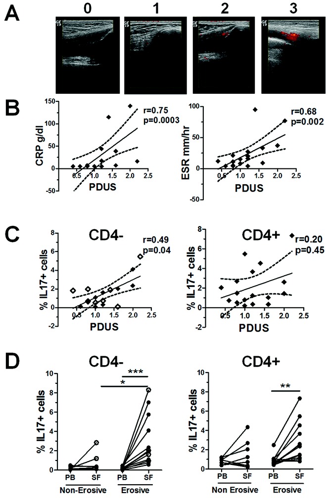
Methods: Mononuclear cells from paired samples of synovial fluid (SF) and peripheral blood (PB) from patients with PsA or patients with RA were stimulated ex vivo, and CD4- T cells were examined by flow cytometry for cytokine expression, cytotoxic markers, and frequencies of γ/δ or mucosal-associated invariant T cells. Clinical measures of arthritis activity (C-reactive protein [CRP] level, erythrocyte sedimentation rate [ESR], Disease Activity Score in 28 joints [DAS28]) and power Doppler ultrasound (PDUS) scores for the presence of active synovitis in the aspirated knee were recorded and assessed for correlations with immunologic markers.

Results: Within the CD3+ T cell compartment, both IL-17+CD4- (predominantly CD8+) and IL-17+CD4+ T cells were significantly enhanced in the SF compared to the PB of patients with PsA (P = 0.0003 and P = 0.002, respectively; n = 21), whereas in patients with RA, only IL-17+CD4+ T cells were increased in the SF compared to the PB (P = 0.008; n = 14). The frequency of IL-17+CD4- T cells in PsA SF was positively correlated with the CRP level (r = 0.52, P = 0.01), ESR (r = 0.59, P = 0.004), and DAS28 (r = 0.52, P = 0.01), and was increased in patients with erosive disease (P < 0.05). In addition, the frequency of IL-17+CD4- T cells positively correlated with the PDUS score, a marker for active synovitis (r = 0.49, P = 0.04).

Conclusion: These results show, for the first time, that the PsA joint, but not the RA joint, is enriched for IL-17+CD8+ T cells. Moreover, the findings reveal that the levels of this T cell subset are correlated with disease activity measures and the radiographic erosion status after 2 years, suggesting a previously unrecognized contribution of these cells to the pathogenesis of PsA.


Figures

Figure 1
Figure 1
Frequency of interleukin-17 (IL-17)–expressing cells in CD3+, CD3+CD4+, CD3+CD4−, and CD3+CD8+ T cell populations in paired peripheral blood (PB) and synovial fluid (SF) samples from patients with psoriatic arthritis (PsA) or rheumatoid arthritis (RA). Mononuclear cells from paired PB and SF samples from patients with PsA (n = 21) and patients with RA (n = 14) were isolated and stimulated as described in Patients and Methods. A, Percentage of IL-17–expressing cells within CD4+ and CD4− T cells (top panels) and within CD8+ and CD8− T cells (bottom panels), as determined by flow cytometry in PB and SF from a representative patient with PsA and a representative patient with RA. B, Percentage of IL-17+ cells in total CD3+, CD4+, CD4−, or CD8+ T cells in paired PB and SF samples from patients with PsA and patients with RA. For the CD8+ subset, the percentage of IL-17+ cells was determined in paired samples from 8 PsA patients and 3 RA patients. Data were analyzed using Wilcoxon's matched pairs signed rank test. Color figure can be viewed in the online issue, which is available at http://onlinelibrary.wiley.com/doi/10.1002/art.38376/abstract.
Figure 2
Figure 2
CD3+CD4− T cells in patients with psoriatic arthritis (PsA) are predominantly CD8+ T cells lacking markers associated with cytolytic activity. Cryopreserved mononuclear cells from paired peripheral blood (PB) and synovial fluid (SF) samples from patients with PsA were thawed and stimulated with phorbol myristate acetate and ionomycin in the presence of GolgiStop for 3 hours, and then stained for the expression of CD3, CD4, CD8, CD161, γ/δ T cell receptor (TCR), Vα7.2 TCR, granzyme B (GrB), perforin (Prf), and CD107a, along with interleukin-17 (IL-17). A, Left, Gating strategy to identify IL-17+CD3+CD4− T cells by flow cytometry. Representative results are shown. A, Right, Percentage of cells within the IL-17+CD3+CD4− T cell population that were either CD8+, CD161+, γ/δ−CD161+Vα7.2+ (mucosal-associated invariant T [MAIT] cells), or γ/δ+. B, Top, Coexpression of IL-17 or interferon-γ (IFNγ) and granzyme B, perforin, or CD107a in CD3+CD8+ T cells from PsA PB and SF, as determined by flow cytometry. Representative dot plots are shown. B, Bottom, Percentage of cells within the IL-17+ CD3+CD8+ T cell population that expressed granzyme B, perforin, or CD107a in PsA PB and SF. Results are the mean ± SEM of 4 samples. Color figure can be viewed in the online issue, which is available at http://onlinelibrary.wiley.com/doi/10.1002/art.38376/abstract.
Figure 3
Figure 3
Frequencies of pro- and antiinflammatory cytokine-expressing T cells in paired peripheral blood (PB) and synovial fluid (SF) samples from patients with psoriatic arthritis (PsA). Mononuclear cells were isolated from paired samples of PsA PB and SF (n = 21), and then stimulated ex vivo with phorbol myristate acetate and ionomycin in the presence of GolgiStop and stained for expression of interferon-γ (IFNγ), tumor necrosis factor α (TNFα), interleukin-10 (IL-10), IL-22, and IL-21. Viable CD3+ T cells were gated and the percentages of cytokine-expressing cells were determined in A, CD3+CD4− T cells (triangles; n = 13) or CD3+CD8+ T cells (circles; n = 8), or in B, CD3+CD4+ T cells. Each symbol joined by a line represents a paired sample from a different patient. Data were analyzed using Wilcoxon's matched pairs signed rank test.
Figure 4
Figure 4
Correlation between the frequency of interleukin-17 (IL-17)–expressing CD4− and CD4+ T cells in psoriatic arthritis (PsA) synovial fluid (SF) and clinical parameters of disease. Clinical measures of disease activity (C-reactive protein [CRP] level, erythrocyte sedimentation rate [ESR], and Disease Activity Score in 28 joints [DAS28]) were determined at the time of PsA SF sampling. Mononuclear cells from the SF of patients with PsA (n = 21) were stimulated as described in Patients and Methods. The percentage of IL-17+ cells within the CD3+CD4− T cell population (including CD3+CD8+ cells [open diamonds]; n = 8) (A) and within the CD3+CD4+ T cell population (B) in PsA SF was plotted against the CRP level, ESR, and DAS28. Regression coefficients (solid lines) with 95% confidence intervals (broken lines) were calculated using Spearman's correlation coefficients for nonparametric data.
Figure 5
Figure 5
Correlation between the frequency of interleukin-17 (IL-17)–expressing CD4− T cells and the power Doppler ultrasound (PDUS) score for the presence of local synovitis in patients with psoriatic arthritis (PsA), and enrichment of this T cell subset in the joints of patients with erosive disease. A, Representative PDUS images illustrating the semiquantitative grading system for active synovitis in the knee joints of patients with PsA, where 0 = no signal, 1 = 1–2 pixels, 2 = <50% signal, and 3 = ≥50% signal. B, Correlations between measures of disease activity (the C-reactive protein level or erythrocyte sedimentation rate) and the mean PDUS score of the aspirated knee joints (n = 17). C, Correlations between the percentage of IL-17+ cells within CD3+CD4− T cells (including CD3+CD8+ T cells [open diamonds]; n = 8) or CD3+CD4+ T cells from PsA synovial fluid (SF) and the mean PDUS score in the same knee joint. In B and C, regression coefficients (solid lines) with 95% confidence intervals (broken lines) are shown for each plot. D, Percentage of IL-17+ cells within the CD3+CD4− T cell (including CD3+CD8+ cells [open circles]) and CD3+CD4+ T cell populations in paired samples of peripheral blood (PB) and SF from patients with erosive PsA (n = 13) compared to patients with nonerosive PsA (n = 8). Each symbol joined by a line represents a paired sample from a different patient. Data were analyzed using one-way analysis of variance for parametric or nonparametric data, where appropriate. ∗ = P < 0.05; ∗∗ = P < 0.01; ∗∗∗ = P < 0.001.

Comment in

Similar articles

Cited by

References

    1. Christophers E. Psoriasis—epidemiology and clinical spectrum. Clin Exp Dermatol. 2001;26:314–20. - PubMed
    1. Scott DL, Smith C, Kingsley G. Joint damage and disability in rheumatoid arthritis: an updated systematic review. Clin Exp Rheumatol. 2003;21:S20–7. - PubMed
    1. Courvoisier N, Dougados M, Cantagrel A, Goupille P, Meyer O, Sibilia J, et al. Prognostic factors of 10-year radiographic outcome in early rheumatoid arthritis: a prospective study. Arthritis Res Ther. 2008;10:R106. - PMC - PubMed
    1. Kane D, Stafford L, Bresnihan B, FitzGerald O. A prospective, clinical and radiological study of early psoriatic arthritis: an early synovitis clinic experience. Rheumatology (Oxford) 2003;42:1460–8. - PubMed
    1. Ho PY, Barton A, Worthington J, Plant D, Griffiths CE, Young HS, et al. Investigating the role of the HLA-Cw*06 and HLA-DRB1 genes in susceptibility to psoriatic arthritis: comparison with psoriasis and undifferentiated inflammatory arthritis. Ann Rheum Dis. 2008;67:677–82. - PMC - PubMed

Publication types

MeSH terms