. 2014 Jan 27;3(1):e000622.
doi: 10.1161/JAHA.113.000622.

Deletion of Krüppel-like factor 4 in endothelial and hematopoietic cells enhances neointimal formation following vascular injury

Affiliations

Deletion of Krüppel-like factor 4 in endothelial and hematopoietic cells enhances neointimal formation following vascular injury

Tadashi Yoshida et al. J Am Heart Assoc. .

Abstract

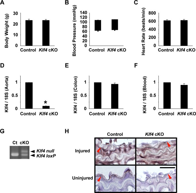
Background: Krüppel-like factor 4 (Klf4) is involved in a variety of cellular functions by activating or repressing the transcription of multiple genes. Results of previous studies showed that tamoxifen-inducible global deletion of the Klf4 gene in mice accelerated neointimal formation following vascular injury, in part via enhanced proliferation of smooth muscle cells (SMCs). Because Klf4 is also expressed in non-SMCs including endothelial cells (ECs), we determined if Tie2 promoter-dependent deletion of Klf4 in ECs and hematopoietic cells affected injury-induced neointimal formation.

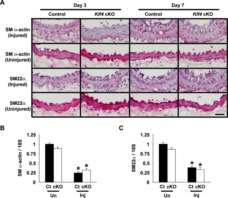
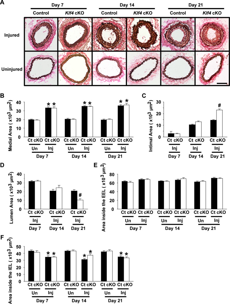
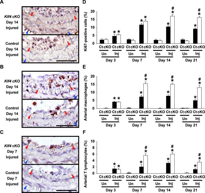
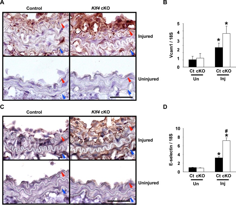
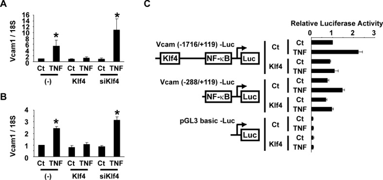
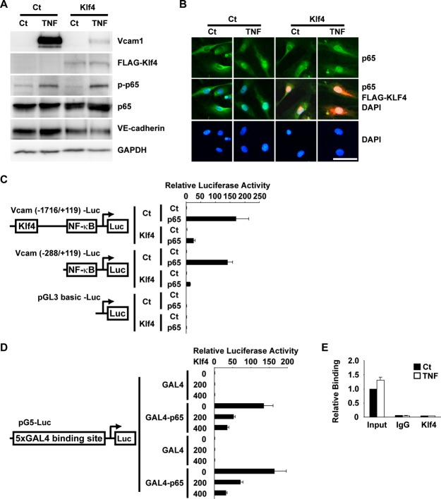
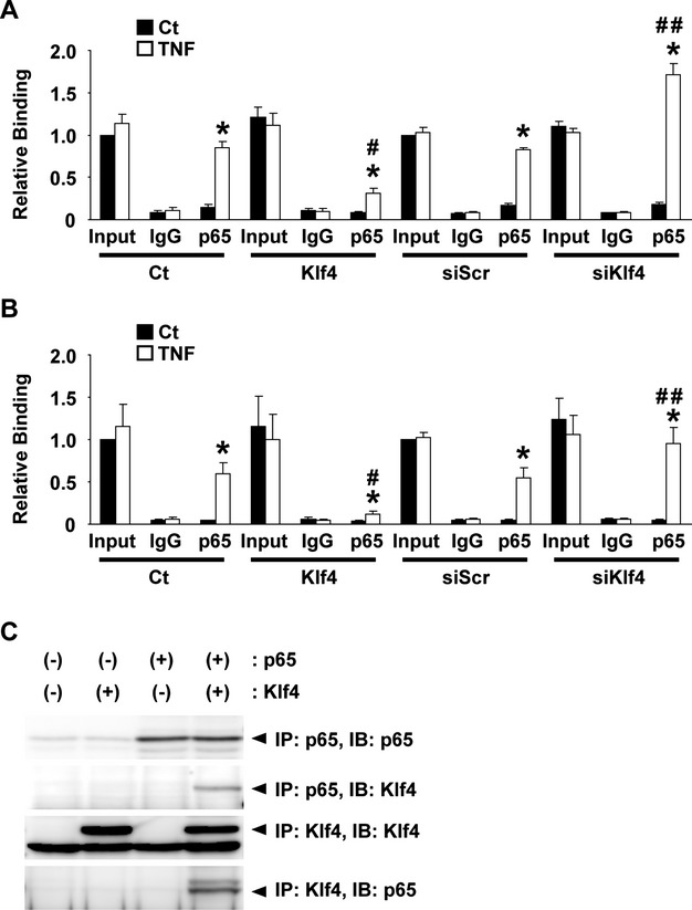
Methods and results: Klf4 conditional knockout (cKO) mice were generated by breeding Tie2-Cre mice and Klf4 floxed mice, and their phenotype was analyzed after carotid ligation injury. Results showed that injury-induced repression of SMC differentiation markers was unaffected by Tie2 promoter-dependent Klf4 deletion. However, of interest, neointimal formation was significantly enhanced in Klf4-cKO mice 21 days following carotid injury. Moreover, Klf4-cKO mice exhibited an augmented proliferation rate, enhanced accumulation of macrophages and T lymphocytes, and elevated expression of cell adhesion molecules including vascular cell adhesion molecule-1 (Vcam1) and E-selectin in injured arteries. Mechanistic analyses in cultured ECs revealed that Klf4 inhibited tumor necrosis factor-α-induced expression of Vcam1 through blocking the binding of nuclear factor-κB to the Vcam1 promoter.

Conclusions: These results provide evidence that Klf4 in non-SMCs such as ECs regulates neointimal formation by repressing arterial inflammation following vascular injury.

Keywords: cell adhesion molecules; endothelium; krüppel‐like factors; smooth muscle; vascular injury.


Figures

Figure 1.
Figure 1.
Klf4‐cKO mice grew up normally to the adulthood. A through C, Body weight (A), systolic and diastolic blood pressure (B), and heart rate (C) of Klf4‐cKO mice and control mice were measured at 11 weeks of age (n=12 for each genotype). B, The tops and bottoms of the squares indicate systolic and diastolic blood pressure, respectively. D through F, Expression of Klf4 in the aorta (D), colon (E), and blood (F) was determined by real‐time RT‐PCR in Klf4‐KO mice and control mice (n=4 for each genotype). *P<0.05 compared with control mice. G, Recombination of the Klf4loxP allele was examined by PCR in the blood of Klf4‐cKO mice (cKO) and control mice (Ct). H, Klf4 expression was examined by immunohistochemistry in the carotid arteries of Klf4‐cKO mice and control mice 3 days after injury. Klf4 expression was visualized by diaminobenzidine, and sections were counterstained with hematoxylin. Representative pictures are shown from 6 mice analyzed per genotype. Bar: 50 μm. Red arrowheads indicate the IEL. cKO indicates conditional knockout; IEL, internal elastic lamina; RT‐PCR, reverse‐transcription polymerase chain reaction.
Figure 2.
Figure 2.
Expression of SMC differentiation markers was decreased in Klf4‐cKO mice after vascular injury. A, Expression of SM α‐actin and SM22α was examined by immunohistochemistry in the carotid arteries of Klf4‐cKO mice and control mice on day 3 and day 7 after ligation injury. Expression of SM α‐actin and SM22α was visualized by Vector Red alkaline phosphatase, and sections were counterstained with hematoxylin. Representative pictures are shown from 6 mice analyzed per genotype and from each time. Bar: 50 μm. B and C, Expression of SM α‐actin (B) and SM22α (C) was determined by real‐time RT‐PCR in the injured (Inj) and uninjured (Un) carotid arteries of Klf4‐cKO mice and control mice on day 7 after injury (n=5 per each genotype). *P<0.05 compared with uninjured arteries. cKO indicates conditional knockout; Ct, control; RT‐PCR, reverse‐transcription polymerase chain reaction; SMC, smooth muscle cell.
Figure 3.
Figure 3.
Injury‐induced neointimal formation was enhanced in Klf4‐cKO mice. Verhoeff‐van Gieson staining (A) was performed in the injured and uninjured carotid arteries of EC‐specific Klf4‐knockout mice and control mice 7, 14, and 21 days after injury. Areas of the media (B), the intima (C), and the lumen (D), as well as the areas within the EEL (E) and the IEL (F), were quantified (n=6 per genotype and each time). Bar: 100 μm. *P<0.05 compared with uninjured arteries; #P<0.05 compared with corresponding control mice. cKO indicates conditional knockout; Ct, control; EC, endothelial cell; EEL, external elastic lamina; IEL, internal elastic lamina; Inj, injured; Un, uninjured.
Figure 4.
Figure 4.
Accumulation of macrophages and T lymphocytes was enhanced in the injured arteries of Klf4‐cKO mice. Left: Representative pictures of injured carotid arteries for Ki67 staining on day 14 (A), Mac2 staining on day 14 (B), and CD3ε staining on day 7 (C) after ligation injury in Klf4‐cKO mice and control mice are shown. Staining for Ki67, Mac2, and CD3ε was visualized by diaminobenzidine, and sections were counterstained with hematoxylin. Bars: 50 μm. Red and blue arrowheads indicate IEL and EEL, respectively. Right: The ratios of Ki67‐positive cells (D), Mac2‐positive cells (E), and CD3ε‐positive cells (F) in the injured and uninjured carotid arteries were calculated in Klf4‐cKO mice and control mice (n=6 per genotype and each time). *P<0.05 compared with uninjured arteries; #P<0.05 compared with corresponding control mice. cKO indicates conditional knockout; Ct, control; EEL, external elastic lamina; IEL, internal elastic lamina; Inj, injured; Un, uninjured.
Figure 5.
Figure 5.
Induction of cell adhesion molecules was augmented in Klf4‐cKO mice following injury. A and C, Representative pictures for staining of Vcam1 (A) and E‐selectin (C) on day 14 after injury in Klf4‐cKO mice and control mice are shown (n=6 per genotype). Staining was visualized by diaminobenzidine, and sections were counterstained with hematoxylin. Bar: 50 μm. Red and blue arrowheads indicate IEL and EEL, respectively. B and D, Expression of Vcam1 (B) and E‐selectin (D) was determined by real‐time RT‐PCR in the carotid arteries of Klf4‐cKO mice and control mice on day 7 after ligation injury (n=5 per each genotype). *P<0.05 compared with uninjured arteries; #P<0.05 compared with corresponding control mice. cKO indicates conditional knockout; Ct, control; EEL, external elastic lamina; IEL, internal elastic lamina; Inj, injured; RT‐PCR, reverse‐transcription polymerase chain reaction; Un, uninjured; Vcam1, vascular cell adhesion molecule‐1.
Figure 6.
Figure 6.
Klf4 attenuated TNF‐α‐induced expression of Vcam1 in ECs. A and B, HUVECs (A) and human aortic ECs (B) were transfected with Klf4 expression plasmid or siRNA for Klf4 (siKlf4), and then treated with TNF‐α. Expression of Vcam1 was determined by real‐time RT‐PCR (n=4). *P<0.05 compared with cells without TNF‐α treatment. C, HUVECs were transfected with the Vcam1 (−1716/+119) promoter‐luciferase construct, the Vcam1 (−288/+119) promoter‐luciferase construct, or pGL3‐basic plasmid with Klf4 expression plasmid. One day after transfection, HUVECs were treated with TNF‐α for an additional 24 hours. Luciferase activity was measured and normalized to protein content (n=4). Ct indicates control; ECs, endothelial cells; HUVECs, human umbilical vein ECs; NF‐κB, nuclear factor‐κB; RT‐PCR, reverse‐transcription polymerase chain reaction; TNF‐α, tumor necrosis factor–α; Vcam1, vascular cell adhesion molecule‐1.
Figure 7.
Figure 7.
Klf4 attenuated TNF‐α‐induced expression of Vcam1 without binding to the Vcam1 promoter. A, HUVECs were transfected with FLAG‐tagged Klf4 expression plasmid, and then treated with TNF‐α. Expression of Vcam1, FLAG, phosphorylated p65 (p‐p65), p65, VE‐cadherin, and GAPDH was examined by Western blotting (n=4). B, HUVECs were transfected with the expression plasmid for FLAG‐tagged Klf4, and treated with TNF‐α. Top: Intracellular localization of endogenous p65 (green) was determined by anti‐p65 antibody. Middle: Merged images for endogenous p65 (green), FLAG‐tagged Klf4 (red), and DAPI nuclear staining (blue) are shown. Bottom: DAPI nuclear staining (blue) is shown. Bar: 50 μm. C, Human aortic ECs were transfected with the Vcam1 (−1716/+119) promoter‐luciferase construct, the Vcam1 (−288/+119) promoter‐luciferase construct, or pGL3‐basic plasmid with Klf4 expression plasmid and p65 expression plasmid. Luciferase activity was measured and normalized to protein content (n=4). D, Human aortic ECs were transfected with Klf4 expression plasmid (0, 200, or 400 ng), expression plasmid for GAL4 or GAL4‐p65 fusion protein, and 5xGAL4 binding site–containing luciferase construct. Luciferase activity was measured and normalized to protein content (n=4). E, HUVECs were treated with TNF‐α. Association of Klf4 with the Vcam1 promoter was determined by chromatin immunoprecipitation assays (n=4). Ct indicates control; DAPI, 4′, 6‐diamidino‐2‐phenylindole; ECs, endothelial cells; GAPDH; glyceraldehyde‐3‐phosphate dehydrogenase; IgG, immunoglobulin G; HUVECs, human umbilical vein ECs; TNF‐α, tumor necrosis factor–α; Vcam1, vascular cell adhesion molecule‐1.
Figure 8.
Figure 8.
Klf4 attenuated TNF‐α‐induced expression of Vcam1 by blocking the binding of NF‐κB to the Vcam1 promoter. A and B, HUVECs were transfected with empty expression plasmid, Klf4 expression plasmid, siRNA for Klf4 (siKlf4), or siRNA for scrambled sequence (siScr) and treated with TNF‐α. Association of p65 with the Vcam1 promoter (A) and the E‐selectin promoter (B) was determined by chromatin immunoprecipitation assays (n=4). *P<0.05 compared with cells without TNF‐α treatment; #P<0.05 compared with cells without transfecting Klf4 expression plasmid; ##P<0.05 compared wutg cells without transfecting siKlf4. C, Expression plasmids for p65 and FLAG‐Klf4 were transfected into COS7 cells. Coimmunoprecipitation assays were performed with anti‐p65 antibody and anti‐FLAG antibody. Ct indicates control; HUVECs, human umbilical vein endothelial cells; IB, immunoblot; IP, immunoprecipitation; TNF‐α, tumor necrosis factor–α; Vcam1, vascular cell adhesion molecule‐1.

Comment in

  • Endothelial KLF4: crippling vascular injury?
    Tugal D, Jain MK, Simon DI. Tugal D, et al. J Am Heart Assoc. 2014 Feb 26;3(1):e000769. doi: 10.1161/JAHA.113.000769. J Am Heart Assoc. 2014. PMID: 24572258 Free PMC article. No abstract available.

Similar articles

Cited by

References

    1. Libby P. Inflammation in atherosclerosis. Nature. 2002; 420:868-874 - PubMed
    1. Segre JA, Bauer C, Fuchs E. Klf4 is a transcription factor required for establishing the barrier function of the skin. Nat Genet. 1999; 22:356-360 - PubMed
    1. Katz JP, Perreault N, Goldstein BG, Lee CS, Labosky PA, Yang VW, Kaestner KH. The zinc‐finger transcription factor Klf4 is required for terminal differentiation of goblet cells in the colon. Development. 2002; 129:2619-2628 - PMC - PubMed
    1. Katz JP, Perreault N, Goldstein BG, Actman L, McNally SR, Silberg DG, Furth EE, Kaestner KH. Loss of Klf4 in mice causes altered proliferation and differentiation and precancerous changes in the adult stomach. Gastroenterology. 2005; 128:935-945 - PubMed
    1. Swamynathan SK, Katz JP, Kaestner KH, Ashery‐Padan R, Crawford MA, Piatigorsky J. Conditional deletion of the mouse Klf4 gene results in corneal epithelial fragility, stromal edema, and loss of conjunctival goblet cells. Mol Cell Biol. 2007; 27:182-194 - PMC - PubMed

Publication types

MeSH terms

LinkOut - more resources