

Angiokeratoma circumscriptum in a young male
- PMID: 24470669
- PMCID: PMC3884937
- DOI: 10.4103/0019-5154.123514
Angiokeratoma circumscriptum in a young male
Abstract
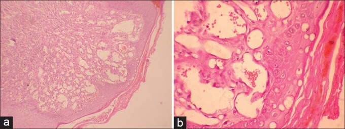
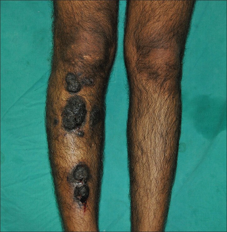
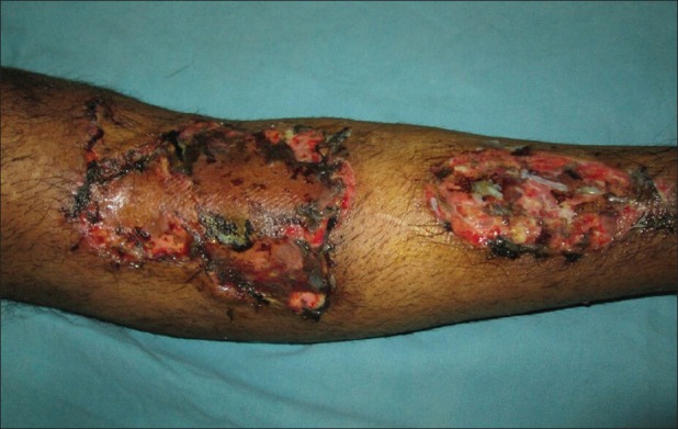
A 20-year-old male presented with multiple eruptions on his right leg since birth; these bled and were painful on trivial trauma. Examination revealed dark brown, hyperkeratotic, indurated, verrucous linear plaques with irregular borders. Histopathological evidence of hyperkeratosis, acanthosis, and extensive vascular proliferation in papillary dermis confirmed clinical suspicion of angiokeratoma circumscriptum (AKC). Excision and skin grafting yielded a cosmetically favorable outcome. Angiokeratomas, first described by Mibeli in 1889, are a group of vascular ectasias involving the papillary dermis. Angiokeratomas are more common in males; however, AKC-the rarest of its five variants-exhibits a female preponderance (F:M:3:1). AKC is an extremely rare nevoid disorder, only 100 of its cases having been reported in the world literature until 2006. Herein, we have reported a typical case of AKC in a young male that was previously misdiagnosed, and the patient wrongly counseled about the likelihood of its spontaneous regression.
Keywords: Angiokeratoma circumscriptum; vascular malformation; verrucous hemangioma.
Conflict of interest statement
Figures
Similar articles
-
Angiokeratoma Circumscriptum Naeviforme Presenting as a Dark Warty Plaque on the Leg.Acta Dermatovenerol Croat. 2021 Dec;29(3):169-170. Acta Dermatovenerol Croat. 2021. PMID: 34990348
-
Angiokeratoma circumscriptum neviforme: An entity, few and far between.Indian Dermatol Online J. 2014 Oct;5(4):472-4. doi: 10.4103/2229-5178.142503. Indian Dermatol Online J. 2014. PMID: 25396132 Free PMC article.
-
Angiokeratoma Circumscriptum.2023 Jul 17. In: StatPearls [Internet]. Treasure Island (FL): StatPearls Publishing; 2025 Jan–. 2023 Jul 17. In: StatPearls [Internet]. Treasure Island (FL): StatPearls Publishing; 2025 Jan–. PMID: 31747176 Free Books & Documents.
-
Angiokeratomas Scroti Associated with Angiokeratomas of the Eyelids: Coincidence or One Entity? A Case Report and Review of the Literature.Dermatology. 2015;231(3):213-6. doi: 10.1159/000435810. Epub 2015 Jul 24. Dermatology. 2015. PMID: 26228617 Review.
-
Angiokeratomas: an update.Dermatology. 1996;193(4):275-82. doi: 10.1159/000246270. Dermatology. 1996. PMID: 8993949 Review.
Cited by
-
Solitary Angiokeratoma: Report of Two Uncommon Cases.J Clin Diagn Res. 2015 May;9(5):WD01-2. doi: 10.7860/JCDR/2015/12163.5946. Epub 2015 May 1. J Clin Diagn Res. 2015. PMID: 26155544 Free PMC article.
-
Localized Angiokeratomas in healthy adolescence responded to topical Timolol.Int J Health Sci (Qassim). 2024 Sep-Oct;18(5):59-61. Int J Health Sci (Qassim). 2024. PMID: 39282129 Free PMC article.
-
Solitary Angiokeratoma in a Young Man: A Rare Case Report.Cureus. 2023 Apr 18;15(4):e37790. doi: 10.7759/cureus.37790. eCollection 2023 Apr. Cureus. 2023. PMID: 37213968 Free PMC article.
References
-
- Enjolras O. Vascular malformations. In: Bolognia JL, Jorizzo JL, Rapini RP, editors. Dermatology. 2nd ed. Vol. 2. Spain: Elsevier limited; 2008. pp. 1581–96.
-
- Jansen T, Bechara FG, Altmeyer P. Angiokeratomas: Symptoms, diagnostics and therapy. TKT Europe-5S AB: Danderyd. 2004
-
- Fabry J. Úber einen Fall von Angiokeratoma circumscriptum am linken Oberschenkel [A case of angiokeratoma circumscriptum] Dermatol Z. 1915;22:1–4.
-
- Mittal R, Aggarwal A, Srivastava G. Angiokeratoma circumscriptum: A case report and review of the literature. Int J Dermatol. 2005;44:1031–4. - PubMed
-
- Schiller PI, Itin PH. Angiokeratomas: An update. Dermatology. 1996;193:275–82. - PubMed
LinkOut - more resources
Full Text Sources
Other Literature Sources