. 2014 Jan 28:15:3.
doi: 10.1186/1471-2199-15-3.

Spdef deletion rescues the crypt cell proliferation defect in conditional Gata6 null mouse small intestine

Affiliations

Spdef deletion rescues the crypt cell proliferation defect in conditional Gata6 null mouse small intestine

Boaz E Aronson et al. BMC Mol Biol. .

Abstract

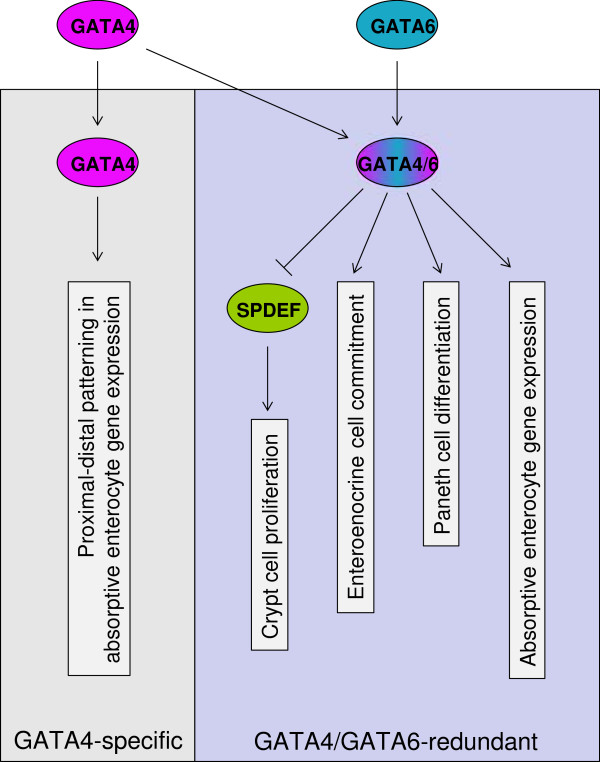
Background: GATA transcription factors are essential for self-renewal of the small intestinal epithelium. Gata4 is expressed in the proximal 85% of small intestine while Gata6 is expressed throughout the length of small intestine. Deletion of intestinal Gata4 and Gata6 results in an altered proliferation/differentiation phenotype, and an up-regulation of SAM pointed domain containing ETS transcription factor (Spdef), a transcription factor recently shown to act as a tumor suppressor. The goal of this study is to determine to what extent SPDEF mediates the downstream functions of GATA4/GATA6 in the small intestine. The hypothesis to be tested is that intestinal GATA4/GATA6 functions through SPDEF by repressing Spdef gene expression. To test this hypothesis, we defined the functions most likely regulated by the overlapping GATA6/SPDEF target gene set in mouse intestine, delineated the relationship between GATA6 chromatin occupancy and Spdef gene regulation in Caco-2 cells, and determined the extent to which prevention of Spdef up-regulation by Spdef knockout rescues the GATA6 phenotype in conditional Gata6 knockout mouse ileum.

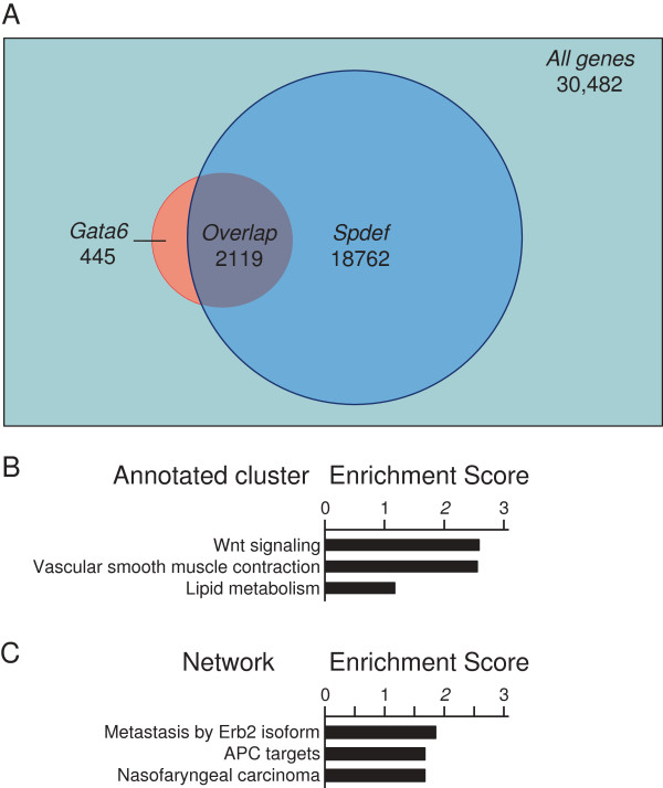
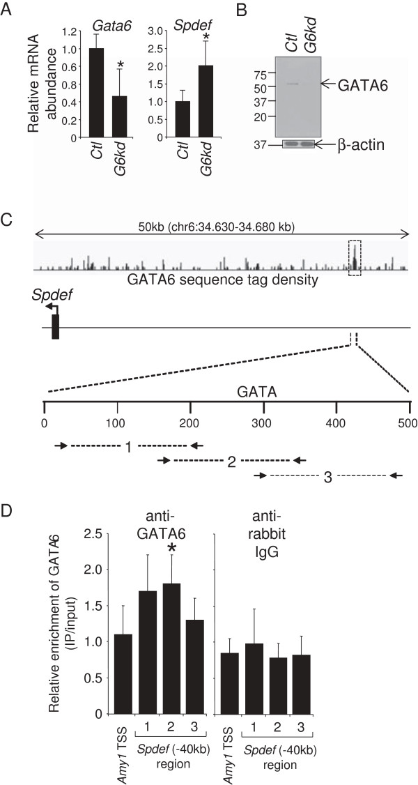
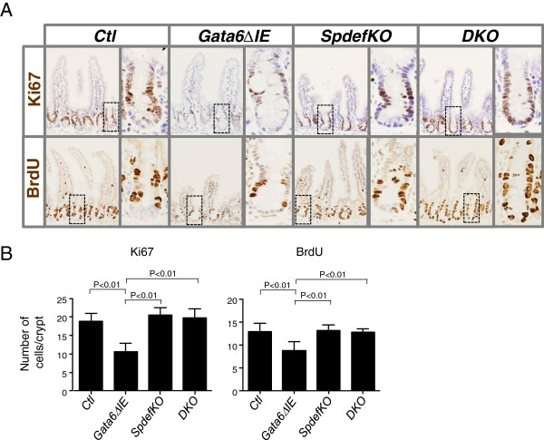
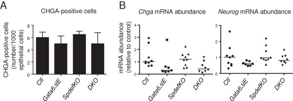
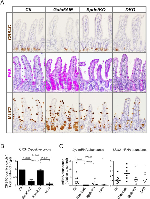
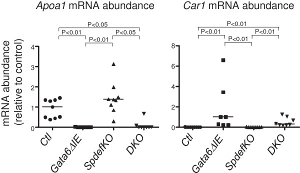
Results: Using publicly available profiling data, we found that 83% of GATA6-regulated genes are also regulated by SPDEF, and that proliferation/cancer is the function most likely to be modulated by this overlapping gene set. In human Caco-2 cells, GATA6 knockdown results in an up-regulation of Spdef gene expression, modeling our mouse Gata6 knockout data. GATA6 occupies a genetic locus located 40 kb upstream of the Spdef transcription start site, consistent with direct regulation of Spdef gene expression by GATA6. Prevention of Spdef up-regulation in conditional Gata6 knockout mouse ileum by the additional deletion of Spdef rescued the crypt cell proliferation defect, but had little effect on altered lineage differentiation or absorptive enterocytes gene expression.

Conclusion: SPDEF is a key, immediate downstream effecter of the crypt cell proliferation function of GATA4/GATA6 in the small intestine.


Figures

Figure 1
Figure 1
Overlap of genes in small intestine modulated by conditional Gata6 or Spdef knockout. (A) Publicly available gene profiling data from conditional Gata6 (GSE22416) and Spdef (GSE14892) knockout mouse intestine show a significant overlap. (B) Functional annotation clustering of pathways shows that the top-enriched cluster, in overlapping Gata6 and Spdef genes, contains the Wnt pathway as its main function (ES score 2.58). (C) GSEA analysis shows, in the overlapping Gata6 and Spdef gene segment, APC targets as the second most highly enriched cluster (ES score 1.57).
Figure 2
Figure 2
GATA6 regulates Spdef expression and occupies an enhancer in the Spdef 5′-flanking region in Caco-2 cells. (A) Quantitative RT-PCR analysis showing reduced expression of Gata6 mRNA, and enhanced expression of Spdef mRNA in Caco-2 cells infected with a lentivirus vector expressing an shRNA for human Gata6 mRNA (G6kd) (mean ± SD, n = 5, *P < 0.05). An shRNA vector for GFP was used as a control (Ctl). (B) Western analysis showing reduced abundance of GATA6 in G6kd Caco-2 cells. Β-actin was used as an internal loading control. (C) Schematic representation of the human Spdef 5′-flanking region showing GATA6 occupancy at a locus ~40 kb upstream of the transcription start site (TSS). GATA6 sequence tag density is shown as a 'wiggle file’, and a statistically significant GATA6-occupied locus was defined by MACS peak analysis [29] (dotted box). (D) ChIP assays on chromatin obtained from Caco-2 cells using a GATA6 antibody and three sets of overlapping primers centered on the GATA motif showing increased GATA6 occupancy (mean ± SD, n = 4, *P < 0.05). The salivary amylase-α1a (Amy-1) TSS was used as a negative control. ChIP assays using anti-rabbit IgG was used as a control for non-specific immunoprecipitation and primer efficiency.
Figure 3
Figure 3
Crypt cell proliferation is decreased in Gata6ΔIE mice. (A) Immunostaining in ileum for the proliferation markers Ki67 (top row) and BrdU (bottom row). (B) Quantification of Ki67- and BrdU-positive cells/crypt in each group. Cells were counted as described in Methods. Gata6ΔIE mice had significantly fewer Ki67- or BrdU-positive cells than each of the other three groups (P < 0.01 in each case), consistent with a decrease in crypt cell proliferation.
Figure 4
Figure 4
Enteroendocrine cell allocation is decreased in Gata6ΔIE and DKO mice. (A) Quantification of CHGA-positive cells in each group. Sections of ileum were stained for CHGA, and the number of positive cells/1000 epithelial cells was determined as described in Methods. (B)Chga and Neurog3 mRNA abundance in each group. Gata6ΔIE and DKO mice had generally lower numbers of CHGA-positive cells and Chga and Neurog3 mRNA abundances than Ctl mice, consistent with a decrease in enteroendocrine lineage allocation.
Figure 5
Figure 5
Paneth cells are decreased in Gata6ΔIE and DKO mice. (A) Immunostaining in ileum for CRS4C (first row) and MUC2 (third row), and chemical staining using the PAS reaction (second row). (B) Quantification of CRS4C-positive cells in each group. Sections of ileum were stained for CRS4C, and the number of positive cells/crypt cross section was determined as described in Methods. (C)Lyz and Muc2 mRNA abundance in each group. Gata6ΔIE and DKO mice had significantly lower numbers of CRS4C-positive cells and Lyz mRNA abundance than Ctl mice, consistent with a decrease in mature Paneth cells.
Figure 6
Figure 6
Absorptive enterocyte gene expression is altered in Gata6ΔIE and DKO mice. Gata6ΔIE and DKO mice had significantly lower Apoa1, and significantly higher Car1 mRNA abundances than Ctl mice.
Figure 7
Figure 7
Model for the known GATA pathways in mature small intestine, and the placement of SPDEF within those pathways. GATA4 (purple), but not GATA6 (blue), activates and represses a subset of absorptive enterocyte genes (GATA4-specific), whereas either GATA4 or GATA6 (purple/blue) promote crypt cell proliferation, enteroendocrine lineage commitment, Paneth cell differentiation, and absorptive enterocyte gene expression (GATA4/GATA6-redundant). GATA4/GATA6 promotes the crypt cell proliferation function by repressing the gene encoding SPDEF (green).

References

    1. Noah TK, Donahue B, Shroyer NF. Intestinal development and differentiation. Exp Cell Res. 2011;317(19):2702–2710. doi: 10.1016/j.yexcr.2011.09.006. - DOI - PMC - PubMed
    1. Gerbe F, van Es JH, Makrini L, Brulin B, Mellitzer G, Robine S, Romagnolo B, Shroyer NF, Bourgaux JF, Pignodel C. et al.Distinct ATOH1 and Neurog3 requirements define tuft cells as a new secretory cell type in the intestinal epithelium. J Cell Biol. 2011;192(5):767–780. doi: 10.1083/jcb.201010127. - DOI - PMC - PubMed
    1. Yeung TM, Chia LA, Kosinski CM, Kuo CJ. Regulation of self-renewal and differentiation by the intestinal stem cell niche. Cell Mol Life Sci. 2011;68(15):2513–2523. doi: 10.1007/s00018-011-0687-5. - DOI - PMC - PubMed
    1. Beuling E, Baffour-Awuah NY, Stapleton KA, Aronson BE, Noah TK, Shroyer NF, Duncan SA, Fleet JC, Krasinski SD. GATA factors regulate proliferation, differentiation, and gene expression in small intestine of mature mice. Gastroenterology. 2011;140(4):1219–1229. doi: 10.1053/j.gastro.2011.01.033. e1211-1212. - DOI - PMC - PubMed
    1. Bosse T, Piaseckyj CM, Burghard E, Fialkovich JJ, Rajagopal S, Pu WT, Krasinski SD. Gata4 is essential for the maintenance of jejunal-ileal identities in the adult mouse small intestine. Mol Cell Biol. 2006;26(23):9060–9070. doi: 10.1128/MCB.00124-06. - DOI - PMC - PubMed

Publication types

Substances

LinkOut - more resources