. 2014 Jan 29:10:8.
doi: 10.1186/1744-8069-10-8.

Impaired neuropathic pain and preserved acute pain in rats overexpressing voltage-gated potassium channel subunit Kv1.2 in primary afferent neurons

Affiliations

Impaired neuropathic pain and preserved acute pain in rats overexpressing voltage-gated potassium channel subunit Kv1.2 in primary afferent neurons

Longchang Fan et al. Mol Pain. .

Abstract

Voltage-gated potassium (Kv) channels are critical in controlling neuronal excitability and are involved in the induction of neuropathic pain. Therefore, Kv channels might be potential targets for prevention and/or treatment of this disorder. We reported here that a majority of dorsal root ganglion (DRG) neurons were positive for Kv channel alpha subunit Kv1.2. Most of them were large and medium, although there was a variety of sizes. Peripheral nerve injury caused by lumbar (L)5 spinal nerve ligation (SNL) produced a time-dependent reduction in the number of Kv1.2-positive neurons in the ipsilateral L5 DRG, but not in the contralateral L5 DRG. Such reduction was also observed in the ipsilateral L5 DRG on day 7 after sciatic nerve axotomy. Rescuing nerve injury-induced reduction of Kv1.2 in the injured L5 DRG attenuated the development and maintenance of SNL-induced pain hypersensitivity without affecting acute pain and locomotor function. This effect might be attributed to the prevention of SNL-induced upregulation of endogenous Kv1.2 antisense RNA, in addition to the increase in Kv1.2 protein expression, in the injured DRG. Our findings suggest that Kv1.2 may be a novel potential target for preventing and/or treating neuropathic pain.


Figures
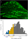
Figure 1
Figure 1
Kv1.2 expression predominantly in the large and medium DRG neurons of normal rats. (A) A representative image from immunohistochemical staining showing the distribution of Kv1.2-positive neurons. Scale bar: 200 μm. (B) Histogram showing the distribution of Kv1.2-positive somata.
Figure 2
Figure 2
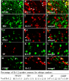
Co-localization of Kv1.2 with NF200, IB4, P2X3, SP, and CGRP in DRG neurons. Double immunohistochemical staining shows that approximately 80.3% of Kv1.2-labeled neurons are positive for NF200 (A–C), 2.45% for IB4 (D–F), 11.11% for P2X3 (G–I), 3.97% for SP (J–L), and 10.7% for CGRP (M–O). Arrows: double-labeled neurons. Scale bar: 70 μm.
Figure 3
Figure 3
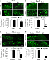
Time-dependent decreases in the number of Kv1.2-positive neurons in the injured DRG after peripheral nerve injury. Representative Kv1.2 immunohistochemical staining (Top) and statistical summary (Bottom) of the percentage of Kv1.2-positive neurons in the contralateral (Contra) and ipsilateral (Ipsi) L5 DRGs on days 3 (A), 7 (B), and 14 (C) after L5 spinal nerve ligation (SNL) or sham surgery and on day 7 after axotomy or sham surgery (D). Scale bar: 100 μm. * P < 0.05, ** P < 0.01 compared to the number of Kv1.2-positive neurons on the contralateral side of sham-operated rats.
Figure 4
Figure 4
In vitro expression and current of Kv1.2 in HEK-293 cells transfected with Kv1.2 vector. (A) Representative Western blot showing Kv1.2 expression at the expected size in HEK-293 cells transfected with full-length Kv1.2 vector. β-actin was used as a loading control. (B) Representative traces of Kv1.2 current recorded by whole-cell voltage patch clamp in HEK-293 cells transfected with full-length Kv1.2 vector before or after bath perfusion of 100 nM maurotoxin (MTX). (C) Current–voltage curves of HEK-293 cells transfected with full-length Kv1.2 before or after treatment with 100 nM MTX. The current density was plotted against each step testing voltage. n = 5 cells.
Figure 5
Figure 5
Overexpressing Kv1.2 in the injured DRG mitigates neuropathic pain. Kv1.2 was delivered into the injured DRG through microinjection of AAV5-Kv1.2. AAV5-EGFP was used as a control. (A-C) Behavioral tests show the effect of overexpressing Kv1.2 on the development of spinal nerve ligation (SNL)-induced pain hypersensitivities. Ipsilateral and contralateral paw withdrawal responses to mechanical (A), cold (B), and thermal (C) stimuli at the times shown before and after SNL. n = 5/group. *P < 0.05, **P < 0.01 vs. the ipsilateral side of the AAV5-EGFP-treated group at the corresponding time point. (D-F) Behavioral tests show the effect of overexpressing Kv1.2 on the maintenance of SNL-induced pain hypersensitivities. Ipsilateral and contralateral paw withdrawal responses to mechanical (D), cold (E), and thermal (F) stimuli at the times shown before and after SNL. n = 5/group. *P < 0.05, **P < 0.01 vs. the ipsilateral side of the AAV5-EGFP-treated group at the corresponding time point.
Figure 6
Figure 6
Overexpressing Kv1.2 in the DRG does not alter capsaicin-induced acute nociceptive pain. (A) The duration of capsaicin-induced paw licking/lifting was recorded for 5 minutes after capsaicin injection in the AAV5-EGFP-injected and AAV5-Kv1.2-injected groups. n = 5/group. (B-D) Paw withdrawal responses of AAV5-EGFP-injected and AAV5-Kv1.2-injected rats to mechanical (B), heat (C), and cold (D) stimuli on the ipsilateral side prior to viral injection (baseline), before capsaicin injection (Pre-Cap; 5 weeks after viral injection), and 30 minutes after capsaicin injection (Post-Cap). n = 5/group.
Figure 7
Figure 7
Overexpressing Kv1.2 rescues SNL-induced Kv1.2 downregulation and blocks SNL-induced Kv1.2 AS RNA upregulation in the injured DRG. (A) Quantitative RT-PCR shows Kv1.2 AS RNA, Kv1.2 mRNA, and Kv1.4 mRNA expression in the ipsilateral and contralateral L5 DRGs on day 14 after spinal nerve ligation (SNL) or sham surgery in the AAV5-EGFP-injected and AAV5-Kv1.2-injected groups. n = 12 rats/group. **P < 0.01 vs. the corresponding AAV5-EGFP-injected group after sham surgery; ##P < 0.01 vs. the corresponding AAV5-EGFP-injected group after SNL. (B) Western blot analysis shows Kv1.2 and Kv1.4 protein expression in the ipsilateral (Ipsi) and contralateral (Con) L5 DRGs on day 14 after SNL in the AAV5-EGFP-injected and AAV5-Kv1.2-injected groups. n = 8 rats/group. **P < 0.01 vs. the contralateral side of the AAV5-EGFP-injected group; #P < 0.05 vs. the ipsilateral side of the AAV5-EGFP-injected group. (C) Western blot analysis shows Kv1.2 and Kv1.4 protein expression in the ipsilateral and contralateral L5 DRGs on day 14 after sham surgery in the AAV5-EGFP-injected and AAV5-Kv1.2-injected groups. n = 8 rats/group. **P < 0.01 vs. the contralateral side of the AAV5-EGFP-injected group.

References

    1. Campbell JN, Meyer RA. Mechanisms of neuropathic pain. Neuron. 2006;52:77–92. doi: 10.1016/j.neuron.2006.09.021. - DOI - PMC - PubMed
    1. Baron R, Binder A, Wasner G. Neuropathic pain: diagnosis, pathophysiological mechanisms, and treatment. Lancet Neurol. 2010;9:807–819. doi: 10.1016/S1474-4422(10)70143-5. - DOI - PubMed
    1. Finnerup NB, Sindrup SH, Jensen TS. The evidence for pharmacological treatment of neuropathic pain. Pain. 2010;150:573–581. doi: 10.1016/j.pain.2010.06.019. - DOI - PubMed
    1. Ji RR, Woolf CJ. Neuronal plasticity and signal transduction in nociceptive neurons: implications for the initiation and maintenance of pathological pain. Neurobiol Dis. 2001;8:1–10. doi: 10.1006/nbdi.2000.0360. - DOI - PubMed
    1. Latremoliere A, Woolf CJ. Central sensitization: a generator of pain hypersensitivity by central neural plasticity. J Pain. 2009;10:895–926. doi: 10.1016/j.jpain.2009.06.012. - DOI - PMC - PubMed

Publication types

MeSH terms