. 2014 Sep;7(5):1139-50.
doi: 10.1038/mi.2014.1. Epub 2014 Jan 29.

Inflammasome activation has an important role in the development of spontaneous colitis

Affiliations

Inflammasome activation has an important role in the development of spontaneous colitis

J Zhang et al. Mucosal Immunol. 2014 Sep.

Abstract

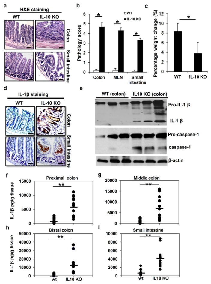
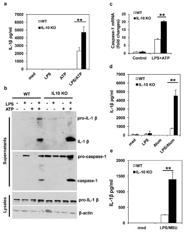
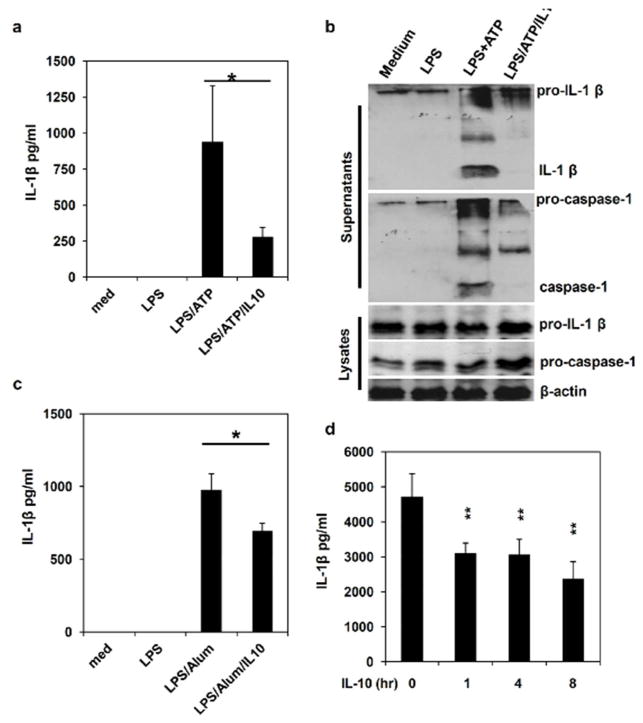
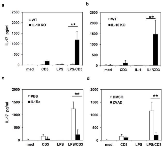
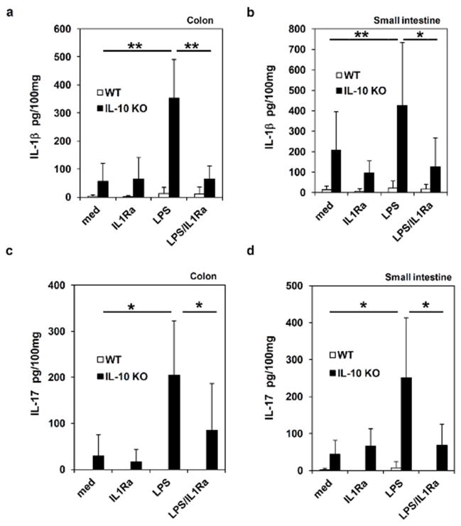
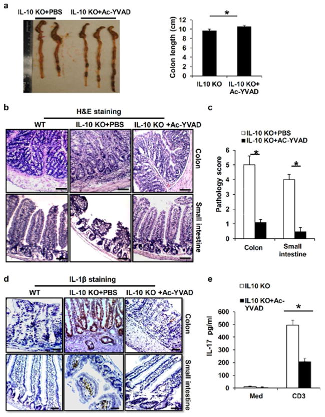
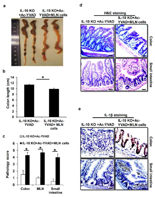
Inflammatory bowel disease (IBD) is characterized for dysregulated intestinal inflammation. Conflicting reports have shown that activation of inflammasome could promote or decrease intestinal inflammation in an acute colitis model, whereas the involvement of inflammasome activation in chronic colitis is poorly understood. In this study, we investigated the role of inflammasome activation in the development of chronic intestinal inflammation by utilizing interleukin-10 (IL-10) knockout (KO) mouse as an animal model, which develops chronic colitis resembling human IBD. We demonstrate the causative link between inflammasome activation and the development of chronic intestinal inflammation. Our results show that mature IL-1β protein levels were significantly increased in all colon sections from IL-10-deficient mice compared with that of wild-type mice. We found that inhibition of inflammasome activities with IL-1 receptor antagonist or caspase-1 inhibitors suppressed IL-1β and IL-17 production from inflamed colon explants. Furthermore, blocking inflammasome activation with caspase-1 inhibitor in vivo significantly ameliorated the spontaneous colitis in IL-10 KO mice. Taken together, these observations demonstrate that inflammasome activation promotes the development of chronic intestinal inflammation.


Conflict of interest statement

Competing interests: The authors have declared that no competing interests exist.

Figures

Figure 1
Figure 1. Spontaneous colitis is associated with increased levels of IL-1β protein in the intestinal system
(a) H&E staining of colon and small intestine sections from 16 weeks old WT and IL-10 KO mice. The marked colitis (mucosal ulceration, loss of villi, and decreased crypts) was observed in IL-10 KO mice. (b) Body weight and (c) pathologic inflammation scores in colon, small intestine and MLN from WT and IL-10 KO at 16 weeks of age. (b) Immunostaining of IL-1β in colon and small intestine sections from 16 weeks old WT and IL-10 KO mice. (e) Colon tissues from WT and IL-10 KO mice at age of 16 weeks were homogenized, and analyzed by Western Blot for pro-caspase-1, mature caspase-1 as well as pro-IL-1β and mature IL-1β proteins. Levels of IL-1β protein in homogenates of proximal (f), middle (g) and distal colon (h), as well as small intestinal (i) from 16 weeks old WT and IL-10 KO mice were analyzed by ELISA. Data represent mean of the pool of two independent experiments (n=12 for WT and IL-10 KO mice), and results for individual mouse are illustrated. Statistical significance is indicated, **P<0.01 (Student’s t test). Scale bar = 100μm.
Figure 2
Figure 2. IL-10 deficiency leads to increased NLRP3 inflammasome activation and IL-1β production
(a) WT and IL-10 deficient bone marrow-derived macrophages (BMDMs) were primed with LPS for 4 hours, and then were treated with ATP for 1 hour to induce inflammasome activation. The concentration of IL-1β in culture supernatants was assayed by ELISA. (b) WT and IL-10 deficient BMDMs were stimulated with LPS and ATP as in (a). Inflammasome activation was determined by Western Blot analysis of activated caspase-1 and processed IL-1β in the culture supernatants of BMDMs stimulated with LPS plus ATP. (c) Caspase-1 mRNA levels in BMDMs from WT and IL-10 KO mice treated as described in (a) were analyzed by RT-PCR. (d) and (e) WT and IL-10 deficient BMDMs were primed with LPS for 4 hours, then cells were treated with Alum or MSU crystal for 4 hours. The concentration of IL-1β in culture supernatants was assayed by ELISA. Representative data from one of at least three independent experiments are shown. **P<0.01 (Student’s t test).
Figure 3
Figure 3. IL-10 inhibits inflammasome activation and IL-1β production
(a) and (b) WT BMDMs were primed with LPS for 4 hours, then treated with ATP for inflammasome induction. 1 hour before adding ATP, cells were treated with 25 ng/ml of recombinant murine IL-10. The concentration of IL-1β in culture supernatants was assayed by ELISA. Caspase 1 and IL-1β processing and releasing were analyzed by Western Blot. (c) WT BMDMs were primed with LPS for 4 hours, then treated with Alum for 4 hours. 1 hour before adding Alum, cells were treated with 25 ng/ml of recombinant murine IL-10. The concentration of IL-1β in culture supernatants was assayed by ELISA. (d) 293T cells were reconstituted with inflammasome components (NLRP3, ASC, caspase 1 and pro-IL-1β plasmids). 36 hours after transfection, cells were pretreated with IL-10 for different time as indicated, and then with ATP for 1 hour. The IL-1β level in the supernatants was determined by ELISA. Results are reported as mean±SD of triplicate samples from one representative experiment of 3 independent experiments. Statistical significance is indicated, * p<0.05; ** p<0.01 (Student’s t test).
Figure 4
Figure 4. Inhibition of inflammasome activities leads to reduced IL-17 production from MLN cells of IL-10 KO mice
(a) and (b) MLN cells from WT and IL-10 KO mice were stimulated with anti-CD3 antibody in combination with LPS (100 ng/ml) or recombinant IL-1β (25 ng/ml) as indicated for 48 hours. The production of IL-17 was measured by ELISA. (c) and (d) MLN cells from IL-10 deficient mice were treated with anti-CD3 antibody or LPS in the presence of IL-1R antagonist (IL-1Ra) or caspase 1 inhibitor Z-VAD-FMK (ZVAD), and IL-17 production was measured 48 hours after treatment. Results are reported as mean ± SD of the pool of two experiments (5–6 mice per group). ** p<0.01 (Student’s t test).
Figure 5
Figure 5. Blocking IL-1R function inhibits IL-1β and IL-17 production from inflamed intestinal tissues of IL-10 KO mice
(a) and (b) Colon and small intestine isolated from WT and IL-10 KO mice were stimulated with LPS in the presence or absence of IL-1Ra. After 24 hours, IL-1β production was determined by ELISA. (c) and (d) Colon and small intestinal explants were treated with LPS or IL-1Ra, and IL-17 production was measured. Results are reported as mean ± SD of the pool of two experiments (5–6 mice per group). * p<0.05; ** p<0.01 (Student’s t test).
Figure 6
Figure 6. Inflammasome blockade ameliorates the spontaneous colitis in IL-10 deficient mice
(a) IL-10 KO mice at 16 weeks old were injected intraperitoneally with caspase-1specific inhibitor Ac-YVAD-cmk for 2 weeks. Macroscopic colon pathology and colon length from IL-10 KO mice treated with or without caspase 1 inhibitor were determined. (b) H&E-stained sections and (c) pathology scores of colon and small intestine sections from 16 weeks old IL-10 KO mice or IL-10 KO mice treated with Ac-YVAD-cmk for two weeks. (d) IL-1β immunostaining of colon and small intestine sections from 16 weeks old IL-10 KO mice treated with caspase-1 inhibitor as in (a). (e) MLN cells from IL-10 KO mice treated with or without caspase 1 inhibitor Ac-YVAD-cmk were stimulated with anti-CD3 antibody for 48 hours. The production of IL-17 was measured by ELISA. Data represent mean of the pool of two independent experiments (n=5–6 for treated or untreated IL-10 KO mice). Statistical significance is indicated, *P<0.05 (Student’s t test). Scale bar = 100μm.
Figure 7
Figure 7. IL-1β-induced IL-17 production enhanced colitis in IL-10 KO mice
(a) and (b) Macroscopic colon pathology and colon length from Ac-YVAD-cmk-treated IL-10 KO mice transferred with MLN cells from IL-10KO mice. (c) Pathologic inflammation scores in colon, small intestine and MLN from Ac-YVAD-cmk-injected IL-10 KO mice treated with or without IL-10 deficient MLN cells. (d) H&E staining and (e) IL-1β immunostaining of colon and small intestine sections from Ac-YVAD-cmk-treated IL-10 KO mice transferred with or without MLN cells of IL-10 KO mice. Data represent mean of the pool of two independent experiments (n=5 for MLN cells treated or untreated Ac-YVAD-cmk-injected IL-10 KO mice). Statistical significance is indicated, *P<0.05 (Student’s t test).

References

    1. Jostins L, et al. Host-microbe interactions have shaped the genetic architecture of inflammatory bowel disease. Nature. 2012;491:119–124. - PMC - PubMed
    1. Abraham C, Cho J. Interleukin-23/Th17 pathways and inflammatory bowel disease. Inflamm Bowel Dis. 2009;15:1090–1100. - PubMed
    1. Cario E. Toll-like receptors in inflammatory bowel diseases: a decade later. Inflamm Bowel Dis. 2010;16:1583–1597. - PMC - PubMed
    1. Franchi L, Munoz-Planillo R, Nunez G. Sensing and reacting to microbes through the inflammasomes. Nat Immunol. 2012;13:325–332. - PMC - PubMed
    1. Fritz T, Niederreiter L, Adolph T, Blumberg RS, Kaser A. Crohn’s disease: NOD2, autophagy and ER stress converge. Gut. 2011;60:1580–1588. - PMC - PubMed

Publication types