. 2014:5:3067.
doi: 10.1038/ncomms4067.

Structure-based discovery of Middle East respiratory syndrome coronavirus fusion inhibitor

Affiliations

Structure-based discovery of Middle East respiratory syndrome coronavirus fusion inhibitor

Lu Lu et al. Nat Commun. 2014.

Abstract

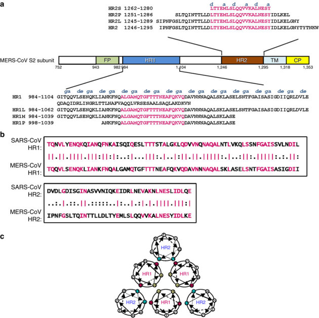
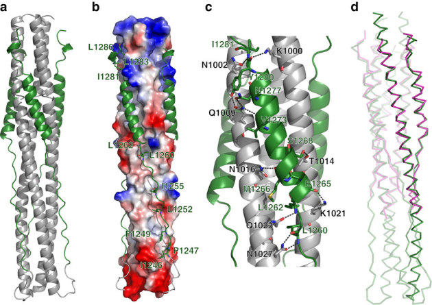
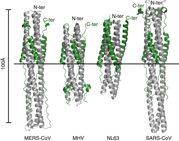
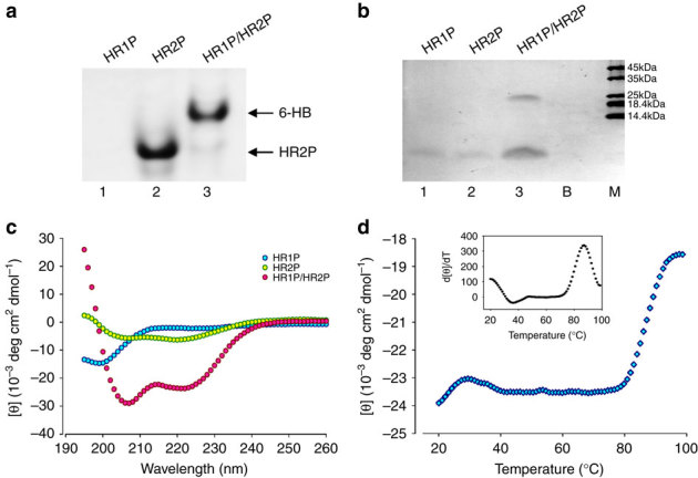
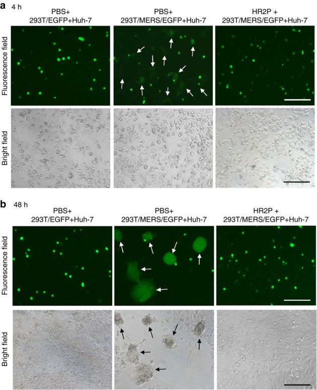
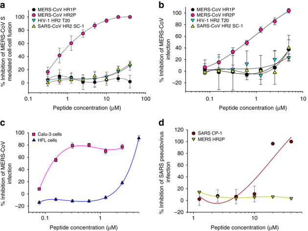
A novel human coronavirus, Middle East respiratory syndrome coronavirus (MERS-CoV), has caused outbreaks of a SARS-like illness with high case fatality rate. The reports of its person-to-person transmission through close contacts have raised a global concern about its pandemic potential. Here we characterize the six-helix bundle fusion core structure of MERS-CoV spike protein S2 subunit by X-ray crystallography and biophysical analysis. We find that two peptides, HR1P and HR2P, spanning residues 998-1039 in HR1 and 1251-1286 in HR2 domains, respectively, can form a stable six-helix bundle fusion core structure, suggesting that MERS-CoV enters into the host cell mainly through membrane fusion mechanism. HR2P can effectively inhibit MERS-CoV replication and its spike protein-mediated cell-cell fusion. Introduction of hydrophilic residues into HR2P results in significant improvement of its stability, solubility and antiviral activity. Therefore, the HR2P analogues have good potential to be further developed into effective viral fusion inhibitors for treating MERS-CoV infection.


Conflict of interest statement

The authors declare no competing financial interests.

Figures

Figure 1
Figure 1. Design of peptides and fusion proteins based on the sequence analysis of the MERS-CoV S protein S2 subunit.
(a) Schematic representation of MERS-CoV S protein S2 subunit. FP, fusion peptide; HR1, heptad repeat 1 domain; HR2, heptad repeat 2 domain; TM, transmembrane domain; CP, cytoplasmic domain. Residue numbers of each region correspond to their positions in S protein of MERS-CoV. The corresponding sequences of the HR1 and HR2 domains and the designed peptides (HR1L, HR1M, and HR1S, HR2L, HR2M, and HR2S) are shown in the diagram. The fragments in the HR1 and HR2 domains highlighted in magenta are the regions involved in the formation of the 6-HB core of MERS-CoV S protein, according to our X-ray crystal structure. (b) Sequence similarities between the N-terminal portion of HR1 domain (residues 986–1,055) in S2 of MERS-CoV and that of SARS-CoV (residues 894–963) and N-terminal portion of the HR2 domain (residues 1,246–1,285) in S2 of MERS-CoV and that of SARS-CoV (residues 1,144–1,183). Identical amino-acid residues are highlighted in magenta. (c) Formation of 6-HB between HR1 and HR2. The internal trimer is formed through the interaction of the residues located at the a and d positions (shown in yellow) in three HR1, and 6-HB is formed through the interaction of the residues located at the e and g positions (shown in magenta) in the HR1 and the a and d positions (shown in blue) in HR2.
Figure 2
Figure 2. Crystal structure of MERS-CoV 6-HB.
(a) Cartoon representation of the MERS-CoV fusion core structure, in which the HR1 and HR2 segments are coloured in grey and green, respectively. (b) Electrostatic potential surface of three central HR1 helices, as calculated using PyMOL (DeLano Scientific, Palo Alto, CA, USA), shows the hydrophobic grooves formed between each of two adjacent HR1 helices. Three HR2 segments in the grooves are shown in cartoon representation. (c) The hydrophilic interactions between HR1 and HR2 helices. The residues involved in forming hydrogen bonds are shown in stick representation and are properly labelled, as well as hydrogen bonds in dark grey dashed lines. (d) Superimposition between fusion core structure of MERS-CoV (green) and SARS-CoV (magenta).
Figure 3
Figure 3. Recapitulation of current structural data on the fusion core of coronavirus S2 protein.
Coloured identically, the three HR1 segments are in grey, while the HR2 segments in forest green. The very left panel displays the structure of MERS-Cov fusion core in this report. The PDB codes of other structures are 1WDF for MHV, 2IEQ for hCoV-NL63 and 1WYY for SARS. The images are all the same scale, with a horizontal bar to provide the alignment means. The N- and C-terminal ends of the constructs have also been marked.
Figure 4
Figure 4. Biophysical analysis of the HR1P and HR2P peptides and their complex.
(a) Determination of the 6-HB formation between HR1P and HR2P by N-PAGE. The mixture of HR1P and HR2P at the final concentration of 35 μM was incubated at 25 °C for 30 min before being loaded into the gel. (b) Molecular mass determined by gel electrophoresis. Samples of each peptide with 50 μM were incubated, diluted 1:1 (v/v) with 2 × Laemmli sample buffer at room temperature and loaded into the gel. B, blank; M, marker. (c) Secondary structures of HR1P, HR2P and HR1P/HR2P complex in phosphate buffer: CD spectra for HR1P (10 μM), HR2P (10 μM) and their complex in phosphate buffer (pH 7.2) at 4 °C. (d) CD signal at 222 nm for the HR1P/HR2P complex as a function of temperature. Insert: curve of the first derivative (d[θ]/dT) against temperature (T), which was used to determine the Tm value.
Figure 5
Figure 5. Images of MERS-CoV S protein-mediated cell–cell fusion and syncytium formation.
(a) MERS-CoV S protein-mediated cell–cell fusion. After Huh-7 cells were co-cultured with 293T/EGFP (left) or 293T/MERS/EGFP cells in the absence (middle) or presence (right) of 10 μM of HR2P at 37 °C for 4 h, cell–cell fusion was photographed under an optical microscope with fluorescence (top) or visible light (bottom). The arrows show the EGFP-labelled cells fused with unlabelled Huh-7 cells (top, middle). (b) MERS-CoV S protein-mediated syncytium formation. After Huh-7 cells were co-cultured with 293T/EGFP (left) or 293T/MERS/EGFP cells in the absence (middle) or presence (right) of 10 μM of HR2P, cell–cell fusion was photographed under an optical microscope with fluorescence or visible light. The arrows show the syncytia formed between 293T/MERS/EGFP and Huh-7 cells (middle). Scale bars, 200 μm.
Figure 6
Figure 6. Inhibition of peptides derived from the HR1 and HR2 regions in S2 subunit of MERS-CoV S protein on MERS-CoV infection.
(a) Inhibitory activity of HP1P and HR2P peptides on MERS-CoV S protein-mediated cell–cell fusion. The HIV-1 HR2-peptide T20 was used as a control. The number of 293T/MERS/EGFP cells fused or unfused with Huh-7 cells were counted under an inverted fluorescence microscope, and the percentage of inhibition was calculated as described in the Methods. (b) Inhibitory activity of HP1P and HR2P peptides on MERS-CoV replication. The HIV-1 HR2-peptide T20 and SARS-CoV HR2 peptide SC-1 were used as controls. (c) Inhibitory activity of HR2P peptide on MERS-CoV replication in Calu-3 and HFL cells. The HIV-1 HR2-peptide T20 and SARS-CoV HR2 peptide SC-1 were used as controls. (d) Inhibitory activity of MERS-CoV HR2 peptide HR2P and SARS-CoV HR2 peptide SC-1 on SARS-CoV infection. The HIV-1 HR2-peptide T20 was used as the control. The cytopathic effect caused by MERS-CoV replication was assessed with MTT assay (b and c), while the SARS-CoV pseudovirus infection in 293T/ACE2 cells was detected by luciferase assay (d). The percentage of inhibition was calculated as described in the Methods. Experiments were performed in triplicate, and the data are expressed as means±s.d. (error bar). The experiment was repeated twice, and similar results were obtained.

References

    1. Peiris JS, et al. Coronavirus as a possible cause of severe acute respiratory syndrome. Lancet. 2003;361:1319–1325. doi: 10.1016/S0140-6736(03)13077-2. - DOI - PMC - PubMed
    1. Drosten C, et al. Identification of a novel coronavirus in patients with severe acute respiratory syndrome. N. Engl. J. Med. 2003;348:1967–1976. doi: 10.1056/NEJMoa030747. - DOI - PubMed
    1. Zaki AM, van Boheemen S, Bestebroer TM, Osterhaus ADME, Fouchier RAM. Isolation of a novel coronavirus from a man with pneumonia in Saudi Arabia. N. Engl. J. Med. 2012;367:1814–1820. doi: 10.1056/NEJMoa1211721. - DOI - PubMed
    1. Gallagher T, Perlman S. Public health: broad reception for coronavirus. Nature. 2013;495:176–177. doi: 10.1038/495176a. - DOI - PMC - PubMed
    1. WHO. Novel coronavirus infection—update (Middle East respiratory syndrome coronavirus), http://www.who.int/csr/don/2013_11_11/en/index.html accessed 12 November (2013).

Publication types

MeSH terms

Associated data

LinkOut - more resources