. 2014 Jan 27;15(2):1983-2002.
doi: 10.3390/ijms15021983.

Hypoxia enhances protective effect of placental-derived mesenchymal stem cells on damaged intestinal epithelial cells by promoting secretion of insulin-like growth factor-1

Affiliations

Hypoxia enhances protective effect of placental-derived mesenchymal stem cells on damaged intestinal epithelial cells by promoting secretion of insulin-like growth factor-1

Lili Du et al. Int J Mol Sci. .

Abstract

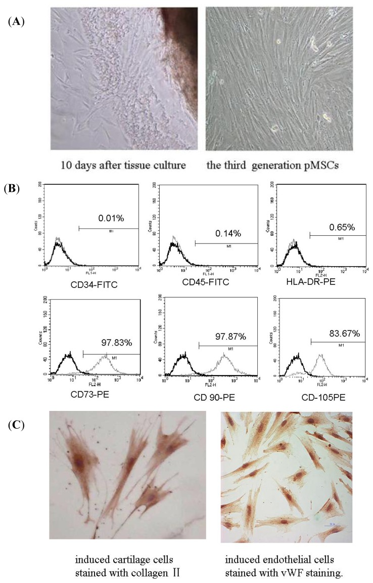
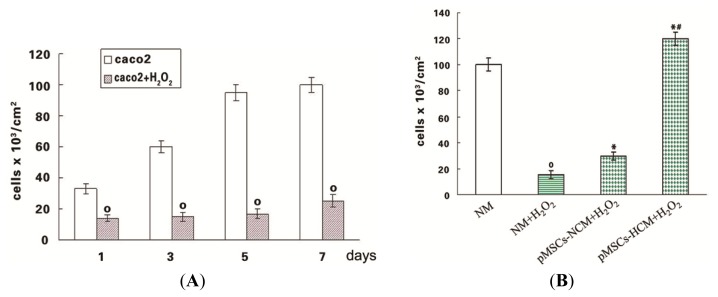
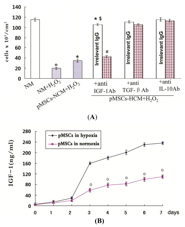
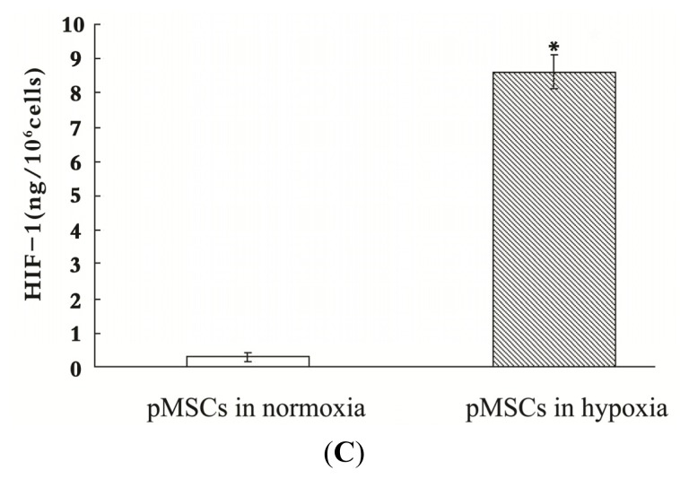
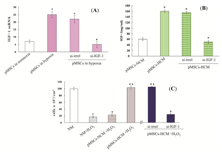
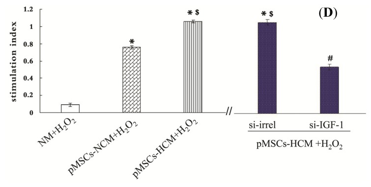
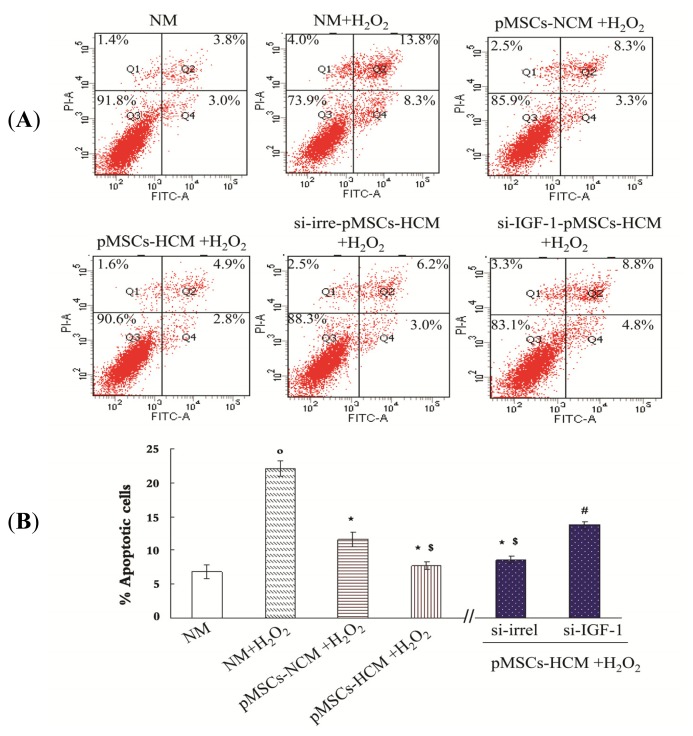
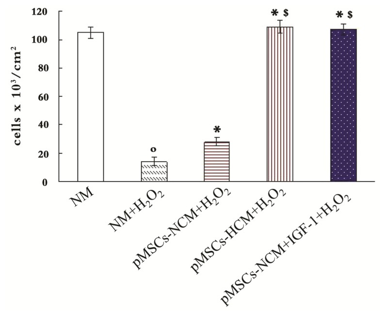
Apoptosis and necrosis of intestinal epithelial cells (IECs), induced by ischemia-reperfusion (I/R) injury, can lead to dysfunction of the intestinal barrier, which could cause multiple organ dysfunction syndromes. Mesenchymal stem cells (MSCs) have the potential of providing protective effects on damaged IECs via paracrine action. This study investigated whether hypoxia can enhance the protective effect of placental-derived MSCs (pMSCs) on H2O2-treated-caco2 cells, and explored the possible mechanism. The pMSCs isolated by tissue culture were fibroblast-like, positive for CD73, CD90 and CD105 and can differentiate into chondrocytes and endothelial cells. Five days after treatment with H2O2, the numbers of living caco2 cells significantly decreased. More live H2O2-treated-caco2 cells were observed in pMSCs hypoxia culture medium (pMSCs-HCM) than pMSCs normoxia culture medium (pMSCs-NCM), and the application of a specific antibody that blocked insulin-like growth factor-1 (IGF-1) leads to a significant decrease of the protective effect of pMSCs-HCM. Hypoxia can promote IGF-1 expression of pMSCs at mRNA and protein levels, and caco2 stably expressed IGF-1 receptor. Knocking down IGF-1 expression in pMSCs by siRNA resulted in a significant attenuation of the increase in apoptosis of H2O2-treated-caco2 cultured in pMSCs-HCM. In conclusion, hypoxia can increase the protective effect of pMSCs on H2O2-treated-caco2 cells via a promotion of their paracrine actions, and the key cytokine involved is IGF-1.


Figures

Figure 1.
Figure 1.
Isolation and identification of pMSCs. (A) Isolation of pMSCs by the method of tissue culture. Some cells appeared around tissues 10 days after culture. The third generation pMSCs had fibroblast-like morphology visualized by phase-contrast microscopy (100× magnification); (B) Flow cytometry assay of pMSCs: most of pMSCs were CD73, CD90 and CD105 positive; CD34, CD45 and HLA-DR negative; and (C) Differentiation ability of pMSCs: pMSCs can differentiate into cartilage cells and endothelial cells (400× magnification).
Figure 2.
Figure 2.
Protective effect of pMSCs-HCM on H2O2-treated-caco2 cells. (A) H2O2 exerted cytotoxic effect on caco2 cell viability. Caco2 cells were incubated with H2O2 (100 μM) for 12 h. After H2O2 removal, cells were maintained in normal medium and viable cells were counted after 1, 3, 5, 7 days; º p < 0.05 versus caco2 cells at the corresponding times (n = 6); and (B) pMSCs-HCM has a better protective effect for H2O2-treated-caco2 cells than that of pMSCs-NCM. Caco2 cells were incubated with 100 μM H2O2 for 12 h; after drug withdrawal, cells were cultured in normal medium, pMSCs-NCM, and pMSCs-HCM. Five days later, viable cells were counted by trypan blue staining; º p < 0.05 versus NM; * p < 0.05 versus NM + H2O2; # p < 0.05 versus pMSCs-NCM + H2O2 (n = 10).
Figure 3.
Figure 3.
The pMSCs hypoxia culture mediums produce a better protective effect on caco2 cells from H2O2 through more IGF-1. (A) Effect of anti-IGF-1, anti-TGF-β and anti-IL-10 Ab on protective effect of pMSCs-HCM. Three hours after culturing H2O2-treated caco2 cells in pMSCs-HCM, cells were exposed to specific functional blocking Ab against IGF-1, TGFβ, L-10 or the corresponding irrelevant IgG. Cell count was evaluated after five days, º p < 0.05 versus NM; * p < 0.05 versus NM + H2O2; $ p < 0.05 versus pMSCs-NCM + H2O2; # p < 0.05 versus corresponding irrelevant IgG. (n = 6); (B) Hypoxia induced more IGF-1 secreted by pMSCs. The secretion of IGF-1 by pMSCs in both normoxia and hypoxia increased with time. There was no difference in the early two days. From the third day on, the secretion by pMSCs cultured in hypoxia was much higher than that in normoxia, and the difference persisted at seven days, º p < 0.05 versus pMSCs in hypoxia; and (C) Activated HIF-1 of pMSCs in hypoxia or normoxia measured by HIF-1 ELISA. pMSCs were cultured in hypoxia or normoxia for seven days and then the amount of activated HIF-1 was detected. Figure 3C shows mean amounts of activated HIF-1 normalized to amounts per million harvested cells. pMSCs in hypoxia have much more activated HIF-1 than that in normoxia; * p < 0.05 versus pMSCs in normoxia.
Figure 3.
Figure 3.
The pMSCs hypoxia culture mediums produce a better protective effect on caco2 cells from H2O2 through more IGF-1. (A) Effect of anti-IGF-1, anti-TGF-β and anti-IL-10 Ab on protective effect of pMSCs-HCM. Three hours after culturing H2O2-treated caco2 cells in pMSCs-HCM, cells were exposed to specific functional blocking Ab against IGF-1, TGFβ, L-10 or the corresponding irrelevant IgG. Cell count was evaluated after five days, º p < 0.05 versus NM; * p < 0.05 versus NM + H2O2; $ p < 0.05 versus pMSCs-NCM + H2O2; # p < 0.05 versus corresponding irrelevant IgG. (n = 6); (B) Hypoxia induced more IGF-1 secreted by pMSCs. The secretion of IGF-1 by pMSCs in both normoxia and hypoxia increased with time. There was no difference in the early two days. From the third day on, the secretion by pMSCs cultured in hypoxia was much higher than that in normoxia, and the difference persisted at seven days, º p < 0.05 versus pMSCs in hypoxia; and (C) Activated HIF-1 of pMSCs in hypoxia or normoxia measured by HIF-1 ELISA. pMSCs were cultured in hypoxia or normoxia for seven days and then the amount of activated HIF-1 was detected. Figure 3C shows mean amounts of activated HIF-1 normalized to amounts per million harvested cells. pMSCs in hypoxia have much more activated HIF-1 than that in normoxia; * p < 0.05 versus pMSCs in normoxia.
Figure 4.
Figure 4.
The pMSCs hypoxia culture medium is more conducive to promote H2O2-treated-caco2 cells proliferation via IGF-1. (A and B) Silence IGF-1 in pMSCs by siRNA: (A) After two-day culture, pMSCs in hypoxia had much more IGF-1 mRNA than that in normoxia, and it is the same with pMSCs in hypoxia transfected with si-irrel. When cultured in hypoxia, si-IGF-1-pMSCs had much less IGF-1 mRNA than si-irrel-pMSCs. º p < 0.05 versus pMSCs in normoxia; * p < 0.05 versus si-irrel-pMSCs in hypoxia (n = 6); (B) IGF-1 protein level in pMSCs-HCM and siirrel-pMSCs-HCM were much higher than that in pMSCs-NCM after four-day culture. IGF1 protein level in si-IGF-1-pMSCs-HCM obviously decreased compared with si-irrelpMSCs-HCM; º p < 0.05 versus pMSCs-NCM; * p < 0.05 versus si-irrel-pMSCs-HCM (n = 6); (C) Trypan blue staining is used to detect viable cell number. After H2O2-treated-caco2 cells were cultured in NM, pMSCs-NCM, pMSCs-HCM, si-irrel-pMSCs-HCM or si-IGF-1pMSCs-HCM for five days, viable cell number were assessed. º p < 0.05 versus NM; * p < 0.05 versus NM + H2O2; $ p < 0.05 versus pMSCs-NCM + H2O2; # p < 0.05 versus si-irrel-pMSCs-HCM + H2O2 (n = 6); and (D) MTT is used to detect cellular metabolic activity. After H2O2-treated caco2 cells were cultured in NM, pMSCs-NCM, pMSCs-HCM, si-irrel-pMSCs-HCM or si-IGF-1pMSCs-HCM for five days, cellular metabolic activity were assessed by MTT. * p < 0.05 versus NM + H2O2; $ p < 0.05 versus pMSCs-NCM + H2O2; # p < 0.05 versus si-irrel-pMSCsHCM + H2O2 (n = 6).
Figure 4.
Figure 4.
The pMSCs hypoxia culture medium is more conducive to promote H2O2-treated-caco2 cells proliferation via IGF-1. (A and B) Silence IGF-1 in pMSCs by siRNA: (A) After two-day culture, pMSCs in hypoxia had much more IGF-1 mRNA than that in normoxia, and it is the same with pMSCs in hypoxia transfected with si-irrel. When cultured in hypoxia, si-IGF-1-pMSCs had much less IGF-1 mRNA than si-irrel-pMSCs. º p < 0.05 versus pMSCs in normoxia; * p < 0.05 versus si-irrel-pMSCs in hypoxia (n = 6); (B) IGF-1 protein level in pMSCs-HCM and siirrel-pMSCs-HCM were much higher than that in pMSCs-NCM after four-day culture. IGF1 protein level in si-IGF-1-pMSCs-HCM obviously decreased compared with si-irrelpMSCs-HCM; º p < 0.05 versus pMSCs-NCM; * p < 0.05 versus si-irrel-pMSCs-HCM (n = 6); (C) Trypan blue staining is used to detect viable cell number. After H2O2-treated-caco2 cells were cultured in NM, pMSCs-NCM, pMSCs-HCM, si-irrel-pMSCs-HCM or si-IGF-1pMSCs-HCM for five days, viable cell number were assessed. º p < 0.05 versus NM; * p < 0.05 versus NM + H2O2; $ p < 0.05 versus pMSCs-NCM + H2O2; # p < 0.05 versus si-irrel-pMSCs-HCM + H2O2 (n = 6); and (D) MTT is used to detect cellular metabolic activity. After H2O2-treated caco2 cells were cultured in NM, pMSCs-NCM, pMSCs-HCM, si-irrel-pMSCs-HCM or si-IGF-1pMSCs-HCM for five days, cellular metabolic activity were assessed by MTT. * p < 0.05 versus NM + H2O2; $ p < 0.05 versus pMSCs-NCM + H2O2; # p < 0.05 versus si-irrel-pMSCsHCM + H2O2 (n = 6).
Figure 5.
Figure 5.
pMSCs hypoxia culture medium has stronger effect on limiting H2O2-treated-caco2 apoptosis via IGF-1 compared with pMSCs normoxia culture medium. (A) After five days culture, untreated caco2 cells in NM, H2O2-treated caco2 cells in NM, pMSCs-NCM, si-irrel-pMSCs-HCM or si-IGF-1-pMSCs-HCM, percentage of apoptotic cells (early apoptosis is FITC-positive, late apoptosis is FITC- and propidium iodide-postive) were analyzed by FACS; and (B) Data was expressed as the mean ± SEM of experiments repeated three times. º p < 0.05 versus NM. * p < 0.05 versus NM + H2O2; $ p < 0.05 versus pMSCs-NCM + H2O2; # p < 0.05 versus si-irrel-pMSCs-HCM + H2O2 (n = 3).
Figure 6.
Figure 6.
Addition of IGF-1 can enhance the protective effect of pMSCs-NCM on H2O2-treated-caco2 cells. The untreated caco2 in NM, H2O2-treated-caco2 cells in NM, pMSCs-NCM, pMSCs-HCM and pMSCs-NCM were cultured with IGF-1 (200 ng/mL). 5 days later, trypan blue staining was used to determine the viable cells. º p < 0.05 versus NM, * p < 0.05 versus NM + H2O2, and $ p < 0.05 versus pMSCs-NCM + H2O2.

Similar articles

Cited by

References

    1. Oldeburg W.A., Lau L.L., Rodenberg T.J., Edmonds H.J., Burger C.D. Acute mesenteric ischemia: A clinical review. Arch. Intern. Med. 2004;164:1054–1062. - PubMed
    1. Abu-Elmagd K., Reyes J., Bond G., Mazariegos G., Wu T., Martin N., Sindhi R. Clinical intestinal transplantation: A decade of experience at a single center. Ann. Surg. 2001;234:416–417. - PMC - PubMed
    1. Quaedackers J.S., Beukm R.J., Bennetm L., Charlton A., Oude Eqbrink M.G., Gunn A.J., Heineman E. An evaluation of methods for grading histologic injury following ischemia/reperfusion of the small bowel. Transplant. Proc. 2000;32:1307–1310. - PubMed
    1. Khanna A., Rossman J., Caty M.G., Fung H.L. Beneficial effects of intraluminal nitroglycerin in intestinal ischemia-reperfusion injury in rats. J. Surg. Res. 2003;114:5–24. - PubMed
    1. Wang H.J., Huang J. Basic and applicative studies of acidic fibroblast growth factor. Anat. Res. 2005;2:308–311.

Publication types

MeSH terms

Substances