. 2014 Jan 15;5(1):150-60.
doi: 10.18632/oncotarget.1430.

Survivin promoter-regulated oncolytic adenovirus with Hsp70 gene exerts effective antitumor efficacy in gastric cancer immunotherapy

Affiliations

Survivin promoter-regulated oncolytic adenovirus with Hsp70 gene exerts effective antitumor efficacy in gastric cancer immunotherapy

Weiguo Wang et al. Oncotarget. .

Abstract

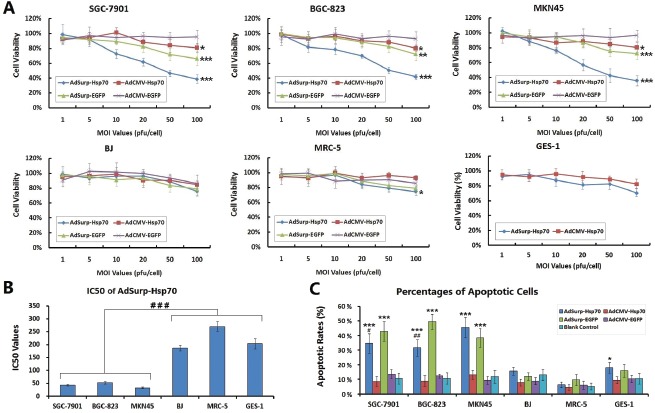
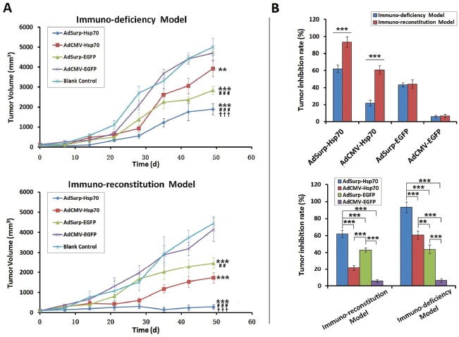
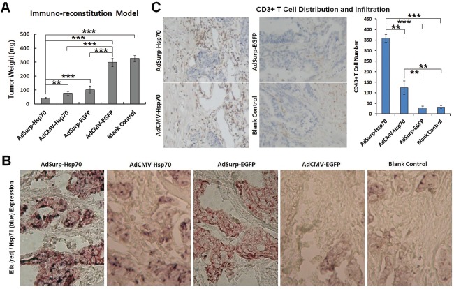
Gene therapy is a promising adjuvant therapeutic strategy for cancer treatment. To overcome the limitations of current gene therapy, such as poor transfection efficiency of vectors, low levels of transgene expression and lack of tumor targeting, the Survivin promoter was used to regulate the selective replication of oncolytic adenovirus in tumor cells, and the heat shock protein 70 (Hsp70) gene was loaded as the anticancer transgene to generate an AdSurp-Hsp70 viral therapy system. The efficacy of this targeted immunotherapy was examined in gastric cancer. The experiments showed that the oncolytic adenovirus can selectively replicate in and lyse the Survivin-positive gastric cancer cells, without significant toxicity to normal cells. AdSurp-Hsp70 reduced viability of cancer cells and inhibited tumor growth of gastric cancer xenografts in immuno-deficient and immuno-reconstruction mouse models. AdSurp-Hsp70 produced dual antitumor effects due to viral replication and high Hsp70 expression. This therapeutic system used the Survivin promoter-regulated oncolytic adenovirus vector to mediate targeted expression of the Hsp70 gene and ensure safety and efficacy for subsequent gene therapy programs against a variety of cancers.


Figures

Figure 1
Figure 1. Survivin expression and oncolytic adenovirus replicative activity in cell lines examined
(A) Cultured gastric cancer and normal cells were collected, and the TRIzol method was used to extract the total RNA from these cells. Survivin was amplified from the extracted RNA through RT-PCR with GAPDH as the internal control. (B) Fluorescence microscopy was used to observe EGFP-positive gastric cancer cells and normal cells 48 h after the cells were infected with AdSurp-EGFP or AdCMV-EGFP at an MOI of 1 pfu/cell, original magnification 200×. (C) Gastric cancer and normal cells were infected with AdSurp-Hsp70 or AdCMV-Hsp70 at an MOI of 1 pfu/cell. Forty-eight hours after infection, the cells were collected and the virus titer was quantified using the TCID50 assay. ** P < 0.01 and *** P < 0.001. (D) Gastric cancer and normal cells were infected with AdSurp-Hsp70 and AdCMV-Hsp70 at an MOI of 1 pfu/cell. Forty-eight hours after infection, the cells were collected, and Western blotting was used to detect E1a expression with GAPDH as the loading control.
Figure 2
Figure 2. Survivin promoter-regulated oncolytic adenoviruses mediated Hsp70 expression
(A) The cells were infected with the AdSurp-Hsp70 or AdCMV-Hsp70 adenovirus at a 10 pfu/cell MOI. Forty-eight hours after infection, the cells were collected, and western blotting was used to detect Hsp70 expression, with GAPDH as the loading control. The densitometry analysis was performed to show Hsp70 expression levels normalized with GAPDH density, ** P < 0.01. (B) The cells were infected with the AdSurp-Hsp70 or AdCMV-Hsp70 adenoviruses at a 10 pfu/cell MOI. Forty-eight hours after infection, the cells were collected, and an ELISA was used to detect Hsp70 expression. ** P < 0.01 and *** P < 0.001.
Figure 3
Figure 3. Specific cytotoxicity of oncolytic adenovirus in gastric cancer cells
(A) The cells were seeded into 96-well plates at a concentration of 104 cells/well and then infected with viruses at MOIs that ranged from 1 to 100 pfu/cell. Forty-eight hours after infection, cell viability was verified using the MTT assay, * P < 0.05, ** P < 0.01 and *** P < 0.001 compared with the control adenovirus AdCMV-EGFP. (B) The IC50 values that produced a 50% cell viability of viral MOI in the AdSurp-Hsp70 group were compared between cancer and normal cell lines, ### P < 0.001. (C) Cells that were infected with viruses at an MOI of 50 pfu/cell were collected and examined the percentages of apoptotic cells by the terminal deoxynucleotidyl transferase-mediated dUTP nick end labeling (TUNEL) assay. The experiment was repeated three times, and the proportions of positive cells in each section were observed under 5 high power fields (×40 objective lens), * P < 0.05 and *** P < 0.001 compared with the blank control group in the same cell line; # P < 0.05 and ## P < 0.01 compared with the AdSurp-EGFP group in the same cell line.
Figure 4
Figure 4. The efficacy of virus treatments in gastric cancer xenograft models
(A) Nude mice were each implanted with 106 SGC-7901 cells, which formed xenograft tumors 12 days thereafter. In the first immuno-defciency model, the mice were directly divided into treatment groups, and the mice in each group received 109 pfu of the appropriate recombinant virus via multisite intratumoral injections. In the second immuno-reconstitution model, 107 CIK cells were infused into each mouse before the mice were grouped and received the aforementioned virus therapies. The tumor diameters were measured weekly and used to calculate tumor volumes. ** P < 0.01 and *** P < 0.001 compared with the blank control group; ## P < 0.01 and ### P < 0.001 compared with the AdCMV-Hsp70 group; ††† P < 0.001 compared with the AdSurp-Hsp70 group. (B) Data collected 49 days after the initial virus treatment were used to compare the tumor inhibition rates from viruses between these two models. ** P < 0.01 and *** P < 0.001.
Figure 5
Figure 5. Pathological examinations on nude mouse xenograft specimens
(A) The nude mouse xenograft tumor weights were compared. ** P < 0.01 and *** P < 0.001. (B) Immunohistochemical double staining to localize E1a (red) and Hsp70 (blue) expression in cancer cells, original magnification 200×. (C) Immunohistochemical staining to indicate the CD3+ T cell number and distribution that infiltrated the tumor stroma (DAB chromogen, yellowish brown), original magnification 200×. The number of positive cells was counted in 5 fields of view for each section using a 20× objective lens. ** P < 0.01 and *** P < 0.001.

References

    1. Huang S, Kamihira M. Development of hybrid viral vectors for gene therapy. Biotechnol Adv. 2013;31:208–23. - PubMed
    1. Kron MW, Kreppel F. Adenovirus vectors and subviral particles for protein and peptide delivery. Curr Gene Ther. 2012;12:362–73. - PubMed
    1. Vacchelli E, Eggermont A, Sautès-Fridman C, Galon J, Zitvogel L, Kroemer G, Galluzzi L. Trial watch: Oncolytic viruses for cancer therapy. Oncoimmunology. 2013;2:e24612. - PMC - PubMed
    1. Meshii N, Takahashi G, Okunaga S, Hamada M, Iwai S, Takasu A, Ogawa Y, Yura Y. Enhancement of systemic tumor immunity for squamous cell carcinoma cells by anoncolytic herpes simplex virus. Cancer Gene Ther. 2013 doi: 10.1038/cgt.2013.45. - PubMed
    1. Li JM, Kao KC, Li LF, Yang TM, Wu CP, Horng YM, Jia WW, Yang CT. MicroRNA-145 regulates oncolytic herpes simplex virus-1 for selective killing of human non-small cell lung cancer cells. Virol J. 2013;10:241. - PMC - PubMed

Publication types

MeSH terms