

Amelanotic metastatic cutaneous melanoma
- PMID: 24474114
- PMCID: PMC3900356
- DOI: 10.1590/abd1806-4841.20132206
Amelanotic metastatic cutaneous melanoma
Abstract
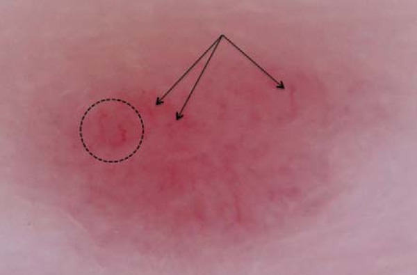
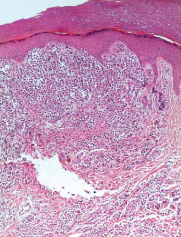
Dermatoscopy of melanocytic lesions has guided the decision of when or not to biopsy a lesion. The use of this tool has increased clinical examination's sensitivity and specificity in 89% and 96% respectively. However, dermatoscopic evaluation of amelanotic or hypomelanotic melanomas, as well as metastases, can be difficult. There is still no standardization for the analysis of these pathologies, which relies mostly on their vascular pattern. We describe the dermatoscopy of acral metastatic amelanotic melanoma.
A dermatoscopia das lesões melanocíticas tem auxiliado na decisão de biopsiar ou não uma lesão. A utilização desta ferramenta aumentou a sensibilidade e a especificidade do diagnóstico para 89% e 96%, respectivamente. No entanto, a avaliação dermatoscópica de melanomas amelanóticos ou hipomelanóticos, bem como a de metástases cutâneas, pode ser difícil. Ainda falta uma padronização para a análise destas patologias, que se baseia, majoritariamente, no seu padrão vascular. Descreve-se a dermatoscopia de melanoma metastático amelanótico acral.
Conflict of interest statement
Conflict of Interests: none
Figures

References
-
- Zalaudek I, Kreusch J, Giacomel J, Ferrara G, Catricalà C, Argenziano G. How to diagnose nonpigmented skin tumors: a review of vascular structures seen with dermoscopy: part I. Melanocytic skin tumors. J Am Acad Dermatol. 2010;63:361–374. - PubMed
-
- Schiffner R, Schiffner-Rohe J, Vogt T, Landthaler M, Wlotzke U, Cognetta AB, et al. Improvement of early recognition of lentigo maligna using dermatoscopy. J Am Acad Dermatol. 2000;42:25–32. - PubMed
-
- Costa MC, Abraham LS, Barcaui CB. Lentigo maligna treated with topical imiquimod: dermatoscopy usefulness in clinical monitoring. An Bras Dermatol. 2011;86:792–794. - PubMed
-
- Grazzini M, Stanganelli I, Rossari S, Gori A, Oranges T, Longo AS, et al. Dermoscopy, confocal laser microscopy, and hi-tech evaluation of vascular skin lesions: diagnostic and therapeutic perspectives. Dermatol Ther. 2012;25:297–303. - PubMed
-
- Jaimes N, Braun RP, Thomas L, Marghoob AA. Clinical and dermoscopic characteristics of amelanotic melanomas that are not of the nodular subtype. J Eur Acad Dermatol Venereol. 2012;26:591–596. - PubMed
Publication types
MeSH terms
LinkOut - more resources
Full Text Sources
Other Literature Sources
Medical