. 2014 Jan 28;111(4):1403-8.
doi: 10.1073/pnas.1311705111. Epub 2014 Jan 13.

Notch signaling regulates cardiomyocyte proliferation during zebrafish heart regeneration

Affiliations

Notch signaling regulates cardiomyocyte proliferation during zebrafish heart regeneration

Long Zhao et al. Proc Natl Acad Sci U S A. .

Abstract

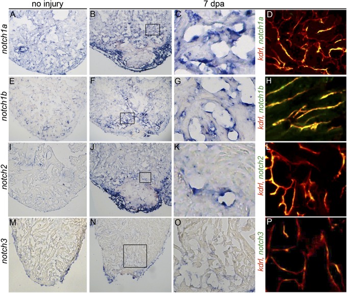
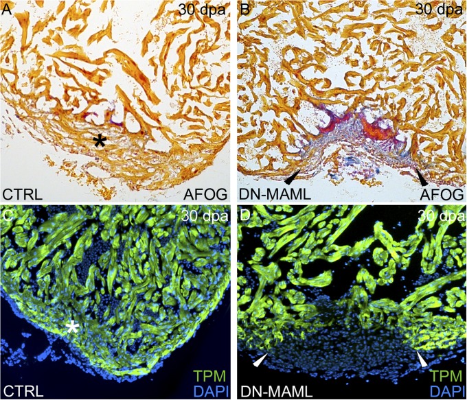
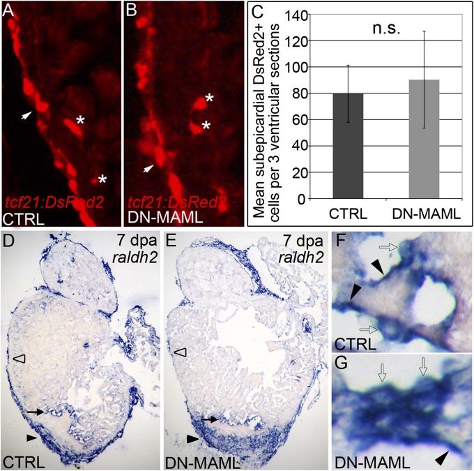
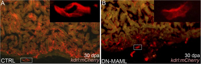
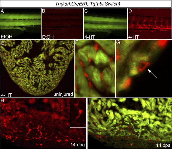
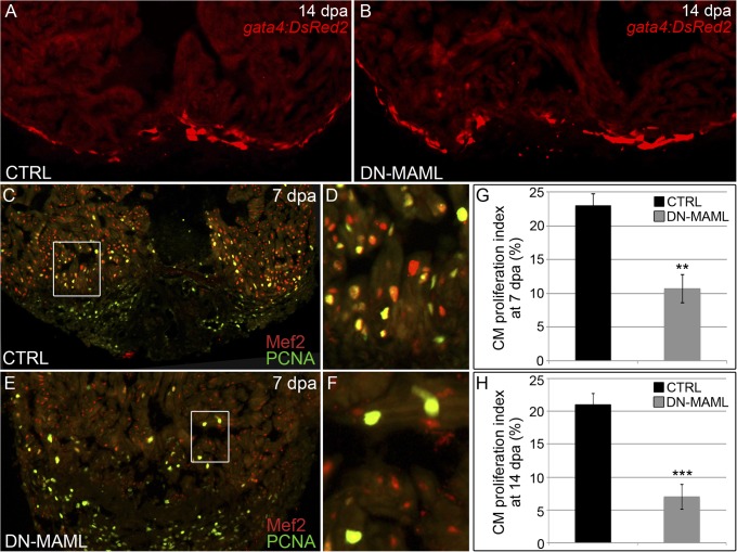
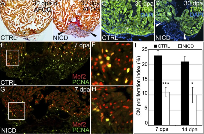
The human heart's failure to replace ischemia-damaged myocardium with regenerated muscle contributes significantly to the worldwide morbidity and mortality associated with coronary artery disease. Remarkably, certain vertebrate species, including the zebrafish, achieve complete regeneration of amputated or injured myocardium through the proliferation of spared cardiomyocytes. Nonetheless, the genetic and cellular determinants of natural cardiac regeneration remain incompletely characterized. Here, we report that cardiac regeneration in zebrafish relies on Notch signaling. Following amputation of the zebrafish ventricular apex, Notch receptor expression becomes activated specifically in the endocardium and epicardium, but not the myocardium. Using a dominant negative approach, we discovered that suppression of Notch signaling profoundly impairs cardiac regeneration and induces scar formation at the amputation site. We ruled out defects in endocardial activation, epicardial activation, and dedifferentiation of compact myocardial cells as causative for the regenerative failure. Furthermore, coronary endothelial tubes, which we lineage traced from preexisting endothelium in wild-type hearts, formed in the wound despite the myocardial regenerative failure. Quantification of myocardial proliferation in Notch-suppressed hearts revealed a significant decrease in cycling cardiomyocytes, an observation consistent with a noncell autonomous requirement for Notch signaling in cardiomyocyte proliferation. Unexpectedly, hyperactivation of Notch signaling also suppressed cardiomyocyte proliferation and heart regeneration. Taken together, our data uncover the exquisite sensitivity of regenerative cardiomyocyte proliferation to perturbations in Notch signaling.

Keywords: model organism; myocardial infarction.


Conflict of interest statement

The authors declare no conflict of interest.

Figures

Fig. 1.
Fig. 1.
Ventricular apex amputation induces up-regulation of Notch receptor expression in endocardial and epicardial cells. Representative histological sections of uninjured (A, E, I, and M) and injured hearts (B, F, J, and N) at 7 d postamputation (dpa) showing up-regulation of three Notch receptors in the endocardium and/or epicardium following amputation injury by colormetric in situ hybridization analyses. Boxed regions of B, F, J, and N are shown at higher magnifications in C, G, K, and O. (D, H, L, and P) Representative cardiac sections from Tg(kdrl:mCherry) zebrafish processed for fluorescent in situ hybridization to visualize expression of the indicated Notch receptors and mCherry immunohistochemistry to visualize endocardial cells. Notch1a, notch1b, notch2, and notch3 transcripts colocalized with endocardial cells without expression in the intervening myocardium. (M and N) Notch3 transcripts were unchanged by injury. Consistent staining patterns were observed in eight hearts per experimental group for the colorimetric analyses and in two hearts for the fluorescent analyses.
Fig. 2.
Fig. 2.
Inhibition of Notch signaling following amputation injury prevents cardiac regeneration. (AD) Inhibition of Notch signaling impairs cardiac regeneration. Representative cardiac sections from heat shocked control (A and C) and Tg(hsp70:DN-MAML) (B and D) animals 30 dpa evaluated by AFOG staining (A and B) or immunofluorescence (C and D) for the myocardial marker TPM. Whereas heat shocked control animals robustly regenerated myocardium (asterisks in A and C), Tg(hsp70:DN-MAML) hearts failed to regenerate new myocardium (arrowheads in B and D) instead showing evidence of residual fibrin (red in B) and collagen deposition (blue in B). Cardiac regeneration failed in 0 of 24 heat shocked control animals and 34 of 35 heat shocked Tg(hsp70:DN-MAML) animals.
Fig. 3.
Fig. 3.
Notch signaling is dispensable for injury-induced epicardial and endocardial activation. (A and B) Representative cardiac sections from 7dpa heat shocked control (CTRL) (A) and Tg(hsp70:DN-MAML) (B) animals carrying the Tg(tcf21:DsRed2) transgene. In regions away from the wound, DsRed2+ epicardial cells (arrows) and EPDCs (asterisks) were grossly normal in Notch suppressed hearts. Seven of seven hearts in each experimental group expressed DsRed2 in epicardium and EPDCs. (C) Graph representing the average numbers of subepicardial DsRed2+ cells quantified in three ventricular sections from four hearts per experimental group. n.s., not significant. (DG) Representative cardiac sections from heat shocked control (D) and Tg(hsp70:DN-MAML) (E) animals at 7 dpa stained for raldh2 transcripts by in situ hybridization. In both experimental groups, prominent raldh2 expression was observed in epicardial cells covering the wound (black arrowheads in D and E), away from the wound (open arrowheads in D and E), and in endocardial cells near the wound edge (black arrows in D and E). Higher magnifications of endocardial raldh2 expression are shown in F and G. Endocardial cells near the wound displayed both flattened (black arrowheads in F and G) and rounded morphologies (open arrows in F and G). Four of four hearts in each experimental group expressed endocardial and epicardial raldh2 transcripts.
Fig. 4.
Fig. 4.
Evidence for regeneration of coronary endothelium in Notch-inhibited hearts. (A and B) Representative cardiac sections from heat shocked control (CTRL) (A) and Tg(hsp70:DN-MAML) (B) animals carrying the Tg(kdrl:mCherry) endothelial reporter at 30 dpa. (Insets) Higher magnification views of boxed regions in A and B. Whereas control animals regenerated mCherry+ endocardium and coronary vessels, Tg(hsp70:DN-MAML) hearts harbored mCherry+ endothelial tubes in the nonregenerated region of the heart. The myocardium is autofluorescent in A and B. Fourteen of 14 control and 11 of 11 Tg(hsp70:DN-MAML) hearts demonstrated these phenotypes.
Fig. 5.
Fig. 5.
Regenerated coronary endothelium derives from preexisting endothelial cells during zebrafish heart regeneration. (AD) Permanent labeling of endothelial cells during zebrafish embryogenesis. Tails of 96-hpf Tg(kdrl:CreER); Tg(ubi:Switch) embryos treated between 24 and 48 hpf with EtOH (A and B) or 4-HT (C and D) and imaged in the green (A and C) and red (B and D) channels. All 4-HT–treated embryos examined expressed mCherry in endothelial cells (n > 100), whereas EtOH-treated embryos failed to report red fluorescence (n > 100). (EG) The adult endocardium and coronary endothelium displayed widespread and permanent mCherry labeling following 4-HT treatment during embryonic stages (n = 3/3). Representative cardiac section (E) and close up views of mCherry-labeled endocardium (F) and coronary endothelium (G). (H and I) Representative cardiac section from a regenerating Tg(kdrl:CreER); Tg(ubi:Switch) animal with mCherry-labeled endocardial and endothelial cells at 14 dpa. Numerous mCherry+ endothelial tubes (Inset) populated the regenerating myocardium. (n = 3/3.)
Fig. 6.
Fig. 6.
Notch signaling is required for cardiomyocyte proliferation during zebrafish heart regeneration. (A and B) Representative cardiac sections from heat shocked control (CTRL) (A) and Tg(hsp70:DN-MAML) (B) animals carrying the Tg(gata4:DsRed2) transgene at 14 dpa. DsRed2 expression was visible in the compact myocardium of both control and Tg(hsp70:DN-MAML) hearts (n = 12/12 in each group). (CF) Representative cardiac sections from heat shocked control (C and D) and Tg(hsp70:DN-MAML) (E and F) animals at 7 dpa. Sections were double immunostained to identify cardiomyocyte nuclei (Mef2+) and nuclei undergoing DNA replication (PCNA+). Boxed regions in C and E are shown at higher zoom in D and F. The percentages of myocardial nuclei undergoing DNA replication near the wound edges were quantified at 7 (G) and 14 (H) dpa and reported as mean proliferation indices ± 1 SD, **P < 0.01 and ***P < 0.001. In the control group, seven and eight hearts were analyzed at 7 and 14 dpa, respectively. In the Tg(hsp70:DN-MAML) group, three and four hearts were analyzed at 7 and 14 dpa, respectively.
Fig. 7.
Fig. 7.
Hyperactivation of Notch signaling impairs cardiomyocyte proliferation and heart regeneration. (AD) Representative cardiac sections from heat shocked control (CTRL) (A and C) and Tg(hsp70:Gal4); Tg(UAS:NICD) (B and D) animals at 30 dpa stained with AFOG (A and B) or an antibody recognizing the myocardial marker TPM (C and D). Whereas heat shocked CTRL animals regenerated myocardium (asterisks in A and C), Tg(hsp70:Gal4); Tg(UAS:NICD) hearts exhibited impaired regeneration (arrowheads in B and D), residual fibrin (red in B), and collagen deposition (blue in B). In the CTRL group, >25 hearts were analyzed and all showed significant myocardial regeneration. In the Tg(hsp70:Gal4); Tg(UAS:NICD) group, 18 hearts were analyzed and 14 showed a significant myocardial deficit, residual fibrin, and collagen deposition. (EH) Representative cardiac sections of heat shocked CTRL (E and F) and Tg(hsp70:NICD) (G and H) animals at 7 dpa double stained with antibodies that recognize cardiomyocyte nuclei (Mef2+) and nuclei undergoing DNA replication (PCNA+). Boxed regions in E and G are shown at higher zoom in F and H. (I) The percentages of cardiomyocyte nuclei undergoing DNA replication were quantified along the wound edge at 7 and 14 dpa and expressed as mean proliferation indices ± 1 SD, ***P < 0.001; *P < 0.05. In the control group, seven and eight hearts were analyzed at 7 and 14 dpa, respectively. In the Tg(hsp70:NICD) group, three and four hearts were analyzed at 7 and 14 dpa, respectively.

References

    1. Laflamme MA, Murry CE. Heart regeneration. Nature. 2011;473(7347):326–335. - PMC - PubMed
    1. Sidney S, Rosamond WD, Howard VJ, Luepker RV. National Forum for Heart Disease and Stroke Prevention The “heart disease and stroke statistics—2013 update” and the need for a national cardiovascular surveillance system. Circulation. 2013;127(1):21–23. - PubMed
    1. Chablais F, Jaźwińska A. Induction of myocardial infarction in adult zebrafish using cryoinjury. J Vis Exp. 2012;(62) - PMC - PubMed
    1. Chablais F, Veit J, Rainer G, Jaźwińska A. The zebrafish heart regenerates after cryoinjury-induced myocardial infarction. BMC Dev Biol. 2011;11:21. - PMC - PubMed
    1. González-Rosa JM, Martín V, Peralta M, Torres M, Mercader N. Extensive scar formation and regression during heart regeneration after cryoinjury in zebrafish. Development. 2011;138(9):1663–1674. - PubMed

Publication types

Substances

LinkOut - more resources