. 2014 Jan 28;111(4):1533-8.
doi: 10.1073/pnas.1316177111. Epub 2014 Jan 13.

Origin of regenerating tubular cells after acute kidney injury

Affiliations

Origin of regenerating tubular cells after acute kidney injury

Katja Berger et al. Proc Natl Acad Sci U S A. .

Abstract

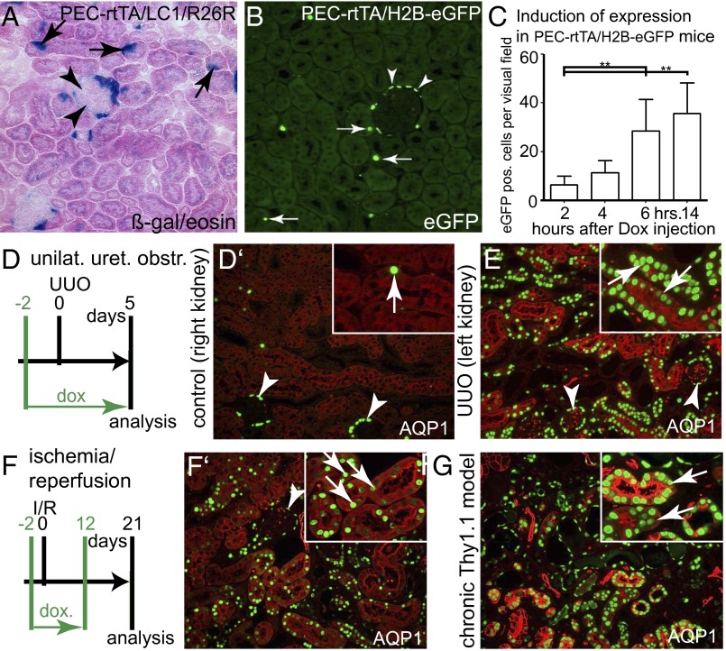
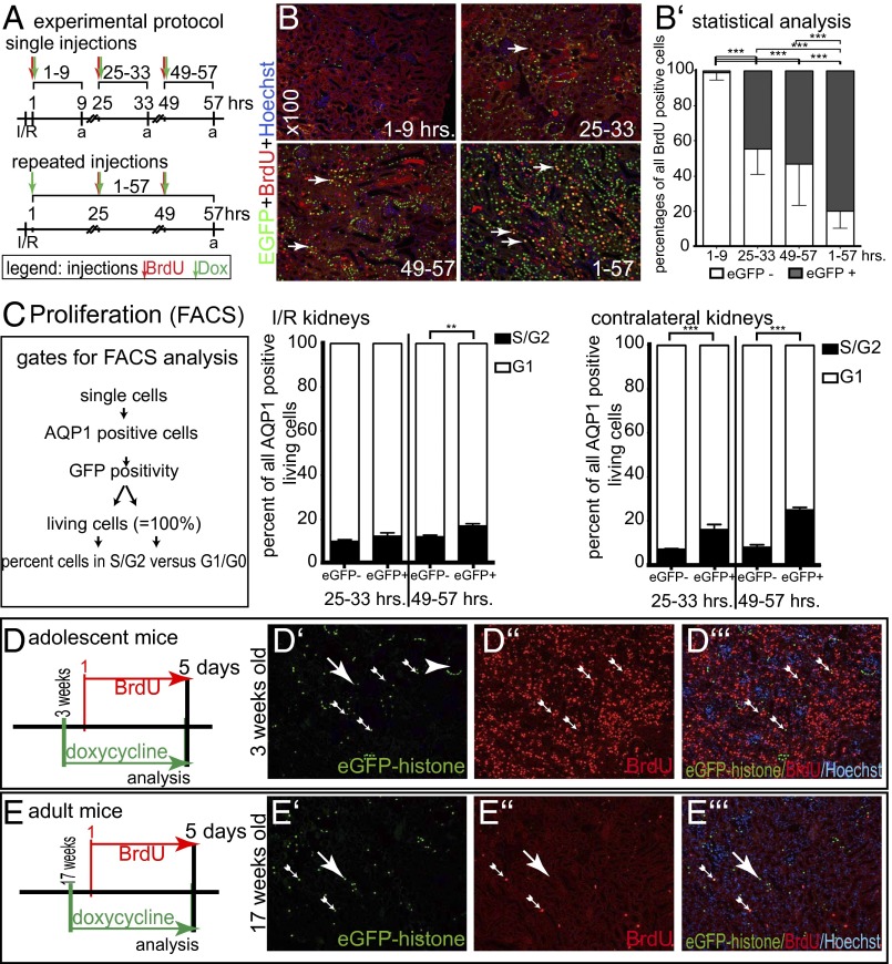
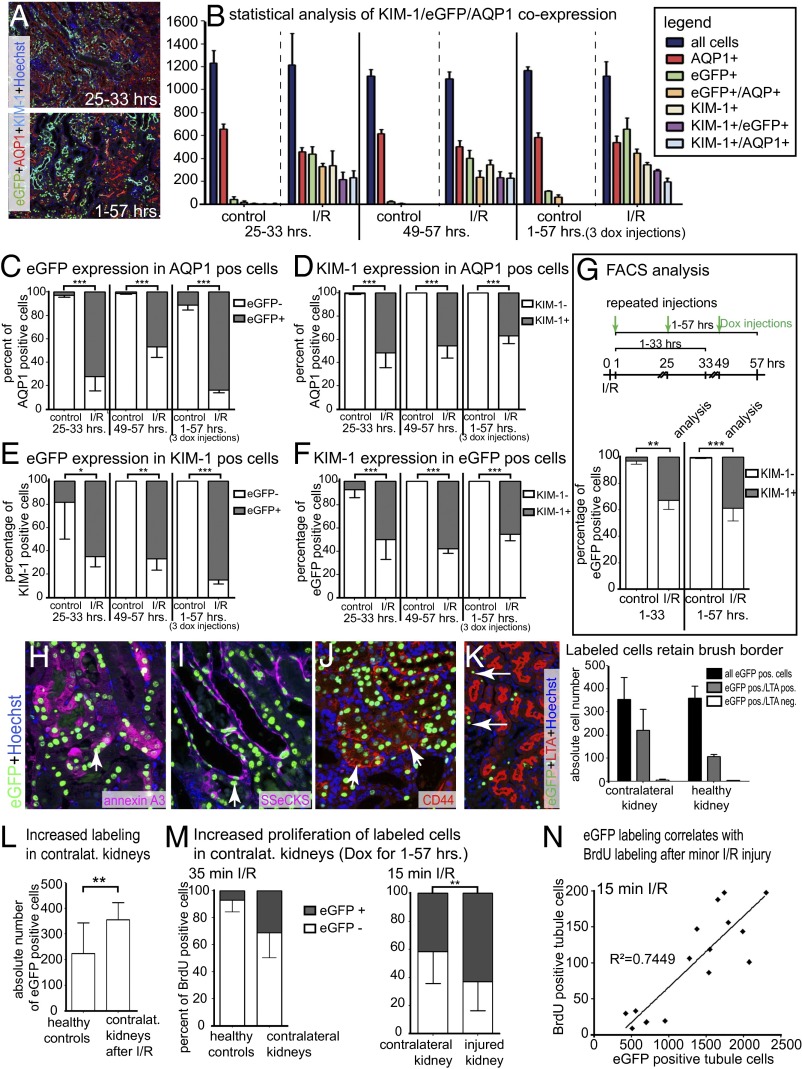
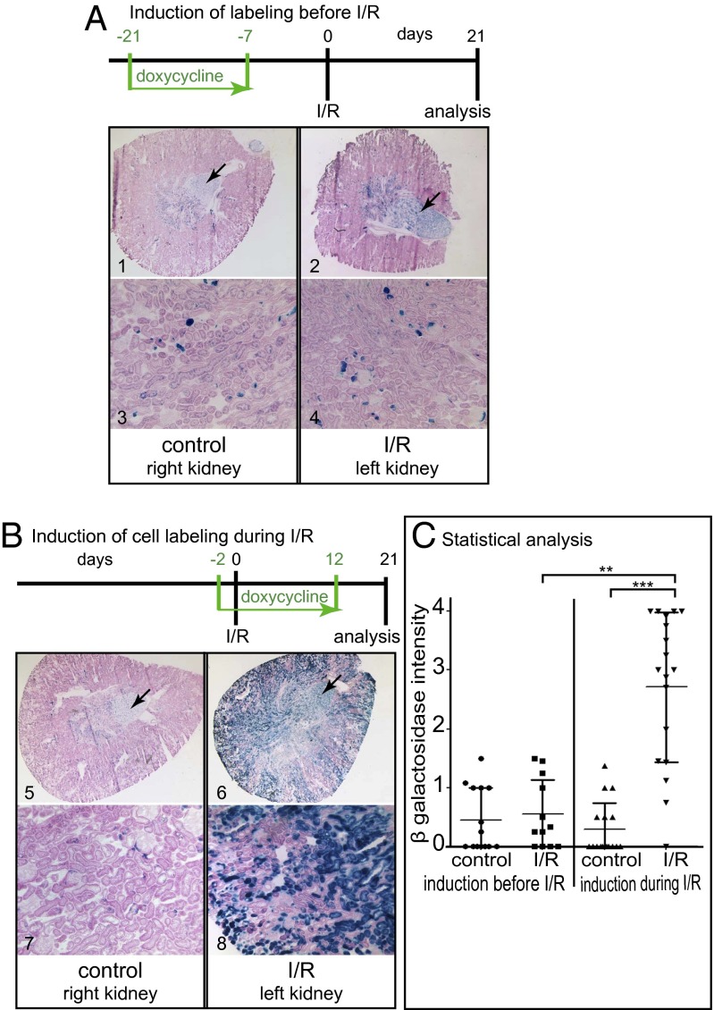
Acute kidney injury (AKI) is associated with high morbidity and mortality. Recent genetic fate mapping studies demonstrated that recovery from AKI occurs from intrinsic tubular cells. It is unresolved whether these intrinsic cells (so-called "scattered tubular cells") represent fixed progenitor cells or whether recovery involves any surviving tubular cell. Here, we show that the doxycycline-inducible parietal epithelial cell (PEC)-specific PEC-reverse-tetracycline transactivator (rtTA) transgenic mouse also efficiently labels the scattered tubular cell population. Proximal tubular cells labeled by the PEC-rtTA mouse coexpressed markers for scattered tubular cells (kidney injury molecule 1, annexin A3, src-suppressed C-kinase substrate, and CD44) and showed a higher proliferative index. The PEC-rtTA mouse labeled more tubular cells upon different tubular injuries but was independent of cellular proliferation as determined in physiological growth of the kidney. To resolve whether scattered tubular cells are fixed progenitors, cells were irreversibly labeled before ischemia reperfusion injury (genetic cell fate mapping). During recovery, the frequency of labeled tubular cells remained constant, arguing against a fixed progenitor population. In contrast, when genetic labeling was induced during ischemic injury and subsequent recovery, the number of labeled cells increased significantly, indicating that scattered tubular cells arise from any surviving tubular cell. In summary, scattered tubular cells do not represent a fixed progenitor population but rather a phenotype that can be adopted by almost any proximal tubular cell upon injury. Understanding and modulating these phenotypic changes using the PEC-rtTA mouse may lead to more specific therapies in AKI.

Keywords: cell fate tracking; lineage tracing; regeneration; stem cells.


Conflict of interest statement

The authors declare no conflict of interest.

Figures

Fig. 1.
Fig. 1.
Transcriptional activity of PEC–rtTA mice in tubular cells. (A and B) Using two alternative reporter mice, PEC–rtTA mice are transcriptionally active in PECs (arrowheads) and also in individual STCs (arrows) in the renal cortex of 20-wk-old normal mice (200× magnification). (C) In a time course experiment, tubular cells could be efficiently labeled with EGFP–histone within 6 h after induction using a single i.p. injection of Dox (absolute numbers of EGFP-positive tubular cells in one visual field at 200× magnification, five fields per time point; **P < 0.01, one-way ANOVA with Bonferroni’s posttest, BPT). (DG) Up-regulation of PEC–rtTA transcriptional activity in tubular cells upon injury. Three different models of tublar injury were induced in PEC–rtTA/H2B–EGFP mice: UUO (D and E), I/R (F and F’), and the chronic Thy1.1 model (resulting in glomerular proteinuria, G). All different modes of tubular injury resulted in significant up-regulation of PEC–rtTA transcriptional activity primarily in proximal tubular cells (marked by AQP1). Arrowheads, EGFP-positive PECs; arrows, EGFP-positive tubular cells.
Fig. 2.
Fig. 2.
Increased proliferation of PEC–rtTA-labeled proximal tubular cells. (A) Experimental setup. Four groups of PEC–rtTA/H2B–EGFP mice received I/R surgery and Dox and/or BrdU injections as indicated. To evaluate the cumulative effects, the 1–57 h group received repeated injections. a, analysis. (B) Representative stainings for EGFP, BrdU, and Hoechst on paraffin sections of each experimental group (100× magnification). Arrows, tubular epithelial cells with nuclear EGFP and BrdU colabeling. (B’) Percentage of EGFP–histone-positive cells costaining for BrdU (n = 3 for each group, four visual fields of 100× magnification were analyzed for each mouse; ***P < 0.001, one-way ANOVA with BPT). (C) Pooled single cell suspensions of the renal cortex of two PEC–rtTA/H2B–EGFP mice described in A were stained for AQP1 and Hoechst and subjected to flow cytometric analysis using the indicated gating protocol. EGFP-positive proximal tubular cells were more likely to be proliferating (i.e., in S or G2 phase) compared with EGFP-negative proximal tubular cells. The difference was even more obvious in the contralateral control kidneys of the same animals (which contained less cellular debris). n = 3 for each time point; error bars mark SD (**P < 0.01; ***P < 0.001, one-way ANOVA BPT). (DE’’’) No increased labeling by PEC–rtTA in developmental renal hypertrophy of mice at 3 wk of age (adolescent mice). In a costaining for EGFP–histone, BrdU and Hoechst significantly increased BrdU incorporation, but no EGFP labeling was detected in tubular cells (D–D’’’). Seventeen-week-old adult mice served as controls (E–E’’’).
Fig. 3.
Fig. 3.
PEC–rtTA-labeled cells coexpress STC markers. For experimental setup, see Fig. 2A. (A) Representative images of EGFP/KIM-1/AQP1/Hoechst stainings of I/R injured kidneys. (B) Absolute numbers of EGFP/KIM-1/AQP1–expressing cortical cells (evaluated in a 200× visual field in three experimental mice at each time point). (C–F) Analysis of marker expression in AQP1- (C and D), KIM-1– (E), or EGFP-positive (F) cells (*P < 0.05; **P < 0.01; ***P < 0.001, one-way ANOVA BPT). (G) FACS analysis of cortical single cell preparations confirmed coexpression of KIM-1 in >30% of EGFP-positive AQP1 proximal tubular cells after I/R. PEC–rtTA/H2B–EGFP mice underwent I/R surgery and received Dox injections after 1, 25, and 49 h after surgery, and all mice were analyzed after 33 or 57 h. For both groups, three independent measurements were made of pooled kidneys of two mice each (total of six mice per group). Error bars mark SD (**P < 0.01, ***P < 0.001, one-way ANOVA). (H and I) Coexpression of EGFP–histone, Hoechst, and annexin A3 (H) or SSeCKS (I) 5 d after I/R (arrows). (J) Coexpression of CD44 with EGFP–histone 5 d after UUO (arrows). (K) No evidence for loss of brush border in PEC–rtTA-labeled cells in healthy and contralateral kidneys. As shown in the triple staining in the Left panel, labeled cells without brush border [i.e., lotus tetragonolobus agglutinin (LTA) negative] were almost always a consequence of the plane of the section (arrows). For statistical analysis, four visual fields at 100× magnification were evaluated in n = 3 animals; error bars mark SD. (L) Absolute numbers of EGFP-positive cells in four visual fields show increased labeling in contralateral control kidneys of animals subjected to I/R and receiving repeated Dox/BrdU injections (1–57 h) versus healthy controls (P < 0.01, unpaired t test). (M) PEC–rtTA-labeled EGFP-positive cells also showed increased BrdU incorporation in contralateral kidneys after I/R injury (n = 3, unpaired t test P = 0.0698). (Right) Similarly labeled cells proliferated more even after only 15 min of ischemia (n = 4). Error bars mark SD; **P < 0.01. (N) Absolute numbers of EGFP-positive tubular cells correlate with BrdU-positive cells in kidneys with different degrees of tubular damage after only 15 min of ischemia; R2 = 0.7449, P < 0.0001.
Fig. 4.
Fig. 4.
PEC–rtTA-labeled cells are not a fixed progenitor population. (A) To test whether the PEC–rtTA mouse labels a fixed tubular progenitor population, irreversible genetic tagging was induced before I/R injury in PEC–rtTA/LC1/R26R mice. There was no increase of labeled tubular cells 21 d after I/R in the injured kidney (beta-gal/eosin–stained cryosections at a magnification of 25× or 100×). Constitutively labeled cells in the medulla serve as positive controls for beta-gal staining (arrows, n = 14). (B) To test whether the PEC–rtTA mouse labels injured and regenerating tubular cells, irreversible genetic labeling was induced during I/R. Compared with the contralateral control kidneys, significantly more tubular cells were genetically labeled in the injured kidney (n = 18). (C) Beta-gal staining intensity was evaluated in a blinded semiquantitative fashion (***P < 0.0001; **P < 0.01, Kruskal Wallis test with Dunn’s posttest). A significant increase in PEC–rtTA-labeled cells was observed only when genetic labeling was induced during I/R and recovery.

References

    1. McCampbell KK, Wingert RA. Renal stem cells: Fact or science fiction? Biochem J. 2012;444(2):153–168. - PMC - PubMed
    1. Duffield JS, Humphreys BD. Origin of new cells in the adult kidney: Results from genetic labeling techniques. Kidney Int. 2011;79(5):494–501. - PubMed
    1. Little MH, Bertram JF. Is there such a thing as a renal stem cell? J Am Soc Nephrol. 2009;20(10):2112–2117. - PubMed
    1. Reule S, Gupta S. Kidney regeneration and resident stem cells. Organogenesis. 2011;7(2):135–139. - PMC - PubMed
    1. Glaumann B, Glaumann H, Trump BF. Studies of cellular recovery from injury. III. Ultrastructural studies on the recovery of the pars recta of the proximal tubule (P3 segment) of the rat kidney from temporary ischemia. Virchows Arch B Cell Pathol Incl Mol Pathol. 1977;25(4):281–308. - PubMed

Publication types