. 2014 Jan 28;111(4):1539-44.
doi: 10.1073/pnas.1317488111. Epub 2014 Jan 13.

Proteolytic cleavage of Ser52Pro variant transthyretin triggers its amyloid fibrillogenesis

Affiliations

Proteolytic cleavage of Ser52Pro variant transthyretin triggers its amyloid fibrillogenesis

P Patrizia Mangione et al. Proc Natl Acad Sci U S A. .

Abstract

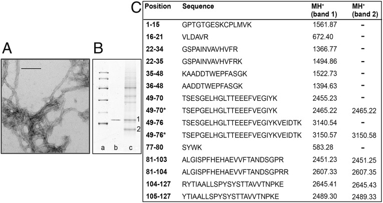
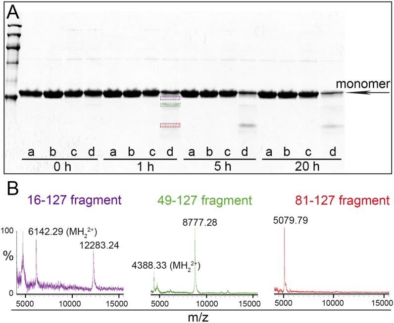
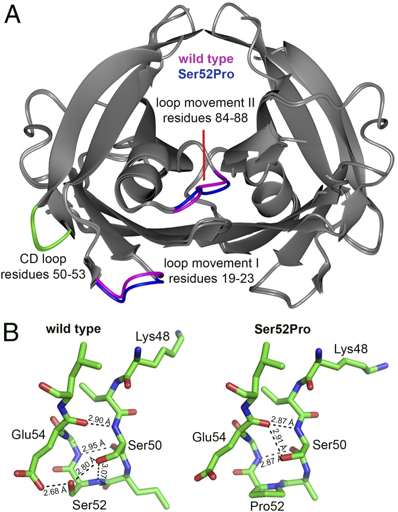
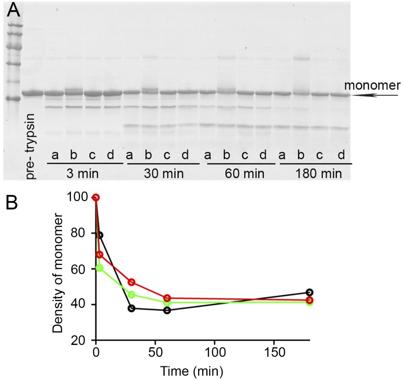
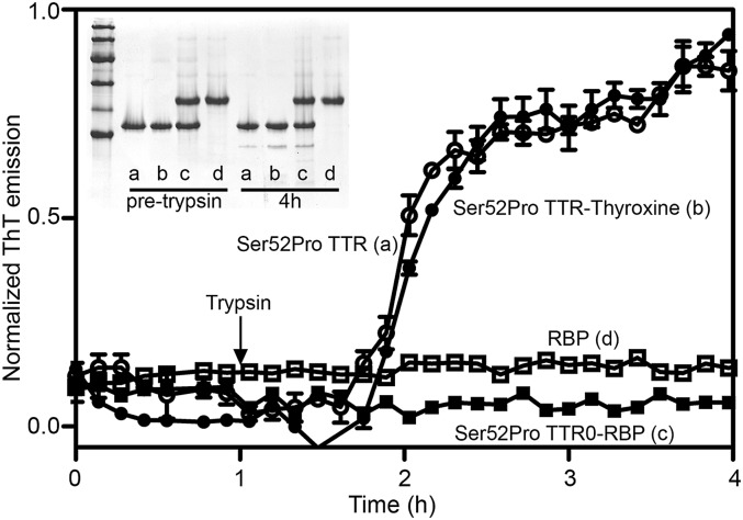
The Ser52Pro variant of transthyretin (TTR) produces aggressive, highly penetrant, autosomal-dominant systemic amyloidosis in persons heterozygous for the causative mutation. Together with a minor quantity of full-length wild-type and variant TTR, the main component of the ex vivo fibrils was the residue 49-127 fragment of the TTR variant, the portion of the TTR sequence that previously has been reported to be the principal constituent of type A, cardiac amyloid fibrils formed from wild-type TTR and other TTR variants [Bergstrom J, et al. (2005) J Pathol 206(2):224-232]. This specific truncation of Ser52Pro TTR was generated readily in vitro by limited proteolysis. In physiological conditions and under agitation the residue 49-127 proteolytic fragment rapidly and completely self-aggregates into typical amyloid fibrils. The remarkable susceptibility to such cleavage is likely caused by localized destabilization of the β-turn linking strands C and D caused by loss of the wild-type hydrogen-bonding network between the side chains of residues Ser52, Glu54, Ser50, and a water molecule, as revealed by the high-resolution crystallographic structure of Ser52Pro TTR. We thus provide a structural basis for the recently hypothesized, crucial pathogenic role of proteolytic cleavage in TTR amyloid fibrillogenesis. Binding of the natural ligands thyroxine or retinol-binding protein (RBP) by Ser52Pro variant TTR stabilizes the native tetrameric assembly, but neither protected the variant from proteolysis. However, binding of RBP, but not thyroxine, inhibited subsequent fibrillogenesis.

Keywords: misfolding; protein aggregation.


Conflict of interest statement

The authors declare no conflict of interest.

Figures

Fig. 1.
Fig. 1.
Ex vivo amyloid fibrils. (A) Transmission electron microscopy of amyloid fibrils extracted from spleen. (Direct magnification: 230,000×; scale bar, 100 nm.) (B) SDS 15% PAGE under reducing conditions. Lane a: marker proteins (14.4, 20.1, 30.0, 45.0, 66.0, and 97.0 kDa, respectively); lane b: 0.5 μg recombinant Ser52Pro TTR; lane c: 60 μg ex vivo amyloid fibrils from spleen. 1 and 2 indicate bands subjected to mass mapping analysis. (C) Tryptic peptides obtained by digestion of SDS/PAGE bands were analyzed by MALDI-MS and nano-LC MS-MS. MH+ monoisotopic values are reported for each peptide; asterisks indicate the presence of the Ser52Pro substitution. Cysteine is carbamidomethylated.
Fig. 2.
Fig. 2.
Limited trypsin proteolysis of wild-type and Ser52Pro TTR. (A) SDS (15%) PAGE under reducing conditions of wild-type and Ser52Pro TTR after incubation at 37 °C with trypsin at an enzyme:substrate ratio of 1:200 for the times shown. Wild-type TTR alone (a) or with trypsin (b), Ser52Pro TTR alone (c) or with trypsin (d). The three main polypeptides released from Ser52Pro TTR after 1 h of digestion are highlighted with purple, green, and red boxes. (B) MALDI-MS spectra of the fragments electroeluted from the bands shown in A and acquired in linear mode.
Fig. 3.
Fig. 3.
Fibrillogenesis of Ser52Pro TTR triggered by release of the residue 49-127 fragment. (A) Normalized ThT fluorescence emission from wild-type TTR (black) and Ser52Pro TTR (red) fibrillogenesis in the presence (solid lines) and absence (dotted lines) of trypsin 5 ng/μL. (B) SDS 15% PAGE under reducing conditions. Lane a: marker proteins (14.4, 20.1, 30.0, 45.0, 66.0, and 97.0 kDa); lane b: recombinant Ser52Pro TTR; lane c: in vitro Ser52Pro fibrils; lane d: ex vivo amyloid fibrils from spleen. (C) Negatively stained transmission electron micrograph of Ser52Pro TTR amyloid fibrils generated in the presence of trypsin and shaking. (Direct magnification: 175,000×; scale bar, 100 nm.) (D) Tapping mode atomic force microscopy image (height data) of Ser52Pro TTR fibrils obtained as in A. Scan size: 2.0 μm; Z range: 10 nm. (E) The X-ray fiber diffraction pattern collected from partially aligned amyloid fibrils formed by Ser52Pro TTR (λ = 1.5417 Å; sample-to-detector distance = 100 nm; exposure time = 30 s) shows a reflection that is stronger on the meridian at 4.7 Å. A reflection also is highlighted at 9.5 Å, and a well-oriented signal is seen at 18 Å.
Fig. 4.
Fig. 4.
3D structure of wild-type and Ser52Pro TTR. (A) Crystal structures of wild-type and Ser52Pro TTR were superposed by secondary structure matching [gray-interpreted and presented by CCP4MG (27)] and show the close similarity of the folds, Rmsd = 0.42Å over 221 residues. Regions of greatest difference are highlighted in blue (Ser52Pro) and magenta (wild type). The CD loops for one subunit of both proteins are shown in green. (B) Stick diagram of the CD loop region (carbon is shown in green, oxygen in red, and nitrogen in blue) for wild-type (Left) and Ser52Pro (Right) TTR showing the diminished hydrogen-bonding network in the vicinity of the proline substitution that may contribute to the enhanced susceptibility of the variant to proteolytic cleavage at Lys48 (28). X-ray data statistics are reported in Table S3.
Fig. 5.
Fig. 5.
Limited proteolysis of Ser52Pro TTR with bound ligands. (A) SDS 15% PAGE under reducing conditions of Ser52Pro TTR alone in presence of DMSO (a) or a fivefold molar excess of epigallocatechin (b), mds84 (c), or Tafamidis (d) after incubation at 37 °C with trypsin at an enzyme:substrate ratio of 1:200 for the times shown. Marker proteins (14.4, 20.1, 30.0, 45.0, 66.0, and 97.0 kDa) and the nontreated Ser52Pro TTR (pretrypsin) are in the first two lanes. (B) Kinetics of proteolytic cleavage of holo Ser52Pro TTR in presence of DMSO (black), mds84 (green), or Tafamidis (red) monitored by density with time of the intact monomeric TTR band (arrow in A).
Fig. 6.
Fig. 6.
Effect of natural interactors, thyroxine and RBP, on proteolysis/fibrillogenesis by Ser52Pro TTR. Normalized ThT fluorescence emission for trypsin-dependent fibrillogenesis of Ser52Pro TTR alone (a) or in the presence of a twofold excess of thyroxine (b), a twofold excess of RBP (c), or RBP alone (d). (Inset) SDS 15% PAGE under reducing conditions of fibrillogenesis samples before and after 4 h of trypsin digestion. Marker proteins (14.4, 20.1, 30.0, 45.0, 66.0, and 97.0 kDa) are included.

References

    1. Pepys MB. Amyloidosis. Annu Rev Med. 2006;57(1):223–241. - PubMed
    1. Merlini G, Bellotti V. Molecular mechanisms of amyloidosis. N Engl J Med. 2003;349(6):583–596. - PubMed
    1. Valleix S, et al. Hereditary systemic amyloidosis due to Asp76Asn variant β2-microglobulin. N Engl J Med. 2012;366(24):2276–2283. - PubMed
    1. Palaninathan SK. Nearly 200 X-ray crystal structures of transthyretin: What do they tell us about this protein and the design of drugs for TTR amyloidoses? Curr Med Chem. 2012;19(15):2324–2342. - PubMed
    1. Hammarström P, Wiseman RL, Powers ET, Kelly JW. Prevention of transthyretin amyloid disease by changing protein misfolding energetics. Science. 2003;299(5607):713–716. - PubMed

Publication types