. 2014 Jan 16:7:54.
doi: 10.3389/fnana.2013.00054. eCollection 2013.

Organization of the human fetal subpallium

Affiliations

Organization of the human fetal subpallium

Marie-Christin Pauly et al. Front Neuroanat. .

Abstract

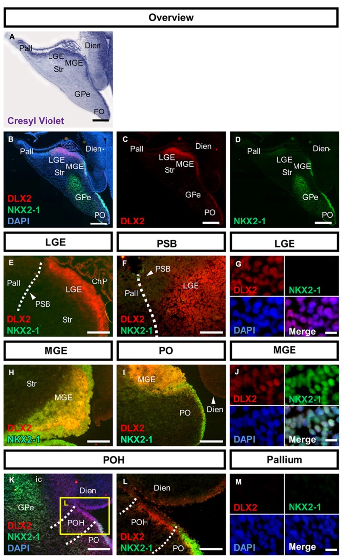
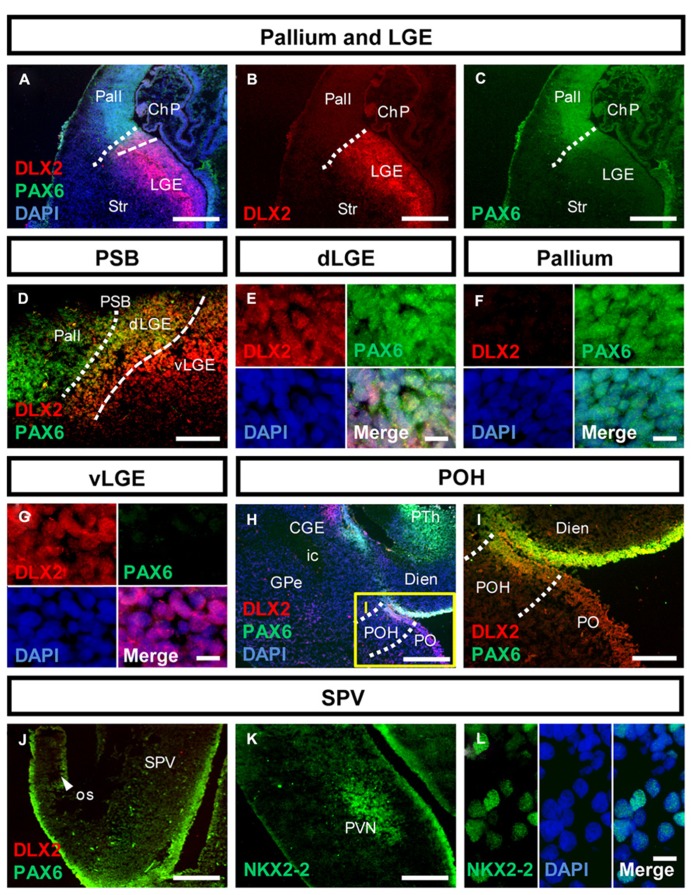
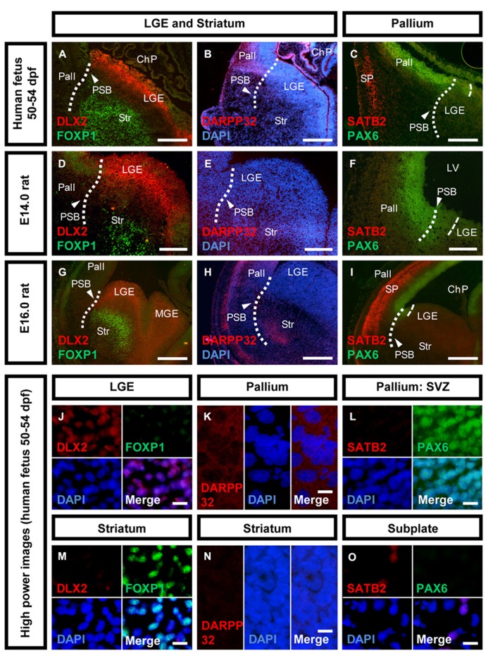
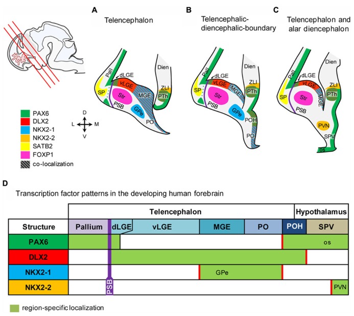
The subpallium comprises large parts of the basal ganglia including striatum and globus pallidus. Genes and factors involved in the development of the subpallium have been extensively studied in most vertebrates, including amphibians, birds, and rodents. However, our knowledge on patterning of the human subpallium remains insufficient. Using double fluorescent immunohistochemistry, we investigated the protein distribution of transcription factors involved in patterning of the subventricular zone (SVZ) in the human forebrain at late embryonic development. Furthermore, we compared the development of cortical and striatal precursors between human fetal brain and E14 and E16 fetal rat brains. Our results reveal that DLX2 marks SVZ precursors in the entire subpallium. Individual subpallial subdomains can be identified based on co-expression of DLX2 with either PAX6 or NKX2-1. SVZ precursors in the dorsal LGE and preopto-hypothalamic boundary are characterized by DLX2/PAX6 co-expression, while precursors in the MGE and preoptic region co-express DLX2/NKX2-1. SVZ precursors in the ventral LGE are DLX2(+)/PAX6(-)/NKX2-1(-). In terms of staging comparisons, the development of the corpus striatum in the human fetal brain during late embryonic stages corresponds well with the development of the striatum observed in E14 fetal rat brains. Our study demonstrates that the pattern underlying the development of the subpallium is highly conserved between rodents and humans and suggests a similar function for these factors in human brain development. Moreover, our data directly influence the application of ganglionic eminence derived human tissue for cell therapeutic approaches in neurodegenerative disorders such as Huntington's disease.

Keywords: development; ganglionic eminence; patterning; striatum; subpallium.


Figures

FIGURE 1
FIGURE 1
Molecular characterization of the human subpallium. (A) Photomicrographs of cresyl-violet coronal human subpallial sections. (B–D) Photomicrographs of sections stained against DLX2 (red) and NKX2-1 (green) and DAPI (blue). (E,F) DLX2-positive cells were detected in the LGE and end abruptly at the PSB. (G) Verification of nuclear DLX2 signal in the LGE and absence of NKX2-1. (H–J) DLX2/NKX2-1 co-expressing cells were detected in MGE and PO. (K,L) Cells within the boundary between subpallium and hypothalamus express DLX2 and lack NKX2-1. (M) Neither DLX2-positive nor NKX2-1 positive cells were detected in the pallium. Abbreviations: CGE, caudal ganglionic eminence; ChP, choroid plexus; DARPP32, dopamine- and cAMP-regulated phosphoprotein, 32 kDa; Dien, diencephalon; dLGE, dorsal part of the lateral ganglionic eminence; DLX2, distal-less homeobox protein 2; FOXP1, forkhead box protein P1; GPe, external segment of the globus pallidus; LGE, lateral ganglionic eminence; LV, lateral ventricle; MGE, medial ganglionic eminence; NKX2-1, homeobox protein NK-2 homolog A; NKX2-2, homeobox protein NK-2 homolog B; os, optic stalk; Pall, pallium; PAX6, paired box protein 6; PO, preoptic area; POH, preopto-hypothalamic boundary; PSB, pallial-subpallial boundary; PTh, prethalamus; PVN, paraventricular nucleus of the hypothalamus; SATB2, special AT-rich sequence-binding protein 2; SP, cortical subplate; SPV, supraopto-paraventricular region of the hypothalamus; Str, striatum; vLGE, ventral part of the lateral ganglionic eminence; ZLI, zona limitans intrathalamica. Scale bars: 1 mm in A–D. 500 μm in E,H,I,K; 200 μm in L; 100 μm in F; 10 μm in G,J,M.
FIGURE 2
FIGURE 2
Molecular identity of the pallial-subpallial boundary and the preopto-hypothalamic boundary in the human fetal brain. (A–C) Photomicrographs of coronal human subpallial sections stained against DLX2 (red) and PAX6 (green); (D) the dLGE co-expresses DLX2 and PAX6. (E–G): co-expressing DLX-2 positive and PAX6-positive cells were detected in the dLGE but not in the Pallium or LGE. (H,I) The boundary between subpallium and hypothalamus is characterized by DLX2/PAX6 co-expression (J–L) photomicrograph of the alar hypothalamus stained against (J) PAX6 (green) and DLX2 (red) and (K,L) NKX2-2 (green). (J) The alar hypothalamus is characterized by PAX6. (K,L) NKX2-2 is expressed in the SVZ of the AB boundary and in post-mitotic cells giving rise to the PVN. Abbreviations see Figure 1. Scale bars: 500 μm in A–C,H,J,K; 200 μm in I; 100 μm in D; 10 μm in E–G,L.
FIGURE 3
FIGURE 3
Neuronal maturation of the striatum versus cortex in human and rat.Photomicrographs of coronal sections of the human and rat telencephalon. (A,D,G,J,M) Expression of DLX2 (red) and FOXP1 (green) in the human, E14.0 and E16.0 rat LGE and striatum. (B,E,H,K,N) Expression of DARPP32 (red) and the nuclear counter stain DAPI (blue) in the human, E14.0 and E16.0 rat pallium, and striatum. (C,F,I,L,O) Expression of SATB2 (red) and PAX6 (green) in the human, E14.0 and E16.0 rat pallium, and cortical subplate. Abbreviations see Figure 1. Scale bars: 500 μm in A–C,G–I; 200 μm in D–F; 10 μm in J–O.
FIGURE 4
FIGURE 4
Schematic representation of the marker expression in the human fetal forebrain. (A–C) Schematic coronal sections of the telenecephalon, and alar dienecephalon. Section planes are indicated by red lines in the localizer on the left side. (D) Transcription factors expressed in subventricular precursors define molecular boundaries in the fetal human brain. Abbreviations see Figure 1.

References

    1. Assimacopoulos S., Grove E. A., Ragsdale C. W. (2003). Identification of a Pax6-dependent epidermal growth factor family signaling source at the lateral edge of the embryonic cerebral cortex. J. Neurosci. 23 6399–6403 - PMC - PubMed
    1. Bachoud-Lévi A., Bourdet C., Brugières P., Nguyen J. P., Grandmougin T., Haddad B., et al. (2000). Safety and tolerability assessment of intrastriatal neural allografts in five patients with Huntington‘s disease. Exp. Neurol. 161 194–202 10.1006/exnr.1999.7239 - DOI - PubMed
    1. Bardet S. M., Ferran J. L. E., Sanchez-Arrones L., Puelles L. (2010). Ontogenetic expression of sonic hedgehog in the chicken subpallium. Front. Neuroanat. 4:28. 10.3389/fnana.2010.00028 - DOI - PMC - PubMed
    1. Bayatti N., Moss J. A., Sun L., Ambrose P., Ward J. F. H., Lindsay S., et al. (2008). A molecular neuroanatomical study of the developing human neocortex from 8 to 17 postconceptional weeks revealing the early differentiation of the subplate and subventricular zone. Cereb. Cortex N. Y. N 1991 18 1536–1548 10.1093/cercor/bhm184 - DOI - PMC - PubMed
    1. Bulfone A., Kim H. J., Puelles L., Porteus M. H., Grippo J. F., Rubenstein J. L. (1993). The mouse Dlx-2 (Tes-1) gene is expressed in spatially restricted domains of the forebrain, face and limbs in midgestation mouse embryos. Mech. Dev. 40 129–140 10.1016/0925-4773(93)90071-5 - DOI - PubMed

LinkOut - more resources