. 2014 Jan 27;9(1):e85843.
doi: 10.1371/journal.pone.0085843. eCollection 2014.

Frequent seizures are associated with a network of gray matter atrophy in temporal lobe epilepsy with or without hippocampal sclerosis

Affiliations

Frequent seizures are associated with a network of gray matter atrophy in temporal lobe epilepsy with or without hippocampal sclerosis

Ana C Coan et al. PLoS One. .

Abstract

Objective: Patients with temporal lobe epilepsy (TLE) with hippocampal sclerosis (HS) have diffuse subtle gray matter (GM) atrophy detectable by MRI quantification analyses. However, it is not clear whether the etiology and seizure frequency are associated with this atrophy. We aimed to evaluate the occurrence of GM atrophy and the influence of seizure frequency in patients with TLE and either normal MRI (TLE-NL) or MRI signs of HS (TLE-HS).

Methods: We evaluated a group of 172 consecutive patients with unilateral TLE-HS or TLE-NL as defined by hippocampal volumetry and signal quantification (122 TLE-HS and 50 TLE-NL) plus a group of 82 healthy individuals. Voxel-based morphometry was performed with VBM8/SPM8 in 3T MRIs. Patients with up to three complex partial seizures and no generalized tonic-clonic seizures in the previous year were considered to have infrequent seizures. Those who did not fulfill these criteria were considered to have frequent seizures.

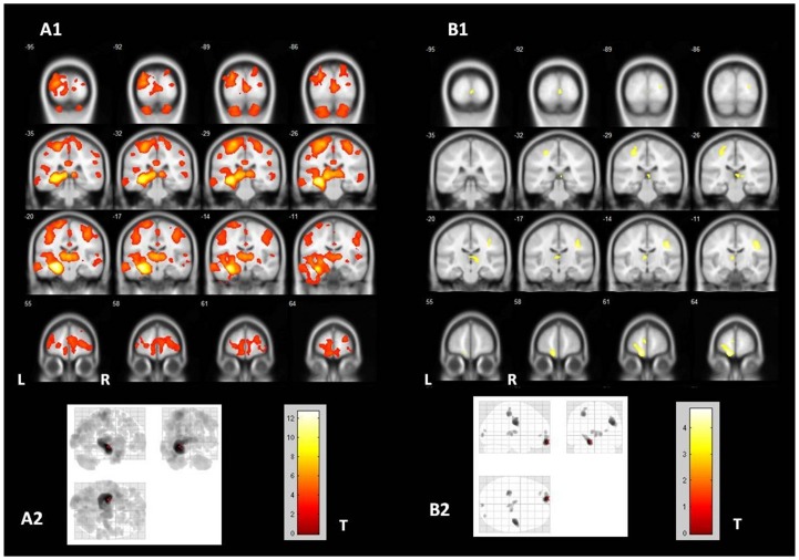
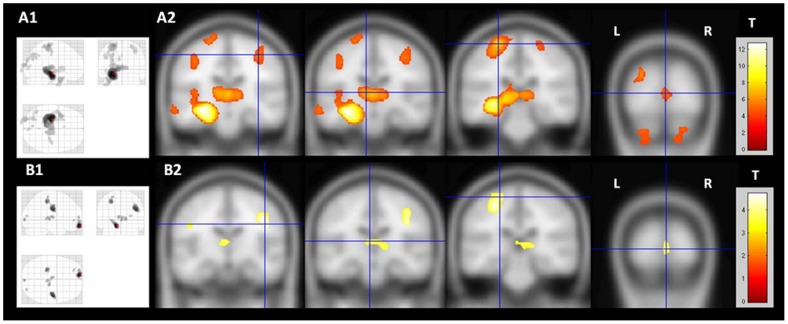
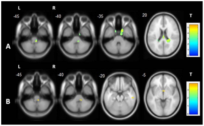
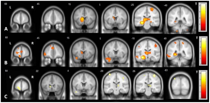
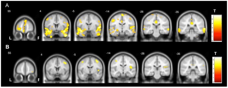
Results: Patients with TLE-HS had more pronounced GM atrophy, including the ipsilateral mesial temporal structures, temporal lobe, bilateral thalami and pre/post-central gyri. Patients with TLE-NL had more subtle GM atrophy, including the ipsilateral orbitofrontal cortex, bilateral thalami and pre/post-central gyri. Both TLE-HS and TLE-NL showed increased GM volume in the contralateral pons. TLE-HS patients with frequent seizures had more pronounced GM atrophy in extra-temporal regions than TLE-HS with infrequent seizures. Patients with TLE-NL and infrequent seizures had no detectable GM atrophy. In both TLE-HS and TLE-NL, the duration of epilepsy correlated with GM atrophy in extra-hippocampal regions.

Conclusion: Although a diffuse network GM atrophy occurs in both TLE-HS and TLE-NL, this is strikingly more evident in TLE-HS and in patients with frequent seizures. These findings suggest that neocortical atrophy in TLE is related to the ongoing seizures and epilepsy duration, while thalamic atrophy is more probably related to the original epileptogenic process.


Conflict of interest statement

Competing Interests: The authors have declared that no competing interests exist.

Figures

Figure 1
Figure 1. Gray matter atrophy in TLE-HS and TLE-NL.
VBM demonstrated significant areas of diffuse gray matter volume loss in TLE-HS and TLE-NL. A1 and A2 (“glass view”) show the areas of gray matter atrophy in TLE-HS (two-sample T-test, p<0.001, uncorrected, minimum threshold cluster of 30 voxels); B1 and B2 (“glass view”) show the areas of gray matter atrophy in TLE-NL (two-sample T-test, p<0.001, uncorrected, minimum threshold cluster of 30 voxels). TLE-HS: temporal lobe epilepsy with MRI signs of hippocampal sclerosis; TLE-NL: temporal lobe epilepsy with normal MRI; VBM: voxel based morphometry; T: t-value; L: left; R: right.
Figure 2
Figure 2. Common areas of gray matter atrophy in TLE-HS and TLE-NL.
The slices demonstrate the common brain areas with gray matter atrophy in TLE-HS and TLE-NL. TLE-HS results are shown with a more stringent statistical significance in order to facilitate the comparison. A1 (“glass view”) and A2 show the areas of gray matter atrophy in TLE-HS (two-sample T-test, p<0.05, FWE corrected, minimum threshold cluster of 30 voxels); B1 (“glass view”) and B2 show the areas of gray matter atrophy in TLE-NL (two-sample T-test, p<0.001, uncorrected, minimum threshold cluster of 30 voxels). TLE-HS: temporal lobe epilepsy with MRI signs of hippocampal sclerosis; TLE-NL: temporal lobe epilepsy with normal MRI; VBM: voxel based morphometry; T: t-value; L: left; R: right.
Figure 3
Figure 3. Gray matter volume increase in TLE-HS and TLE-NL.
VBM demonstrated areas of gray matter increase in TLE-HS and TLE-NL. A: shows the areas of gray matter volume increase in TLE-HS (two-sample T-test, p<0.001, uncorrected, minimum threshold cluster of 30 voxels); B: shows the areas of gray matter volume increase in TLE-NL (two-sample T-test, p<0.001, uncorrected, minimum threshold cluster of 30 voxels). TLE-HS: temporal lobe epilepsy with MRI signs of hippocampal sclerosis; TLE-NL: temporal lobe epilepsy with normal MRI; VBM: voxel based morphometry; T: t-value; L: left; R: right.
Figure 4
Figure 4. Patterns of gray matter atrophy according to seizure frequency in TLE-HS and TLE-NL.
VBM demonstrated significant areas of diffuse gray matter atrophy in TLE-HS patients with infrequent and frequent seizures but only in TLE-NL with frequent seizures. A: areas of gray matter atrophy in TLE-HS with infrequent seizures (two-sample T-test, p<0.001, uncorrected, minimum threshold cluster of 30 voxels); B: areas of gray matter atrophy in TLE-HS with frequent seizures (two-sample T-test, p<0.001, uncorrected, minimum threshold cluster of 30 voxels); C: areas of gray matter atrophy in TLE-NL with frequent seizures (two-sample T-test, p<0.001, uncorrected, minimum threshold cluster of 30 voxels). TLE-HS: temporal lobe epilepsy with MRI signs of hippocampal sclerosis; TLE-NL: temporal lobe epilepsy with normal MRI; VBM: voxel based morphometry; T: t-value; L: left; R: right.
Figure 5
Figure 5. Areas of gray matter atrophy associated with epilepsy duration in TLE-HS and TLE-NL.
Both groups of patients had areas of GM atrophy associated with epilepsy duration. In TLE-HS, the duration of epilepsy was correlated with GM atrophy, mainly in the bilateral anterior and lateral temporal lobes (A), while in TLE-NL it was more pronounced in bilateral precentral areas and contralateral insula (B) (Multiple regression, p<0.001, uncorrected, minimum threshold cluster of 30 voxels). TLE-HS: temporal lobe epilepsy with MRI signs of hippocampal sclerosis; TLE-NL: temporal lobe epilepsy with normal MRI; T: t-value; L: left; R: right.

References

    1. Bonilha L, Rorden C, Castellano G, Pereira F, Rio PA, et al. (2004) Voxel-based morphometry reveals gray matter network atrophy in refractory medial temporal lobe epilepsy. Arch Neurol 61: 1379–1384. - PubMed
    1. Spencer SS (2002) Neural networks in human epilepsy: Evidence of and implications for treatment. Epilepsia 43: 219–227. - PubMed
    1. Hauser WA (1992) The natural history of temporal lobe epilepsy. In: Lüders, H.O., ed. Epilepsy Surgery. New York: Raven Press, p. 133–141.
    1. Cohen-Gadol AA, Bradley CC, Williamson A, Kim JH, Westerveld M, et al. (2005) Normal magnetic resonance imaging and medial temporal lobe epilepsy: the clinical syndrome of paradoxical temporal lobe epilepsy. J Neurosurg 102: 902–909. - PubMed
    1. Bell ML, Rao S, So EL, Trenerry M, Kazemi N, et al. (2009) Epilepsy surgery outcomes in temporal lobe epilepsy with a normal MRI. Epilepsia 50: 2053–2060. - PMC - PubMed

Publication types