. 2014 Jan 24;9(1):e86117.
doi: 10.1371/journal.pone.0086117. eCollection 2014.

Flos Lonicera ameliorates obesity and associated endotoxemia in rats through modulation of gut permeability and intestinal microbiota

Affiliations

Flos Lonicera ameliorates obesity and associated endotoxemia in rats through modulation of gut permeability and intestinal microbiota

Jing-Hua Wang et al. PLoS One. .

Abstract

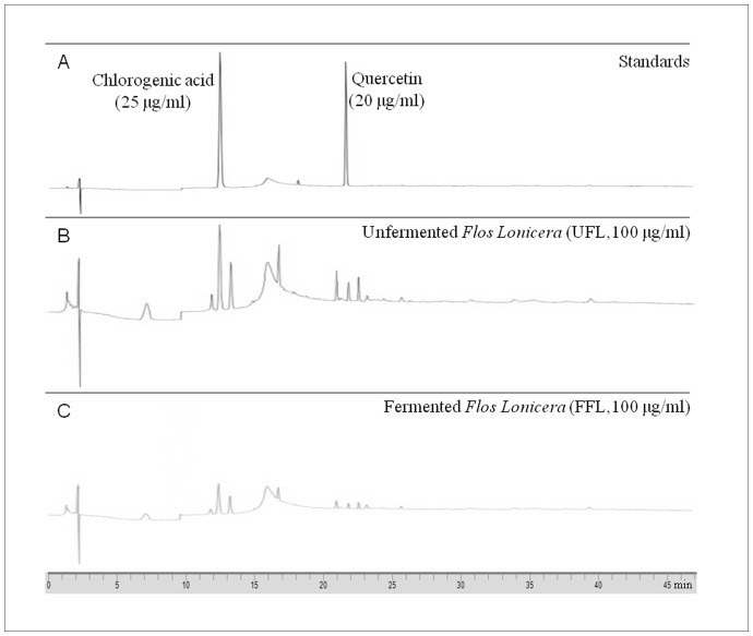
Background and aim: Increasing evidence has indicated a close association of host-gut flora metabolic interaction with obesity. Flos Lonicera, a traditional herbal medicine, is used widely in eastern Asia for the treatment of various disorders. The aim of this study was to evaluate whether unfermented or fermented formulations of Flos Lonicera could exert a beneficial impact to combat obesity and related metabolic endotoxemia.

Methods: Obesity and metabolic endotoxemia were induced separately or together in rats through feeding a eight-week high fat diet either alone (HFD control group) or in combination with a single LPS stimulation (intraperitoneal injection, 0.75 mg/kg) (LPS control group). While, the mechanism of action of the Lonicera formulations was explored in vitro using RAW 264.7 and HCT 116 cell lines as models.

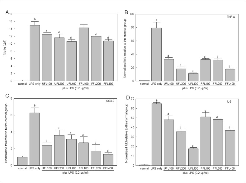
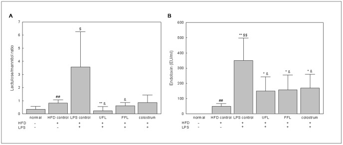
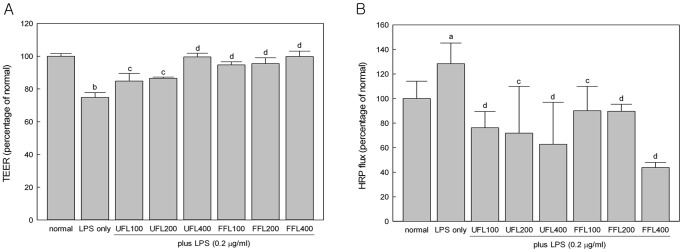
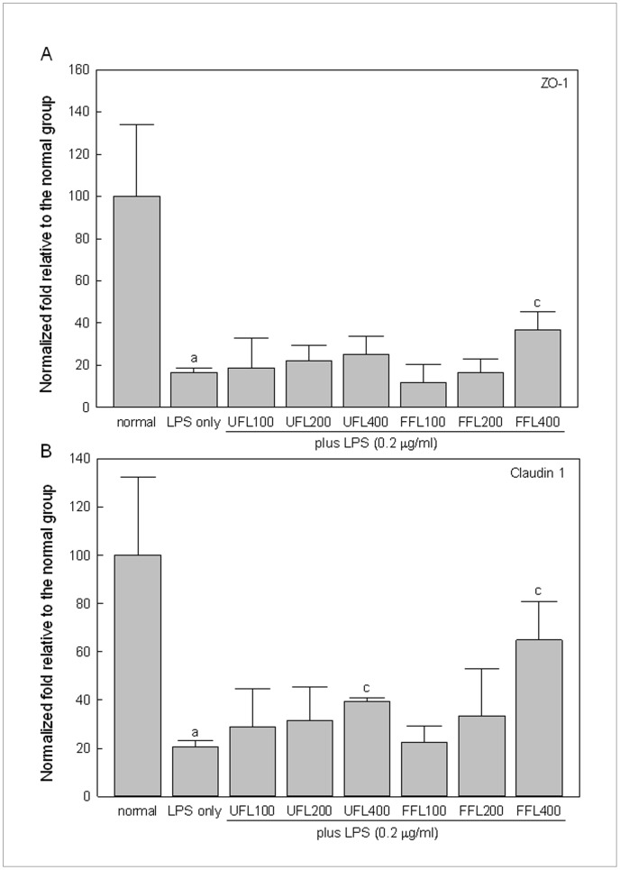
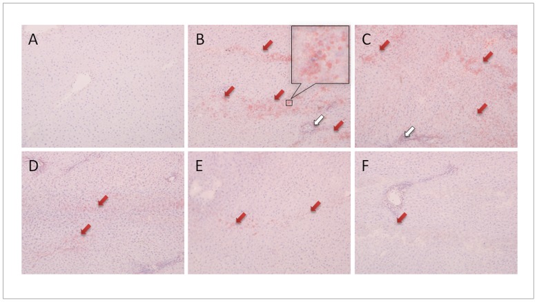
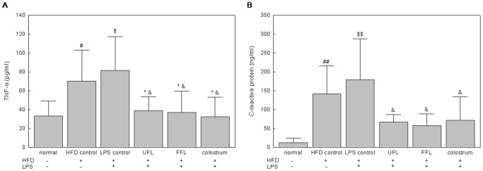
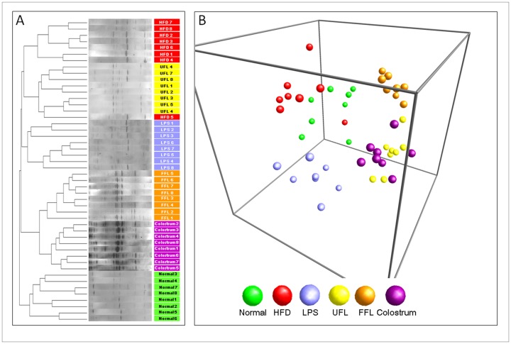
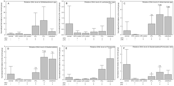
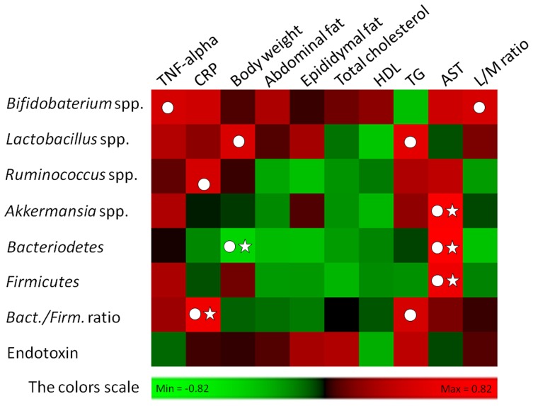
Results: In cell-based studies, treatment with both unfermented Flos Lonicera (UFL) and fermented Flos Lonicera (FFL) formulations resulted in suppression of LPS-induced NO production and gene expression of vital proinflammatory cytokines (TNF-α, COX-2, and IL-6) in RAW 264.7 cells, reduced the gene expression of zonula occludens (ZO)-1 and claudin-1, and normalized trans epithelial electric resistance (TEER) and horseradish peroxidase (HRP) flux in LPS-treated HCT-116 cells. In an animal study, treatment of HFD as well as HFD+LPS groups with UFL or FFL resulted in a notable decrease in body and adipose tissue weights, ameliorated total cholesterol, HDL, triglyceride, aspartate transaminase and endotoxin levels in serum, reduced the urinary lactulose/mannitol ratio, and markedly alleviated lipid accumulation in liver. In addition, exposure of HFD as well as HFD+LPS groups with UFL or FFL resulted in significant alteration of the distribution of intestinal flora, especially affecting the population of Akkermansia spp. and ratio of Bacteroidetes and Firmicutes.

Conclusion: This evidence collectively demonstrates that Flos Lonicera ameliorates obesity and related metabolic endotoxemia via regulating distribution of gut flora and gut permeability.


Conflict of interest statement

Competing Interests: The authors have declared that no competing interests exist.

Figures

Figure 1
Figure 1. The in vitro anti-inflammatory activities of UFL and FFL in terms of their ability to suppress the LPS-induced production of NO and proinflammatory cytokines.
(A) Immediately after pretreatment with UFL or FFL at 0 (normal, received DMEM instead of herbal extraxt), 100, 200, 400 µg/ml doses for 4 h, RAW 264.7 cells were exposed to DMEM (normal and LPS-alone) or 0.2 µg/ml LPS for 24 h following which NO measurement was performed as described in Materials and Methods section. b P<0.01 compared to the normal and c P<0.05, d P<0.01 compared to the LPS alone. (B–D) Immediately after pretreatment with UFL or FFL at 0 (normal, received DMEM instead of herbal extraxt), 100, 200, 400 µg/ml doses for 4 h, RAW 264.7 cells were exposed to DMEM (normal and LPS-alone) or 0.2 µg/ml LPS for 24 h following which the gene expression profile of proinflammatory cytokines (TNF-α, COX-2 and IL-6) were determined as described in Materials and Methods section. The results are expressed as normalized fold values relative to the normal. b P<0.01 compared to the normal and c P<0.05, d P<0.01 compared to the LPS alone.
Figure 2
Figure 2. UFL and FFL attenuate HFD+LPS-induced increase in gut permeability and serum endotoxin level.
(A) Following the termination of treatment schedule, the animals were fasted for 12 h with access to water ad libitum. Subsequently, 1.0 ml of lactulose-mannitol solution (containing 66 mg/ml lactulose and 50 mg/ml mannitol) was administered to the animals by oral gavage. After another 20 h of fasting, the urine samples were collected to determine the level of lactulose and mannitol as described in Materials and Methods section. The data are expressed as a ratio of lactulose to mannitol. (B) After the termination of experimental schedule, the blood was collected from the animals following which the serum endotoxin level was determined as described in Materials and Methods section. ## P<0.01, compared to the normal group; * P<0.05, ** P<0.01 compared to the HFD control group; $ P<0.05, $$ P<0.01, compared to the normal group; & P<0.05 compared to the LPS control group (n = 8).
Figure 3
Figure 3. UFL and FFL treatment reduce the elevation of membrane permeability in vitro.
(A) Immediately after pretreatment with UFL or FFL at 0 (normal, received McCoy’s 5A medium instead of herbal extraxt), 100, 200, 400 µg/ml doses for 24 h, HCT-116 cells were exposed to McCoy’s 5A medium (normal and LPS-alone) or 10 µg/ml LPS for 24 h following which measurement of transepithelial electrical residence (TEER) was performed as described in Materials and Methods section. (B) Immediately after pretreatment with UFL or FFL at 0 (normal, received McCoy’s 5A medium instead of herbal extract), 100, 200, 400 µg/ml doses for 24 h, HCT-116 cells cells were exposed to McCoy’s 5A medium (normal and LPS-alone) or 10 µg/ml LPS for 24 h following which horseradish peroxidase (HRP) flux assay was performed as described in Materials and Methods section. The experimental results in both cases are expressed as a percentage of the normal. a P<0.05, b P<0.01 compared to the normal and c P<0.05, d P<0.01 compared to the LPS alone.
Figure 4
Figure 4. UFL and FFL positively regulate tight junction protein genes expression in HCT 116 cells.
Immediately after pretreatment with UFL or FFL at 0 (normal, received McCoy’s 5A medium instead of herbal extraxt), 100, 200, 400 µg/ml doses for 2 h, HCT-116 cells were exposed to McCoy’s 5A medium (normal and LPS-alone) or 10 µg/ml LPS for 6 h following which expression of claudin-1 and zonula occludens-1 (ZO-1) genes was determined as described in Materials and Methods section. The experimental results are expressed as normalized fold change relative to the normal group. a P<0.05 compared to the normal and c P<0.05 compared to the LPS treatment alone.
Figure 5
Figure 5. UFL and FFL attenuate HFD and HFD+LPS-induced inflammation and obesity-related histological changes in liver.
Following the termination of experimental schedule, the animals were sacrificed and livers were collected and stored in −70°C. After embedding and making frozen sections of the tissues, the slides were stained with Oil red O solution and then counter-stained with hematoxylin. The pathophysiological examination of the tissue sections was performed under light microscopy with 400×magnification as described in Materials and Methods section. (A): normal, (B): HFD control, (C): LPS control, (D): HFD+LPS+UFL, (E): HFD+LPS+FFL, (F): HFD+LPS+colostrum. The red arrowheads indicate lipid droplets stained by Oil red O, and the white arrowheads indicate the infiltration of inflammatory cells.
Figure 6
Figure 6. UFL and FFL diminish the vital inflammatory markers in the serum of HFD-fed plus LPS-treated rats.
After the termination of experimental schedule, the blood was collected from the animals following which the serum endotoxin level was determined as described in Materials and Methods section. # P<0.05, ## P<0.01, compared to the normal group; * P<0.05 compared to the HFD control group; $ P<0.05, $$ P<0.01, compared to the normal group; & P<0.05, && P<0.01 compared to the LPS control group (n = 8).
Figure 7
Figure 7. PCR-denaturing gradient gel electrophoresis (PCR-DGGE) fingerprinting and principal coordinate analysis (PCA) of rat fecal samples.
(A) Following the termination of treatment schedule, the animals were fasted for 12 h with access to water ad libitum. After another 20 h of fasting, the stool samples were collected following which the fecal microbial communities were analyzed by DGGE as described in Materials and Methods section. (B) PCA of the data was performed based on distance matrix (two-dimensional array) to further evaluate the similarity between bacterial communities.
Figure 8
Figure 8. The relative DNA (gene-encoding 16S rRNA) content of related microbes in rat fecal samples.
Following the termination of treatment schedule, the animals were fasted for 12ad libitum. After another 20 h of fasting, the stool samples were collected following which the abundance of 16S rRNA gene of the bacterial strains were determined as described in Materials and Methods section. The results are expressed as normalized fold values relative to the normal group. * P<0.05, ** P<0.01 compared to the HFD control group; & P<0.05, && P<0.01 compared to the LPS control group (n = 8).
Figure 9
Figure 9. HPLC fingerprint of UFL and FFL.
The HPLC elution profile of extracted samples of unfermented and fermented Flos Lonicera and standards (chlorogenic acid and quercetin). The extracted samples and standards were filtrated and subjected to HPLC analysis as described in Materials and Methods section. A: standard: Chlorogenic acid (left) 25 µg/ml and Quercetin (right) 20 µg/ml, B: UFL (Unfermented Flos Lonicera) 100 µg/mL, C: FFL (fermented Flos Lonicera) 100 µg/mL. All chromatograms were produced at a wavelength of 238 nm.
Figure 10
Figure 10. Analyses of correlation between gut flora composition and host metabolic parameters.
The entire data of all experimental groups except normal were gathered and analyzed by SPSS software (17.0 version) using two-tailed Pearson’s correlation test. The scores of Pearson’s correlation were figured by PermutMatrix software (Version 1.9.3 EN) using heatmap plots. As the colors scale shown, green color indicates a positive correlation, while red color shows a negative correlation. (A symbol of ○ indicates absolute value of Pearson r >0.4; a symbol of ☆ indicates statistical significance of P<0.05).

References

    1. Kopelman PG (2000) Obesity as a medical problem. Nature 404: 635–643. - PubMed
    1. National Institutes of Health (1998) Clinical Guidelines on the Identification, Evaluation, and Treatment of Overweight and Obesity in Adults. Bethesda: National Institutes of Health.
    1. World Health Organization (2005) Preventing Chronic Diseases: a vital investment: WHO global report. Geneva: World Health Organization. 54–55 p.
    1. Rocha VZ, Folco EJ (2011) Inflammatory concepts of obesity. Int J Inflam 2011: 529061 10.4061/2011/529061 - DOI - PMC - PubMed
    1. Fei N, Zhao L (2013) An opportunistic pathogen isolated from the gut of an obese human causes obesity in germfree mice. ISME J 7: 880–884. - PMC - PubMed

Publication types

MeSH terms