. 2014 Jan 27;9(1):e86553.
doi: 10.1371/journal.pone.0086553. eCollection 2014.

Farnesoid X receptor ligand prevents cisplatin-induced kidney injury by enhancing small heterodimer partner

Affiliations

Farnesoid X receptor ligand prevents cisplatin-induced kidney injury by enhancing small heterodimer partner

Eun Hui Bae et al. PLoS One. .

Abstract

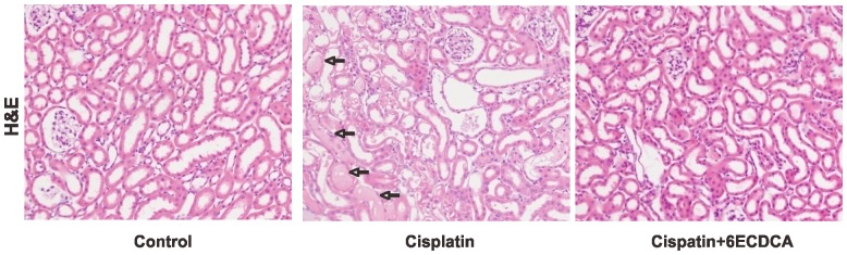
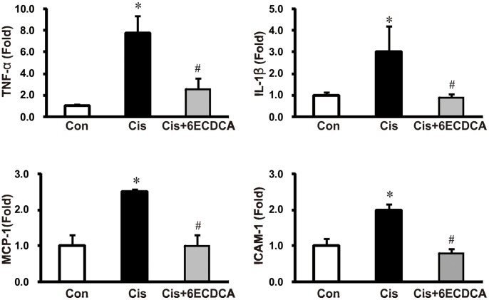
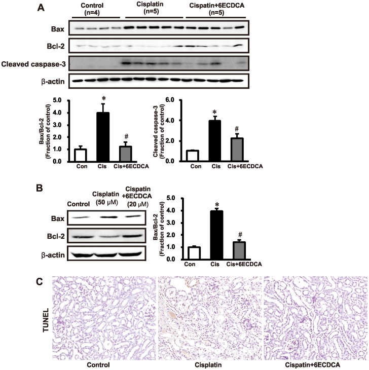
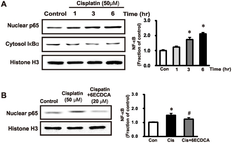
The farnesoid X receptor (FXR) is mainly expressed in liver, intestine and kidney. We investigated whether 6-ethyl chenodeoxycholic acid (6ECDCA), a semisynthetic derivative of chenodeoxycholic aicd (CDCA, an FXR ligand), protects against kidney injury and modulates small heterodimer partner (SHP) in cisplatin-induced kidney injury. Cisplatin inhibited SHP protein expression in the kidney of cisplatin-treated mice and human proximal tubular (HK2) cells; this effect was counteracted by FXR ligand. Hematoxylin and eosin staining revealed the presence of tubular casts, obstructions and dilatations in cisplatin-induced kidney injury, which was attenuated by FXR ligand. FXR ligand also attenuated protein expression of transforming growth factor-β1 (TGF-β1), Smad signaling, and the epithelial-to-mesenchymal transition process, inflammatory markers and cytokines, and apoptotic markers in cisplatin-treated mice. Cisplatin induced NF-κB activation in HK2 cell; this effect was attenuated by pretreatment with FXR ligand. In SHP knockdown by small interfering RNA, cisplatin-induced activation of TGF-β1, p-JNK and Bax/Bcl-2 ratio was not attenuated, while SHP overexpression and FXR ligand inhibited expression of these proteins in cisplatin-pretreated HK2 cells. In conclusion, FXR ligand, 6ECDCA prevents cisplatin-induced kidney injury, the underlying mechanism of which may be associated with anti-fibrotic, anti-inflammatory, and anti-apoptotic effects through SHP induction.


Conflict of interest statement

Competing Interests: The authors have declared that no competing interests exist.

Figures

Figure 1
Figure 1. Time- and dose-dependent SHP mRNA and protein expression after FXR ligand 6ECDCA treatment in HK-2 cells.
(A) mRNA expression of SHP was increased with 6ECDCA treatment. (B) SHP protein expression was increased by treatment with 6ECDCA. Each column represents mean ± SEM. 6ECDCA, 6-Ethyl chenodeoxycholic acid; SHP, small heterodimer partner. GAPDH and β-actin protein levels were analyzed as internal controls. *p<0.05, compared with the control. Data are representative of at least 3 independent experiments.
Figure 2
Figure 2. Immunoblotting of SHP in cisplatin-induced renal injury.
SHP protein expression was reduced in cisplatin-induced renal injury mice, and it was attenuated by FXR ligand treatment (A). In HK-2 cells, SHP protein exhibited significantly decreased expression after cisplatin (50 µM, 16 h) treatment, and FXR ligand (20 µM, 1 h) pretreatment prevented this cisplatin-mediated decrease in SHP expression (B). Each column represents mean ± SEM. 6ECDCA, 6-ethyl chenodeoxycholic acid; SHP, small heterodimer partner. β-actin protein served as an internal control. Each column represents mean ± SEM. *p<0.05, compared with the control. #p<0.05, compared with the cisplatin treatment.
Figure 3
Figure 3. Effects of FXR ligand treatment on morphological changes in cisplatin-induced kidney injury.
Hematoxylin and eosin (H&E) staining revealed the presence of tubular casts and obstructions (arrow) in the kidney of cisplatin-treated mice, which was attenuated by FXR ligand co-treatment. Magnification,×200.
Figure 4
Figure 4. Effects of FXR ligand on TGF β1-Smad signaling in cisplatin-induced kidney injury.
(A) TGF-β1 expression was increased in the cisplatin group. Cisplatin also increased the expression of Smad-4, while it reduced the level of inhibitory Smad-6. These cisplatin-induced changes were abolished or significantly attenuated in the FXR ligand-co-treated group. Each column represents mean ± SEM. *p<0.05, compared with the control. #p<0.05, compared with the cisplatin treatment. (B) Immunoperoxidase microscopy of TGF-β1 in the kidney cortex. Increased immunolabeling was evident in the cisplatin-treated mouse kidneys; the increase was prevented by FXR ligand treatment. Magnification,×200.
Figure 5
Figure 5. Effects of FXR ligand on epithelial-mesenchymal transition and fibrosis in cisplatin-induced kidney injury.
(A) Protein expression of the epithelial receptor E-cadherin and the myofibroblast molecular marker α-SMA was also assessed. In cisplatin-treated mice, E-cadherin expression decreased and α-SMA increased; this effect was prevented in the FXR ligand-co-treated group. (B) In vitro (HK-2 cells) studies showed that FXR ligand pretreatment (20 µM, 1h) also reduced fibronectin and connective tissue growth factor (CTGF) expression induced by cisplatin (50 µM, 16 h). Each column represents mean ± SEM. *p<0.05, compared with the control. #p<0.05, compared with the cisplatin treatment.
Figure 6
Figure 6. Effects of FXR ligand on inflammatory cytokines and adhesion molecules in cisplatin-induced kidney injury.
Cisplatin treatment significantly induced renal TNF-α, IL-1β, MCP-1, and ICAM-1 mRNA expression. FXR ligand co-treatment suppressed the overexpression of these inflammatory cytokines and adhesion molecules. Each column represents mean ± SEM. *p<0.05 compared with the control animals. #p<0.05 compared with the cisplatin-treated mice.
Figure 7
Figure 7. Effects of FXR ligand on inflammatory proteins and MAPK pathway in cisplatin-induced kidney injury.
(A) ED-1 and COX2 protein expression in the kidney was significantly higher in cisplatin-treated mice than in the controls, which was ameliorated by FXR ligand treatment. Each column represents mean ± SEM. *p<0.05 compared with the control animals. #p<0.05 compared with cisplatin-treated mice. (B) The infiltration of ED-1-positive macrophages in cisplatin-treated mice was significantly higher than in control mice. FXR ligand co-treatment abrogated inflammatory cell infiltration in cisplatin-treated kidneys. Magnification, 200×. (C) HK-2 cells were incubated with cisplatin (50 µM. 3 h) and pERK and pJNK expression were determined in cells pretreated with FXR ligand (20 µM, 1 h) before cisplatin administration. In this experiment, pERK and pJNK overexpression induced by cisplatin was significantly repressed by FXR ligand pretreatment.
Figure 8
Figure 8. Effects of FXR ligand on apoptosis in cisplatin-induced kidney injury.
(A) Cisplatin treatment increased expression of the pro-apoptotic marker Bax, cleaved caspase-3 and decreased expression of the antiapoptotic protein Bcl-2, resulting in an overall increase in the Bax/Bcl-2 ratio. FXR ligand treatment attenuated the increase in Bax/Bcl-2 ratio and cleaved caspase-3 in cisplatin-treated mice. *p<0.05 compared with the control animals. #p<0.05 compared with cisplatin-treated mice. (B) Bax/Bcl-2 increased in cisplatin (50 µM, 24 h)-treated HK-2 cells, and the effect was attenuated by FXR ligand (20 µM, 1 h) pretreatment. *p<0.05 compared with the control. #p<0.05 compared with cisplatin-treated HK-2 cells. (C) The terminal deoxynucleotidyl transferase-mediated dUTP nick end-labeling (TUNEL) assay showed increased apoptosis in response to cisplatin, whereas FXR ligand co-treatment significantly reduced the number of TUNEL-positive cells. Magnification,×200.
Figure 9
Figure 9. Effects of FXR ligand on NF-κB expression in HK 2 cells.
(A) Expression of the p65 subunit of nuclear factor κB (NF-κB) and cytosol IκBα was assessed by semiquantitative immunoblotting in HK-2 cells incubated with cisplatin (50 µM). Nuclear p65 subunit expression was increased by cisplatin (50 µM, 3h). (B) FXR ligand (20 µM) was used to pretreat HK-2 cells 1 h before cisplatin exposure. The cisplatin-induced overexpression of the nuclear p65 subunit of NF-κB was ameliorated with FXR ligand (20 µM, 1h) pretreatment. *p<0.05 compared with the control. #p<0.05 compared with cisplatin-treated HK-2 cells.
Figure 10
Figure 10. Effects of SHP knockdown or overexpression on TGF β1, Bax/bcl-2 and pJNK expression in HK 2 cells.
The expression of SHP protein was decreased in SHP knockdown cells by siRNA. HK2 cells were transfected with Flag-tagged-SHP expression vector. Two days after transfection, cell lysates were immunoblotted using SHP and Flag antibodies, and their expressions were increased in SHP overexpression cells by SHP transfection (A). In HK-2 cells, cisplatin (50 µM, 3h) increased the expression of TGF-ß1, Bax/Bcl-2, and pJNK, which was partially blocked by pretreatment with FXR ligand (20 µM, 1h). In SHP knockdown cells, FXR ligand did not block cisplatin-induced overexpression of TGF-β1, Bax/Bcl-2, and pJNK (B). In SHP-overexpressing cells, FXR ligand intensified inhibition of cisplatin-induced overexpression of TGF-ß1, Bax/Bcl-2, and pJNK (C). Data are the mean ± SEM of 3 independent measurements. *p<0.05 compared with controls. #p<0.05 compared with cisplatin-treated HK-2 cells. & p<0.05 compared FXR ligand treatment with or without SHP overexpression.

Similar articles

Cited by

References

    1. Lebwohl D, Canetta R (1998) Clinical development of platinum complexes in cancer therapy: an historical perspective and an update. Eur J Cancer 34: 1522–1534. - PubMed
    1. Park JW, Cho JW, Joo SY, Kim CS, Choi JS, et al. (2012) Paricalcitol prevents cisplatin-induced renal injury by suppressing apoptosis and proliferation. Eur J Pharmacol 683: 301–309. - PubMed
    1. Yamate J, Tatsumi M, Nakatsuji S, Kuwamura M, Kotani T, et al. (1995) Immunohistochemical observations on the kinetics of macrophages and myofibroblasts in rat renal interstitial fibrosis induced by cis-diamminedichloroplatinum. J Comp Pathol 112: 27–39. - PubMed
    1. Yamate J, Machida Y, Ide M, Kuwamura M, Kotani T, et al. (2005) Cisplatin-induced renal interstitial fibrosis in neonatal rats, developing as solitary nephron unit lesions. Toxicol Pathol 33: 207–217. - PubMed
    1. Jiang M, Wei Q, Wang J, Du Q, Yu J, et al. (2006) Regulation of PUMA-alpha by p53 in cisplatin-induced renal cell apoptosis. Oncogene 25: 4056–4066. - PubMed

Publication types

MeSH terms