. 2014 Jan 24;9(1):e86755.
doi: 10.1371/journal.pone.0086755. eCollection 2014.

Effects of vitamin D on airway epithelial cell morphology and rhinovirus replication

Affiliations

Effects of vitamin D on airway epithelial cell morphology and rhinovirus replication

Rebecca A Brockman-Schneider et al. PLoS One. .

Abstract

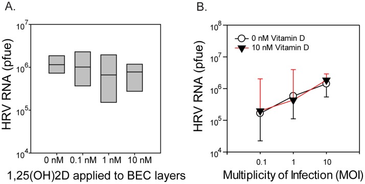
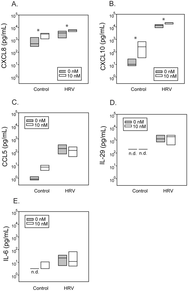
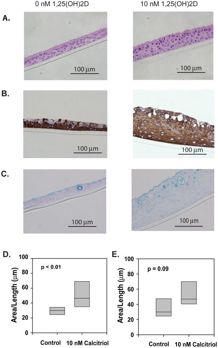
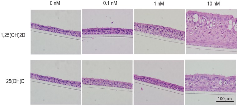
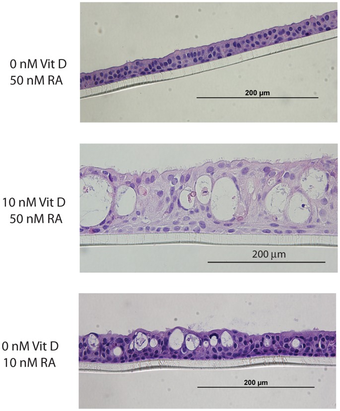
Vitamin D has been linked to reduced risk of viral respiratory illness. We hypothesized that vitamin D could directly reduce rhinovirus (RV) replication in airway epithelium. Primary human bronchial epithelial cells (hBEC) were treated with vitamin D, and RV replication and gene expression were evaluated by quantitative PCR. Cytokine/chemokine secretion was measured by ELISA, and transepithelial resistance (TER) was determined using a voltohmmeter. Morphology was examined using immunohistochemistry. Vitamin D supplementation had no significant effects on RV replication, but potentiated secretion of CXCL8 and CXCL10 from infected or uninfected cells. Treatment with vitamin D in the form of 1,25(OH)2D caused significant changes in cell morphology, including thickening of the cell layers (median of 46.5 µm [35.0-69.0] vs. 30 µm [24.5-34.2], p<0.01) and proliferation of cytokeratin-5-expressing cells, as demonstrated by immunohistochemical analysis. Similar effects were seen for 25(OH)D. In addition to altering morphology, higher concentrations of vitamin D significantly upregulated small proline-rich protein (SPRR1β) expression (6.3 fold-induction, p<0.01), suggestive of squamous metaplasia. Vitamin D treatment of hBECs did not alter repair of mechanically induced wounds. Collectively, these findings indicate that vitamin D does not directly affect RV replication in airway epithelial cells, but can influence chemokine synthesis and alters the growth and differentiation of airway epithelial cells.


Conflict of interest statement

Competing Interests: The authors have read the journal′s policy and have the following conflicts: Dr. Gern is a consultant for GlaxoSmithKline, Merck Inc., AstraZeneca, Gilead, and Boehringer Ingelheim, and has had investigator initiated research grants from Merck Inc, GlaxoSmithKline, and AstraZeneca. Co-author Raymond J.Pickles is a PLOS ONE Editorial Board member. This does not alter the authors′ adherence to all the PLOS ONE policies on sharing data and materials.

Figures

Figure 1
Figure 1. Effects of 1,25(OH)2D on RV replication.
(A) Replication of RV16 at 24 hours in differentiated hBEC cultured at air-liquid interface for 24 days in the presence of various concentrations of 1,25(OH)2D (n = 7). (B) Replication of RV16 (MOI = 0.1, 1, or 10) at 24 hours in differentiated hBEC cultured at air-liquid interface for 27 days in the presence of 10 nM of 1,25(OH)2D (n = 3).
Figure 2
Figure 2. Effects of 1,25(OH)2D on cytokine secretion.
(A,B) 1,25(OH)2D enhanced RV-induced CXCL8 (n = 6), and CXCL10 (n = 5) secretion. (C–E) Production of CCL5, IL-29, and IL-6 (n = 6) in these same cells, with “n.d.” indicating non-detectable levels. *p≤0.05 for cells treated with 1,25(OH)2D compared to untreated cells.
Figure 3
Figure 3. Effects of 1,25(OH)2D on cell morphology.
(A) Hematoxylin and eosin stained paraffin cross-sections of primary hBEC layers cultured at an air-liquid interface for 24 days in the presence of 10 nM 1,25(OH)2D. (B) Staining of paraffin cross-sections from 1,25(OH)2D-treated primary hBEC cell layers with antibody against cytokeratin 5/6 (CK5/6). (C) Alcian blue stained paraffin cross-sections of primary hBEC layers cultured at an air-liquid interface for 24 days in the presence of 10 nM 1,25(OH)2D. (D) Average thicknesses of primary hBEC layers differentiated in the presence of 10 nM 1,25(OH)2D for 4 to 6 weeks (n = 8). (E) Average thicknesses of fully differentiated primary hBEC layers after treatment with 10 nM 1,25(OH)2D for 6 weeks (n = 5).
Figure 4
Figure 4. 1,25(OH)2D and 25(OH)D have similar effects on cell growth and differentiation.
Hematoxylin and eosin stained paraffin cross-sections of primary hBEC layers cultured at an air-liquid interface for 28 days in the presence of various concentrations of 1,25(OH)2D or 25(OH)D.
Figure 5
Figure 5. Similar effects of supplemental vitamin D vs. retinoic acid deficiency on cell morphology.
Hematoxylin and eosin-stained paraffin cross-sections of primary hBEC layers cultured at an air-liquid interface for 28 days in the presence of 10 nM (deficient) or 50 nM (normal) all-trans retinoic acid, with 0 nM or 10 nM 1,25(OH)2D.

References

    1. Hansdottir S, Monick MM, Hinde SL, Lovan N, Look DC, et al. (2008) Respiratory epithelial cells convert inactive vitamin D to its active form: potential effects on host defense. J Immunol 181: 7090–7099. - PMC - PubMed
    1. Schauber J, Gallo RL (2009) Antimicrobial peptides and the skin immune defense system. J Allergy Clin Immunol 124: R13–R18. - PubMed
    1. Lin R, White JH (2004) The pleiotropic actions of vitamin D. Bioessays. 26: 21–28. - PubMed
    1. Beard JA, Bearden A, Striker R (2011) Vitamin D and the anti-viral state. J Clin Virol 50: 194–200. - PMC - PubMed
    1. Schwalfenberg GK (2011) A review of the critical role of vitamin D in the functioning of the immune system and the clinical implications of vitamin D deficiency. Mol Nutr Food Res 55: 96–108. - PubMed

Publication types

MeSH terms