. 2014 Jan 27;9(1):e86886.
doi: 10.1371/journal.pone.0086886. eCollection 2014.

Mycobacterium tuberculosis subverts the TLR-2-MyD88 pathway to facilitate its translocation into the cytosol

Affiliations

Mycobacterium tuberculosis subverts the TLR-2-MyD88 pathway to facilitate its translocation into the cytosol

Aejazur Rahman et al. PLoS One. .

Abstract

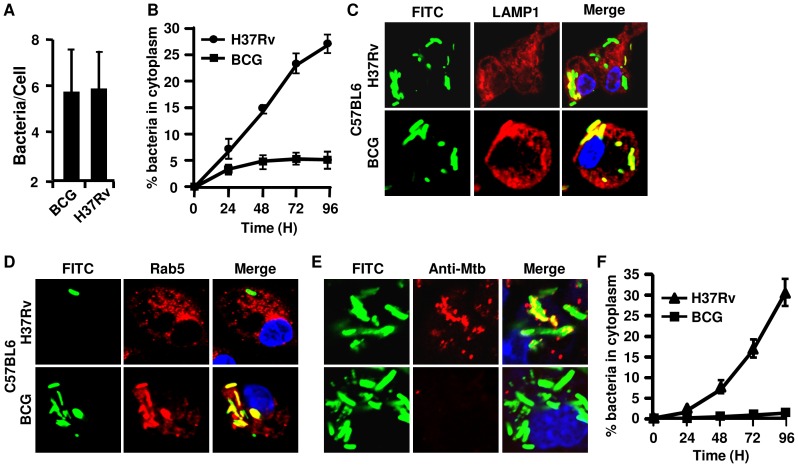
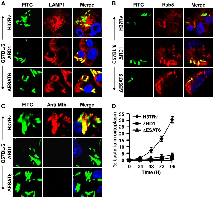
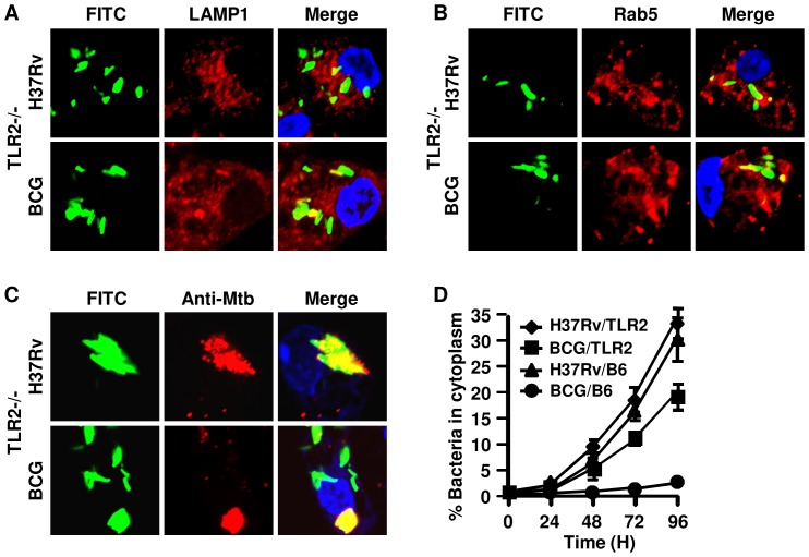
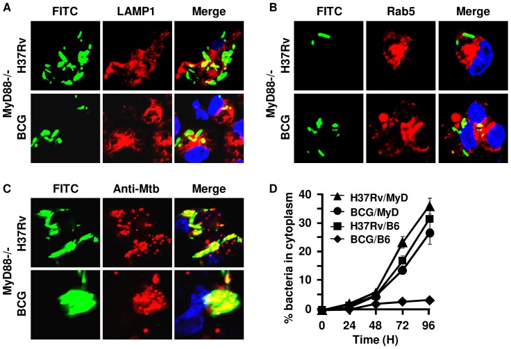
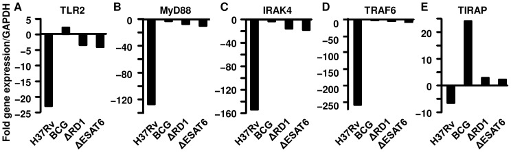
Mycobacterium tuberculosis (M.tb) has evolved mechanisms to evade its destruction in phagolysosomes, where it successfully survives and replicates within phagocytes. Recent studies have shown that virulent strains of M.tb can translocate from the phagosome into the cytosol of dendritic cells (DC). The molecular mechanisms by which virulent M.tb strains can escape the phagosome remain unknown. Here we show that the virulent M.tb strain H37Rv, but not the vaccine strain Bacille Calmette-Guérin (BCG), escapes from the phagolysosome and enters the cytosol by interfering with the TLR-2-MyD88 signaling pathway. Using H37Rv mutants, we further demonstrate that the region of difference-1 (RD-1) locus and ESAT-6, a gene within the RD-1 locus, play an important role in the capacity of M.tb to migrate from the phagosome to the cytosol of macrophages. H37Rv, BCG, H37RvΔRD1, and H37RvΔESAT6 were able to translocate to the cytosol in macrophages derived from TLR-2- and MyD88-deficient animals, whereas only virulent H37Rv was able to enter the cytosol in macrophages from wild type mice. Therefore, signaling through the TLR-2-MyD88 pathway in macrophages plays an important role in confining M.tb within phagolysomes. Virulent strains of M.tb have evolved mechanisms to subvert this pathway, thus facilitating their translocation to the cytosol and to escape the toxic microenvironment of the phagosome or phagolysosome.


Conflict of interest statement

Competing Interests: The author Gobardhan Das, a co-author in this manuscript, serves as an Academic Editor for PLOS ONE. This does not alter the authors' adherence to all the PLOS ONE policies on sharing data and materials.

Figures

Figure 1
Figure 1. Mycobacterium tuberculosis H37Rv, but not M. bovis BCG, translocates into the cytosol of murine peritoneal macrophages.
Bacteria were labelled green with FITC and used to infect peritoneal macrophages from C57BL/6 mice at an MOI of 5∶1. ( A ) Average number of bacteria present per cell after 5 hr of infection. ( B ) Percentage of bacteria that did not co-localize with LAMP-1 and/or Rab5 and, therefore considered to be in the cytosol, among the total number of bacteria at different time points after infection. Representative confocal images shown for the 72 hr time point after infection indicate that M. bovis BCG is completely co-localized with ( C ) LAMP-1 (red) or ( D ) Rab5 (red) but that some of the H37Rv organisms do not. ( E ) Infected cells were permeabilized with digitonin and stained with rabbit anti-Mtb antibody followed by anti-rabbit IgG-Alexa 594 (red). The M.tb organisms in the cytosol accessible to these antibodies stained red and yellow after merge, respectively. Bacteria that stained green in merged pictures were localized in the phagosome or phagolysosome. The nucleus of the cells was stained with DAPI (blue). The upper row of each section shows macrophages infected with H37Rv and the lower row shows macrophages infected with M. bovis BCG. ( F ) Kinetics of bacterial translocation to the cytosol of infected macrophages from 0 to 96 hr after infection. This was calculated from confocal studies with digitonin-permeabilized cells, as in panel ( E ). Bacteria in the cytoplasm of macrophages were counted from an average of 50 infected cells, which were accessible to the anti-Mtb antibody (red) and turned yellow after merging pictures. Experiments were run in triplicates and repeated three times. Representative data are shown.
Figure 2
Figure 2. Mycobacterium tuberculosis H37Rv mutants ΔESAT6 and ΔRD1 fail to enter the cytosol of wild type (C57BL/6) macrophages.
Bacteria were labelled green with FITC and used to infect peritoneal macrophages from C57BL/6 mice at an MOI of 5∶1. The average number of bacteria present per cell after 5 hr of infection was the same. Representative confocal images show data at the 72 hr time point after infection. M.tb mutants H37RvΔRD1 and H37RvΔESAT6 (green) are completely co-localized (yellow after merge) with (A) LAMP-1 (red) or (B) Rab5 (red), but some of the H37Rv organisms do not co-localize with these markers. (C) Infected cells were permeabilized with digitonin and stained with rabbit anti-Mtb antibody followed by anti-rabbit IgG-Alexa 594 (red). The M.tb in the cytosol accessible to these antibodies stained red or yellow after merging of pictures. Bacteria that were green in merged pictures are localized in phagolysosomes. The nucleus of the cells was stained with DAPI (blue). The upper row of each section shows H37Rv, the middle row shows H37RvΔRD1 and the lower row shows H37RvΔESAT6. (D) Kinetics of bacterial translocation to the cytosol of macrophages from 0 to 96 hr after infection. This was calculated from confocal studies of digitonin-permeabilized cells, as in panel (C). Bacteria in the cytoplasm of macrophages were counted from an average of 50 infected cells, which were accessible to the anti-Mtb antibody (red) and turned yellow after merging pictures. Data showed that around ∼30% of H37Rv (♦) bacteria escaped to the cytosol after 96 hr, whereas ΔRD1 (▪) and ΔESAT6 (▴) were unable to enter the cytoplasm. Experiments were run in triplicates and repeated thrice. Representative data are shown.
Figure 3
Figure 3. TLR-2-deficient macrophages fail to restrict M. bovis BCG to phagolysosomes.
Bacteria were labelled green with FITC and used to infect peritoneal macrophages from TLR-2−/− or wild type C57BL/6 mice at an MOI of 5∶1. The average number of bacteria present per cell after 5 hr of infection was the same. Representative confocal images show data at the 72 hr time point after infection in macrophages. In cells from TLR-2−/− mice, a population of H37Rv and M. bovis BCG organisms are not co-localized (green after merging pictures) with either (A) LAMP-1 (red) or (B) Rab5 (red). (C) Infected cells were permeabilized with digitonin and stained with rabbit anti-Mtb antibody followed by anti-rabbit IgG-Alexa 594 (red). The M.tb organisms in the cytosol accessible to these antibodies stained red and yellow after merging pictures. Bacteria that were green in merged pictures were localized to phagolysosomes. The nucleus of the cells was stained with DAPI (blue). The upper row of each section shows H37Rv and the lower row shows BCG. (D) The kinetics of bacterial translocation to the cytosol of macrophages from 0 to 96 hr after infection. This was calculated from confocal studies of digitonin-permeabilized cells (♦ H37Rv- and ▪ BCG-infected macrophages from TLR-2−/− mice; ▴ H37Rv- and • BCG-infected macrophage from C57BL/6 mice), as in panel (C). Bacteria in the cytoplasm of macrophages were counted from an average of 50 infected cells that were accessible to the anti-Mtb antibody (red) and turned yellow after merging pictures. Experiments were run in triplicates and repeated three times. Representative data are shown.
Figure 4
Figure 4. M. bovis BCG enters the cytosol in MyD88-deficient macrophages.
Bacteria were labelled green with FITC and used to infect peritoneal macrophages from MyD88−/− or wild type C57BL/6 mice at an MOI of 5∶1. The average number of bacteria present per cell after 5 hr of infection was the same. Representative confocal images are shown at the 72 hr time point after infection of macrophages. In macrophages from MyD88−/− mice, a population of H37Rv and M. bovis BCG are not co-localized (green after merging pictures) with either (A) LAMP-1 (red) or (B) Rab5 (red). (C) Infected cells were permeabilized with digitonin and stained with rabbit anti-Mtb antibody followed by anti-rabbit IgG-Alexa 594 (red). The M.tb in the cytosol accessible to these antibodies stained red and yellow after merging pictures. Bacteria that were green in merged pictures are localized in phagosomes. The nucleus of the cells was stained with DAPI (blue). The upper row of each section shows H37Rv and the lower row shows BCG. (D) The kinetics of bacterial translocation to the cytosol of the macrophages from 0 to 96 hr after infection (▴ H37Rv- and • BCG-infected macrophages from MyD88−/− mice; ▪ H37Rv- and ♦ BCG-infected in macrophages from C57BL/6 mice). This was calculated from confocal studies of digitonin-permeabilized cells, as in panel (C). Bacteria in the cytoplasm of macrophages were counted from an average of 50 infected cells that were accessible to the anti-Mtb antibody (red) and turned yellow after merging pictures. Experiments were run in triplicates and repeated three times. Representative data are shown.
Figure 5
Figure 5. Mycobacterium tuberculosis H37Rv down-regulates the expression of key genes involved in the TLR-2 - MyD88 signaling pathway.
Macrophages form wild type (C57BL/6) mice were infected with the indicated mycobacterial strains and 48 hr later cells were harvested for either analysis of relative mRNA expression of the indicated genes by quantitative RT-PCR or protein expression using immune-blotting. The gene expression of ( A ) TLR-2, ( B ) MyD88, ( C ) IRAK4, ( D ) TRAF6 and ( E ) TIRAP was down-regulated in macrophages infected with M.tb H37Rv. In contrast, no changes were observed in the expression of these genes in macrophages infected with BCG or the H37RvΔRD-1 and H37RvΔESAT6 mutants of H37Rv. The mRNA expression profiles were normalized with respect to expression of the GAPDH gene for each sample. The relative expression of genes in infected to uninfected macrophages is shown in each panel. Fold increase in the expression of each gene was calculated with respect to uninfected control at the same time point using the 2−ΔΔCt method. Data shown here are representative of three independent experiments and qPCR assays were set up in triplicate for each target and the GAPDH gene.

Similar articles

Cited by

References

    1. Vergne I, Chua J, Singh SB, Deretic V (2004) Cell biology of mycobacterium tuberculosis phagosome. Annu Rev Cell Dev Biol 20: 367–394. - PubMed
    1. Rohde K, Yates RM, Purdy GE, Russell DG (2007) Mycobacterium tuberculosis and the environment within the phagosome. Immunol Rev 219: 37–54. - PubMed
    1. Sturgill-Koszycki S, Schlesinger PH, Chakraborty P, Haddix PL, Collins HL, et al. (1994) Lack of acidification in Mycobacterium phagosomes produced by exclusion of the vesicular proton-ATPase. Science 263: 678–681. - PubMed
    1. Sinai AP, Joiner KA (1997) Safe haven: the cell biology of nonfusogenic pathogen vacuoles. Annu Rev Microbiol 51: 415–462. - PubMed
    1. van der Wel N, Hava D, Houben D, Fluitsma D, van Zon M, et al. (2007) M. tuberculosis and M. leprae translocate from the phagolysosome to the cytosol in myeloid cells. Cell 129: 1287–1298. - PubMed

Publication types

MeSH terms

LinkOut - more resources