Clinical Trial
. 2014 Jan 27;9(1):e87334.
doi: 10.1371/journal.pone.0087334. eCollection 2014.

Effect of maraviroc intensification on HIV-1-specific T cell immunity in recently HIV-1-infected individuals

Collaborators, Affiliations
Clinical Trial

Effect of maraviroc intensification on HIV-1-specific T cell immunity in recently HIV-1-infected individuals

Ai Kawana-Tachikawa et al. PLoS One. .

Abstract

Background: The effect of maraviroc on the maintenance and the function of HIV-1-specific T cell responses remains unknown.

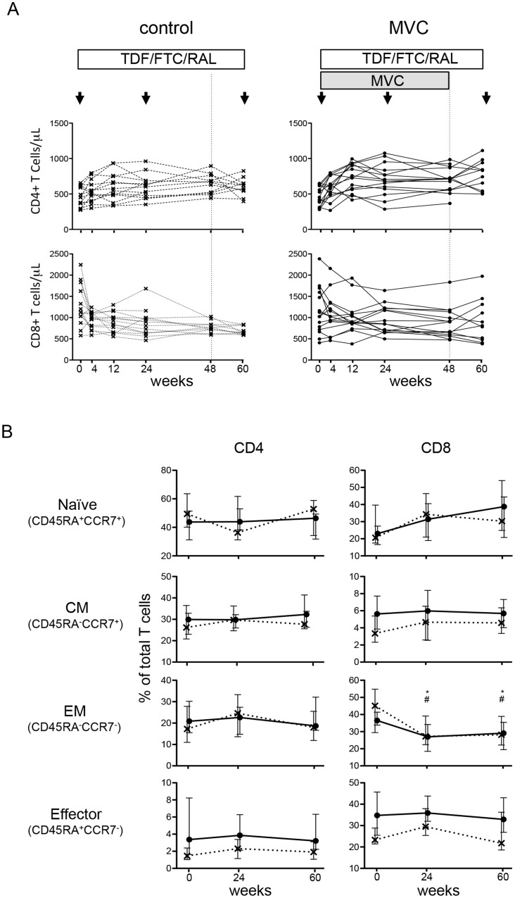
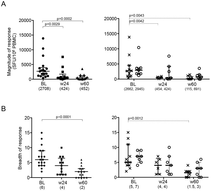
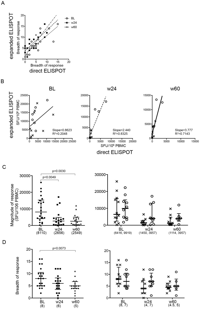
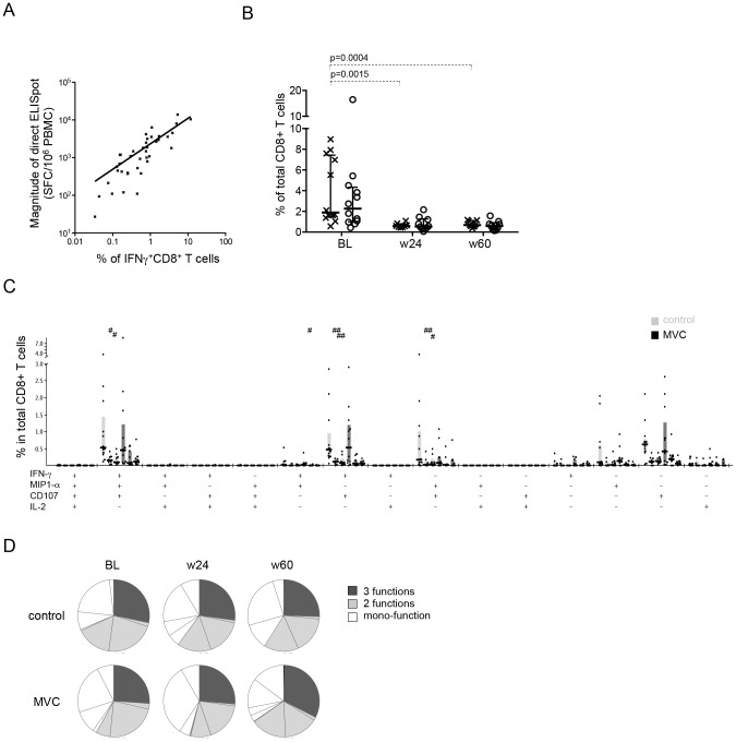
Methods: Subjects recently infected with HIV-1 were randomized to receive anti-retroviral treatment with or without maraviroc intensification for 48 weeks, and were monitored up to week 60. PBMC and in vitro-expanded T cells were tested for responses to the entire HIV proteome by ELISpot analyses. Intracellular cytokine staining assays were conducted to monitor the (poly)-functionality of HIV-1-specific T cells. Analyses were performed at baseline and week 24 after treatment start, and at week 60 (3 months after maraviroc discontinuation).

Results: Maraviroc intensification was associated with a slower decay of virus-specific T cell responses over time compared to the non-intensified regimen in both direct ex-vivo as well as in in-vitro expanded cells. The effector function profiles of virus-specific CD8⁺ T cells were indistinguishable between the two arms and did not change over time between the groups.

Conclusions: Maraviroc did not negatively impact any of the measured parameters, but was rather associated with a prolonged maintenance of HIV-1-specific T cell responses. Maraviroc, in addition to its original effect as viral entry inhibitor, may provide an additional benefit on the maintenance of virus-specific T cells which may be especially important for future viral eradication strategies.


Conflict of interest statement

Competing Interests: This work was partly funded by Pfizer Inc and ViiV Healthcare. This does not alter the authors' adherence to all the PLOS ONE policies on sharing data and materials.

Figures

Figure 1
Figure 1. Differentiation status in CD4+ and CD8+ T cells.
A. Changes of CD4+ and CD8+ T cell count in each subject. B. The proportion of naïve (CD45RA+/CCR7+), central memory (CM, CD45RA/CCR7+), effector memory (EM, CD45RA/CCR7), and Terminal effector memory (TEMRA, CD45RA+/CCR7) cells among CD4+ and CD8+ T cells in the control (cross and hatched line) and MVC arm (circle and solid line). The median and interquartile range (vertical line) are shown. Stars (control) and hatches (MVC arm) above the lines indicate significant differences relative to baseline values (p<0.05).
Figure 2
Figure 2. Longitudinal analyses of HIV-1-specific T cell responses in PBMC.
The total magnitude (A) and breadth (B) of ELISpot responses at baseline (BL), week 24 (w24) and week 60 (w60) are shown for all subjects together (left panels) and for each study arm separately (right panels, crossed lines for control arm, circles for MVC arm). Horizontal lines represent median values of Spot-forming cells (SFC)/106 PBMC and the IQR, respectively. Mann-Whitney test was used in all statistical analysis. Only p values with significance after Bonferroni correction was shown. The numbers in parenthesis below the x-axis represent the median value.
Figure 3
Figure 3. Longitudinal assessment of HIV-specific T cell responses with in vitro expanded T cells.
A. Relationship of the breadth between responses detected by direct ELISpot and ELISpot using in vitro expanded cells. Responses on the x-axes represent the total HIV-1-specific responses in direct ELISpot, and the y-axes indicate total HIV-1-specific responses in expanded ELISpot for samples taken at baseline (circle), week 24 (square), and week 60 (triangle). B. Relationship of the magnitude between direct ELISpot and expanded ELISpot at each time point. cross: control arm, circle: MVC arm. The lines in A and B show linear regression lines. C, D. Changes in magnitude and breadth of total HIV-specific T cell responses in expanded ELISpot are shown as in Figure 2.
Figure 4
Figure 4. Longitudinal assessment in functional profile of HIV-specific CD8+ T cells during cART with MVC intensification.
A. Correlation between the total HIV-specific responses determined by direct ex-vivo ELISpot analysis (as spot-forming cells (SFC)/106 PBMC) and by ICS analysis (% of IFN-γ+ CD8+ T cells). Linear regression line, and correlation coefficient and p-values (Spearman's rank correlation test) are shown. B. The change in total HIV-specific CD8+ T cell frequency over time by ICS analysis. Horizontal lines indicate median values of all positive responses. P values were determined by Mann-Whitney tests and shown if the significance remains after Bonferroni correction. C, D. Effector function profiles of HIV-specific CD8+ T cells over time in controls and MVC treated subjects. (left, baseline; middle, week 24; right, week 60). The median and IQR are indicated by horizontal lines and boxes, respectively. Differences relative to baseline values in each arm were tested for statistic significance by Mann-Whitney tests, and shown as # for p<0.05, ## for p<0.01.

Similar articles

Cited by

References

    1. Samson M, Libert F, Doranz BJ, Rucker J, Liesnard C, et al. (1996) Resistance to HIV-1 infection in caucasian individuals bearing mutant alleles of the CCR-5 chemokine receptor gene. Nature 382: 722–725. - PubMed
    1. Liu R, Paxton WA, Choe S, Ceradini D, Martin SR, et al. (1996) Homozygous defect in HIV-1 coreceptor accounts for resistance of some multiply-exposed individuals to HIV-1 infection. Cell 86: 367–377. - PubMed
    1. Paxton WA, Liu R, Kang S, Wu L, Gingeras TR, et al. (1998) Reduced HIV-1 infectability of CD4+ lymphocytes from exposed-uninfected individuals: association with low expression of CCR5 and high production of beta-chemokines. Virology 244: 66–73. - PubMed
    1. Glass WG, Lim JK, Cholera R, Pletnev AG, Gao JL, et al. (2005) Chemokine receptor CCR5 promotes leukocyte trafficking to the brain and survival in West Nile virus infection. J Exp Med 202: 1087–1098. - PMC - PubMed
    1. Glass WG, McDermott DH, Lim JK, Lekhong S, Yu SF, et al. (2006) CCR5 deficiency increases risk of symptomatic West Nile virus infection. J Exp Med 203: 35–40. - PMC - PubMed

Publication types