

Structural characterization of the mechanosensitive channel candidate MCA2 from Arabidopsis thaliana
- PMID: 24475319
- PMCID: PMC3903776
- DOI: 10.1371/journal.pone.0087724
Structural characterization of the mechanosensitive channel candidate MCA2 from Arabidopsis thaliana
Abstract
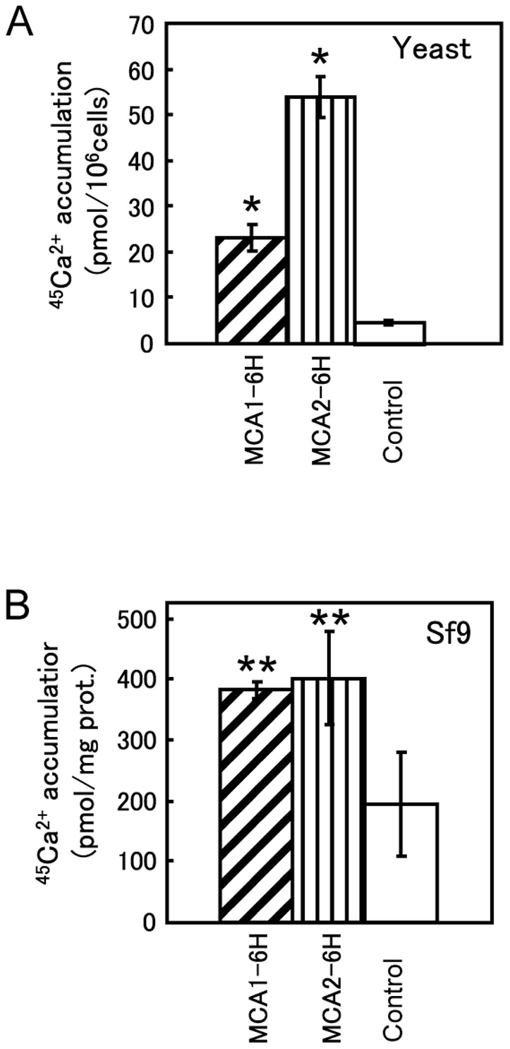
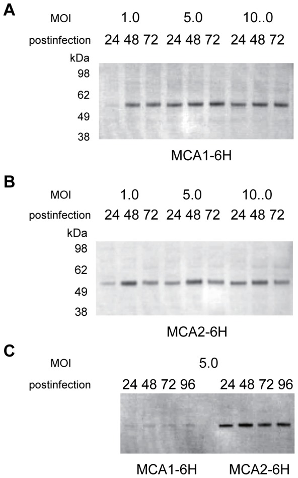
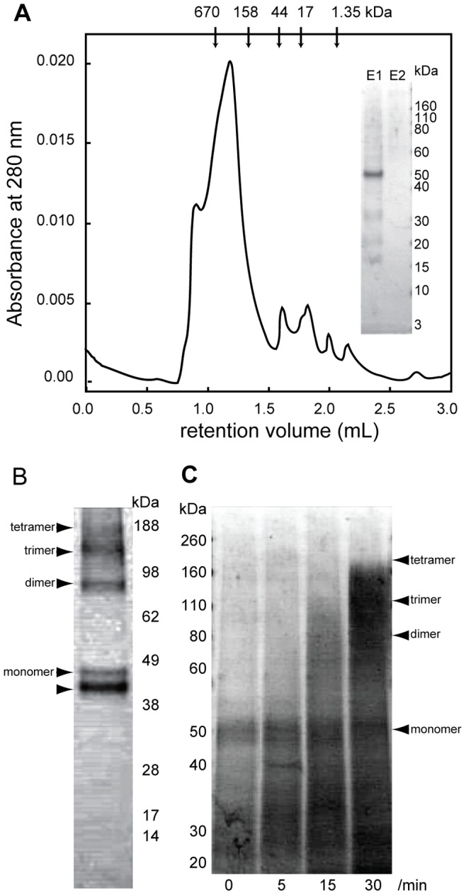
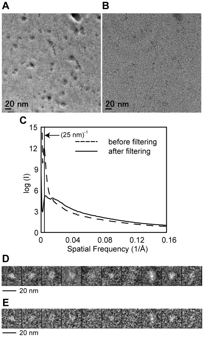
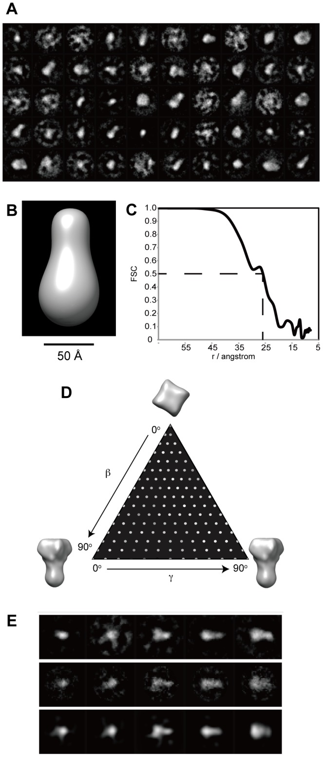
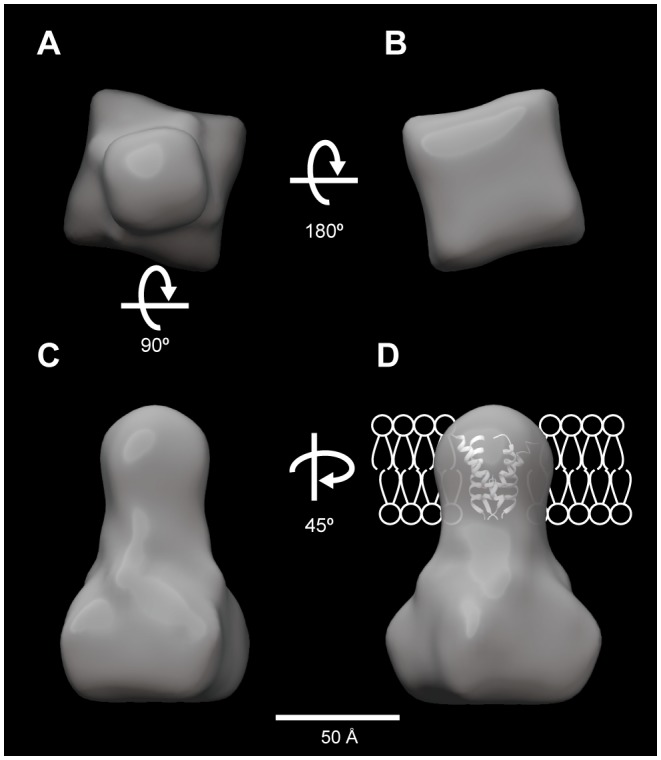
Mechanosensing in plants is thought to be governed by sensory complexes containing a Ca²⁺-permeable, mechanosensitive channel. The plasma membrane protein MCA1 and its paralog MCA2 from Arabidopsis thaliana are involved in mechanical stress-induced Ca²⁺ influx and are thus considered as candidates for such channels or their regulators. Both MCA1 and MCA2 were functionally expressed in Sf9 cells using a baculovirus system in order to elucidate their molecular natures. Because of the abundance of protein in these cells, MCA2 was chosen for purification. Purified MCA2 in a detergent-solubilized state formed a tetramer, which was confirmed by chemical cross-linking. Single-particle analysis of cryo-electron microscope images was performed to depict the overall shape of the purified protein. The three-dimensional structure of MCA2 was reconstructed at a resolution of 26 Å from 5,500 particles and appears to comprise a small transmembrane region and large cytoplasmic region.
Conflict of interest statement
Figures
Similar articles
-
Transmembrane Topologies of Ca2+-permeable Mechanosensitive Channels MCA1 and MCA2 in Arabidopsis thaliana.J Biol Chem. 2015 Dec 25;290(52):30901-9. doi: 10.1074/jbc.M115.692574. Epub 2015 Nov 10. J Biol Chem. 2015. PMID: 26555262 Free PMC article.
-
MCA1 and MCA2 that mediate Ca2+ uptake have distinct and overlapping roles in Arabidopsis.Plant Physiol. 2010 Mar;152(3):1284-96. doi: 10.1104/pp.109.147371. Epub 2010 Jan 22. Plant Physiol. 2010. PMID: 20097794 Free PMC article.
-
Determination of structural regions important for Ca(2+) uptake activity in Arabidopsis MCA1 and MCA2 expressed in yeast.Plant Cell Physiol. 2011 Nov;52(11):1915-30. doi: 10.1093/pcp/pcr131. Epub 2011 Sep 24. Plant Cell Physiol. 2011. PMID: 21949028
-
Physiological roles of Arabidopsis MCA1 and MCA2 based on their dynamic expression patterns.J Plant Res. 2024 Sep;137(5):785-797. doi: 10.1007/s10265-024-01575-8. Epub 2024 Aug 28. J Plant Res. 2024. PMID: 39196431 Free PMC article. Review.
-
New candidates for mechano-sensitive channels potentially involved in gravity sensing in Arabidopsis thaliana.Plant Biol (Stuttg). 2014 Jan;16 Suppl 1:39-42. doi: 10.1111/plb.12044. Epub 2013 Jun 4. Plant Biol (Stuttg). 2014. PMID: 23731064 Review.
Cited by
-
Plant signaling networks involving Ca(2+) and Rboh/Nox-mediated ROS production under salinity stress.Front Plant Sci. 2015 Jun 10;6:427. doi: 10.3389/fpls.2015.00427. eCollection 2015. Front Plant Sci. 2015. PMID: 26113854 Free PMC article. Review.
-
Entanglement of Arabidopsis Seedlings to a Mesh Substrate under Microgravity Conditions in KIBO on the ISS.Plants (Basel). 2022 Mar 31;11(7):956. doi: 10.3390/plants11070956. Plants (Basel). 2022. PMID: 35406935 Free PMC article.
-
United in diversity: mechanosensitive ion channels in plants.Annu Rev Plant Biol. 2015;66:113-37. doi: 10.1146/annurev-arplant-043014-114700. Epub 2014 Dec 8. Annu Rev Plant Biol. 2015. PMID: 25494462 Free PMC article. Review.
-
The calcium connection: exploring the intricacies of calcium signaling in plant-microbe interactions.Front Plant Sci. 2023 Oct 2;14:1248648. doi: 10.3389/fpls.2023.1248648. eCollection 2023. Front Plant Sci. 2023. PMID: 37849843 Free PMC article. Review.
-
Transmembrane Topologies of Ca2+-permeable Mechanosensitive Channels MCA1 and MCA2 in Arabidopsis thaliana.J Biol Chem. 2015 Dec 25;290(52):30901-9. doi: 10.1074/jbc.M115.692574. Epub 2015 Nov 10. J Biol Chem. 2015. PMID: 26555262 Free PMC article.
References
-
- Trewavas A, Knight M (1994) Mechanical signalling, calcium and plant form. Plant Mol Biol 26: 1329–1341. - PubMed
Publication types
MeSH terms
Substances
LinkOut - more resources
Full Text Sources
Other Literature Sources
Molecular Biology Databases
Miscellaneous
