Placental thrombosis in acute phase abortions during experimental Toxoplasma gondii infection in sheep
- PMID: 24475786
- PMCID: PMC3931317
- DOI: 10.1186/1297-9716-45-9
Placental thrombosis in acute phase abortions during experimental Toxoplasma gondii infection in sheep
Abstract
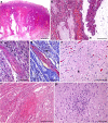
After oral administration of ewes during mid gestation with 2000 freshly prepared sporulated oocysts of T. gondii isolate M4, abortions occurred between days 7 and 11 in 91.6% of pregnant and infected ewes. Afterwards, a further infection was carried out at late gestation in another group of sheep with 500 sporulated oocysts. Abortions happened again between days 9 and 11 post infection (pi) in 58.3% of the infected ewes. Classically, abortions in natural and experimental ovine toxoplasmosis usually occur one month after infection. Few experimental studies have reported the so-called acute phase abortions as early as 7 to 14 days after oral inoculation of oocysts, and pyrexia was proposed to be responsible for abortion, although the underline mechanism was not elucidated. In the present study, all placentas analysed from ewes suffering acute phase abortions showed infarcts and thrombosis in the caruncullar villi of the placentomes and ischemic lesions (periventricular leukomalacia) in the brain of some foetuses. The parasite was identified by PCR in samples from some placentomes of only one sheep, and no antigen was detected by immunohistochemical labelling. These findings suggest that the vascular lesions found in the placenta, and the consequent hypoxic damage to the foetus, could be associated to the occurrence of acute phase abortions. Although the pathogenesis of these lesions remains to be determined, the infectious dose or virulence of the isolate may play a role in their development.
Figures


Similar articles
-
Early dynamics of Toxoplasma gondii infection in sheep inoculated at mid-gestation with archetypal type II oocysts.Vet Res. 2025 Jul 1;56(1):134. doi: 10.1186/s13567-025-01557-1. Vet Res. 2025. PMID: 40598310 Free PMC article.
-
Acute phase toxoplasma abortions in sheep.Vet Rec. 1998 May 2;142(18):480-2. doi: 10.1136/vr.142.18.480. Vet Rec. 1998. PMID: 9612913
-
Experimental ovine toxoplasmosis: influence of the gestational stage on the clinical course, lesion development and parasite distribution.Vet Res. 2016 Mar 16;47:43. doi: 10.1186/s13567-016-0327-z. Vet Res. 2016. PMID: 26983883 Free PMC article.
-
Ovine toxoplasmosis.Parasitology. 2009 Dec;136(14):1887-94. doi: 10.1017/S0031182009991636. Parasitology. 2009. PMID: 19995468 Review.
-
[Problems and limitations of conventional and innovative methods for the diagnosis of Toxoplasmosis in humans and animals].Parassitologia. 2004 Jun;46(1-2):177-81. Parassitologia. 2004. PMID: 15305712 Review. Italian.
Cited by
-
Advances in vaccine development and the immune response against toxoplasmosis in sheep and goats.Front Vet Sci. 2022 Aug 25;9:951584. doi: 10.3389/fvets.2022.951584. eCollection 2022. Front Vet Sci. 2022. PMID: 36090161 Free PMC article. Review.
-
Molecular survey for cyst-forming coccidia (Toxoplasma gondii, Neospora caninum, Sarcocystis spp.) in Mediterranean periurban micromammals.Parasitol Res. 2020 Aug;119(8):2679-2686. doi: 10.1007/s00436-020-06777-2. Epub 2020 Jun 25. Parasitol Res. 2020. PMID: 32588173
-
Virulence in Mice of a Toxoplasma gondii Type II Isolate Does Not Correlate With the Outcome of Experimental Infection in Pregnant Sheep.Front Cell Infect Microbiol. 2019 Jan 4;8:436. doi: 10.3389/fcimb.2018.00436. eCollection 2018. Front Cell Infect Microbiol. 2019. PMID: 30662874 Free PMC article.
-
Factors Associated With Toxoplasma gondii IgG and IgM Antibodies, and Placental Histopathological Changes Among Women With Spontaneous Abortion in Mwanza City, Tanzania.East Afr Health Res J. 2017;1(2):86-94. doi: 10.24248/EAHRJ-D-16-00408. Epub 2017 Jul 1. East Afr Health Res J. 2017. PMID: 34308163 Free PMC article.
-
Report of an unsual case of anophthalmia and craniofacial cleft in a newborn with Toxoplasma gondii congenital infection.BMC Infect Dis. 2017 Jul 3;17(1):459. doi: 10.1186/s12879-017-2565-8. BMC Infect Dis. 2017. PMID: 28673238 Free PMC article.
References
-
- Buxton D, Maley SW, Wright SE, Rodger S, Bartley P, Innes EA. Ovine toxoplasmosis: transmission, clinical outcome and control. Parassitologia. 2007;49:219–221. - PubMed
-
- Dubey JP. Toxoplasmosis of Animals and Humans. 2. Boca Raton, Florida: CRC Press; 2010.
-
- Buxton D. Protozoan infections (Toxoplasma gondii, Neospora caninum and Sarcocystis spp.) in sheep and goats: recent advances. Vet Res. 1998;29:289–310. - PubMed
Publication types
MeSH terms
LinkOut - more resources
Full Text Sources
Other Literature Sources
Medical
Research Materials
Miscellaneous

