. 2014 Apr;271(1):220-9.
doi: 10.1148/radiol.13130625. Epub 2013 Nov 18.

Bone metastases in castration-resistant prostate cancer: associations between morphologic CT patterns, glycolytic activity, and androgen receptor expression on PET and overall survival

Affiliations

Bone metastases in castration-resistant prostate cancer: associations between morphologic CT patterns, glycolytic activity, and androgen receptor expression on PET and overall survival

Hebert Alberto Vargas et al. Radiology. 2014 Apr.

Abstract

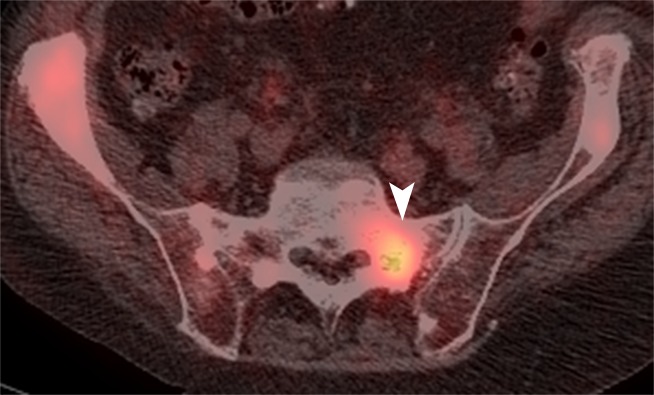
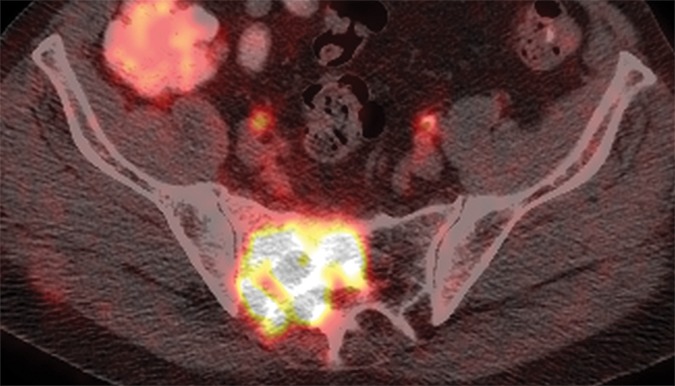
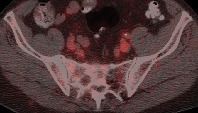
Purpose: To compare the features of bone metastases at computed tomography (CT) to tracer uptake at fluorine 18 fluorodeoxyglucose (FDG) positron emission tomography (PET) and fluorine 18 16β-fluoro-5-dihydrotestosterone (FDHT) PET and to determine associations between these imaging features and overall survival in men with castration-resistant prostate cancer.

Materials and methods: This is a retrospective study of 38 patients with castration-resistant prostate cancer. Two readers independently evaluated CT, FDG PET, and FDHT PET features of bone metastases. Associations between imaging findings and overall survival were determined by using univariate Cox proportional hazards regression.

Results: In 38 patients, reader 1 detected 881 lesions and reader 2 detected 867 lesions. Attenuation coefficients at CT correlated inversely with FDG (reader 1: r = -0.3007; P < .001; reader 2: r = -0.3147; P < .001) and FDHT (reader 1: r = -0.2680; P = .001; reader 2: r = -0.3656; P < .001) uptake. The number of lesions on CT scans was significantly associated with overall survival (reader 1: hazard ratio [HR], 1.025; P = .05; reader 2: HR, 1.021; P = .04). The numbers of lesions on FDG and FDHT PET scans were significantly associated with overall survival for reader 1 (HR, 1.051-1.109; P < .001) and reader 2 (HR, 1.026-1.082; P ≤ .009). Patients with higher FDHT uptake (lesion with the highest maximum standardized uptake value) had significantly shorter overall survival (reader 1: HR, 1.078; P = .02; reader 2: HR, 1.092; P = .02). FDG uptake intensity was not associated with overall survival (reader 1, P = .65; reader 2, P = .38).

Conclusion: In patients with castration-resistant prostate cancer, numbers of bone lesions on CT, FDG PET, and FDHT PET scans and the intensity of FDHT uptake are significantly associated with overall survival.


Figures

Figure 1a:
Figure 1a:
Representative examples of the different CT lesion morphologies (arrowhead) evaluated in this study. (a) Osteoblastic dense, (b) osteoblastic ground glass, (c) osteoblastic mixed dense and ground glass, (d) miliary, (e) osteolytic, and (f) mixed osteolytic and osteoblastic.
Figure 1b:
Figure 1b:
Representative examples of the different CT lesion morphologies (arrowhead) evaluated in this study. (a) Osteoblastic dense, (b) osteoblastic ground glass, (c) osteoblastic mixed dense and ground glass, (d) miliary, (e) osteolytic, and (f) mixed osteolytic and osteoblastic.
Figure 1c:
Figure 1c:
Representative examples of the different CT lesion morphologies (arrowhead) evaluated in this study. (a) Osteoblastic dense, (b) osteoblastic ground glass, (c) osteoblastic mixed dense and ground glass, (d) miliary, (e) osteolytic, and (f) mixed osteolytic and osteoblastic.
Figure 1d:
Figure 1d:
Representative examples of the different CT lesion morphologies (arrowhead) evaluated in this study. (a) Osteoblastic dense, (b) osteoblastic ground glass, (c) osteoblastic mixed dense and ground glass, (d) miliary, (e) osteolytic, and (f) mixed osteolytic and osteoblastic.
Figure 1e:
Figure 1e:
Representative examples of the different CT lesion morphologies (arrowhead) evaluated in this study. (a) Osteoblastic dense, (b) osteoblastic ground glass, (c) osteoblastic mixed dense and ground glass, (d) miliary, (e) osteolytic, and (f) mixed osteolytic and osteoblastic.
Figure 1f:
Figure 1f:
Representative examples of the different CT lesion morphologies (arrowhead) evaluated in this study. (a) Osteoblastic dense, (b) osteoblastic ground glass, (c) osteoblastic mixed dense and ground glass, (d) miliary, (e) osteolytic, and (f) mixed osteolytic and osteoblastic.
Figure 2a:
Figure 2a:
(a) Axial CT, (b) fused FDG PET/CT, and (c) fused FDHT PET/CT images in an 81-year-old patient with an FDHT-predominant phenotype. A ground-glass lesion in the left sacral ala (white arrowheads) has an attenuation of 352 HU and uptake on FDG (SUVmax, 8) and FDHT (SUVmax, 22) PET images. A subtle ground-glass lesion in the right ilium (black arrowheads) demonstrates uptake at FDHT PET (SUVmax, 10) but no abnormal uptake at FDG PET. An osteoblastic dense lesion (839 HU) in the right ilium (arrows) shows increased uptake at FDHT (SUVmax, 13) but no uptake at FDG PET. All measurements are from reader 1.
Figure 2b:
Figure 2b:
(a) Axial CT, (b) fused FDG PET/CT, and (c) fused FDHT PET/CT images in an 81-year-old patient with an FDHT-predominant phenotype. A ground-glass lesion in the left sacral ala (white arrowheads) has an attenuation of 352 HU and uptake on FDG (SUVmax, 8) and FDHT (SUVmax, 22) PET images. A subtle ground-glass lesion in the right ilium (black arrowheads) demonstrates uptake at FDHT PET (SUVmax, 10) but no abnormal uptake at FDG PET. An osteoblastic dense lesion (839 HU) in the right ilium (arrows) shows increased uptake at FDHT (SUVmax, 13) but no uptake at FDG PET. All measurements are from reader 1.
Figure 2c:
Figure 2c:
(a) Axial CT, (b) fused FDG PET/CT, and (c) fused FDHT PET/CT images in an 81-year-old patient with an FDHT-predominant phenotype. A ground-glass lesion in the left sacral ala (white arrowheads) has an attenuation of 352 HU and uptake on FDG (SUVmax, 8) and FDHT (SUVmax, 22) PET images. A subtle ground-glass lesion in the right ilium (black arrowheads) demonstrates uptake at FDHT PET (SUVmax, 10) but no abnormal uptake at FDG PET. An osteoblastic dense lesion (839 HU) in the right ilium (arrows) shows increased uptake at FDHT (SUVmax, 13) but no uptake at FDG PET. All measurements are from reader 1.
Figure 3a:
Figure 3a:
(a) Axial CT, (b) fused FDG PET/CT, and (c) fused FDHT PET/CT images in a 66-year-old patient with a predominantly lytic lesion in the sacrum (arrow) that shows marked FDG uptake (SUVmax, 16) but no abnormal FDHT uptake on PET images.
Figure 3b:
Figure 3b:
(a) Axial CT, (b) fused FDG PET/CT, and (c) fused FDHT PET/CT images in a 66-year-old patient with a predominantly lytic lesion in the sacrum (arrow) that shows marked FDG uptake (SUVmax, 16) but no abnormal FDHT uptake on PET images.
Figure 3c:
Figure 3c:
(a) Axial CT, (b) fused FDG PET/CT, and (c) fused FDHT PET/CT images in a 66-year-old patient with a predominantly lytic lesion in the sacrum (arrow) that shows marked FDG uptake (SUVmax, 16) but no abnormal FDHT uptake on PET images.
Figure 4a:
Figure 4a:
Scatterplots show the relationship between FDG SUVmax and FDHT SUVmax for each bone lesion according to (a) reader 1 and (b) reader 2.
Figure 4b:
Figure 4b:
Scatterplots show the relationship between FDG SUVmax and FDHT SUVmax for each bone lesion according to (a) reader 1 and (b) reader 2.
Figure 5a:
Figure 5a:
Scatterplots show the relationships of mean Hounsfield units to SUVmax at FDG PET and SUVmax at FDHT PET for each bone lesion for (a, b) reader 1 and (c, d) reader 2.
Figure 5b:
Figure 5b:
Scatterplots show the relationships of mean Hounsfield units to SUVmax at FDG PET and SUVmax at FDHT PET for each bone lesion for (a, b) reader 1 and (c, d) reader 2.
Figure 5c:
Figure 5c:
Scatterplots show the relationships of mean Hounsfield units to SUVmax at FDG PET and SUVmax at FDHT PET for each bone lesion for (a, b) reader 1 and (c, d) reader 2.
Figure 5d:
Figure 5d:
Scatterplots show the relationships of mean Hounsfield units to SUVmax at FDG PET and SUVmax at FDHT PET for each bone lesion for (a, b) reader 1 and (c, d) reader 2.

Comment in

References

    1. Schrecengost R, Knudsen KE. Molecular pathogenesis and progression of prostate cancer. Semin Oncol 2013;40(3):244–258. - PMC - PubMed
    1. Scher HI, Beer TM, Higano CS, et al. . Antitumour activity of MDV3100 in castration-resistant prostate cancer: a phase 1-2 study. Lancet 2010;375(9724):1437–1446. - PMC - PubMed
    1. Scher HI, Fizazi K, Saad F, et al. . Increased survival with enzalutamide in prostate cancer after chemotherapy. N Engl J Med 2012;367(13):1187–1197. - PubMed
    1. Lee RJ, Saylor PJ, Smith MR. Treatment and prevention of bone complications from prostate cancer. Bone 2011;48(1):88–95. - PMC - PubMed
    1. Larson SM, Morris M, Gunther I, et al. . Tumor localization of 16beta-18F-fluoro-5alpha-dihydrotestosterone versus 18F-FDG in patients with progressive, metastatic prostate cancer. J Nucl Med 2004;45(3):366–373. - PubMed

Publication types

MeSH terms