. 2014 Jan 24;6(1):e00135.
doi: 10.1042/AN20130022.

Gas6 enhances axonal ensheathment by MBP+ membranous processes in human DRG/OL promyelinating co-cultures

Affiliations

Gas6 enhances axonal ensheathment by MBP+ membranous processes in human DRG/OL promyelinating co-cultures

Kathleen N O'Guin et al. ASN Neuro. .

Abstract

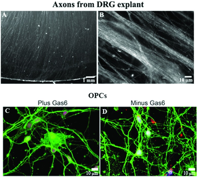
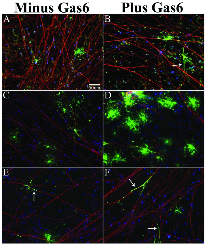
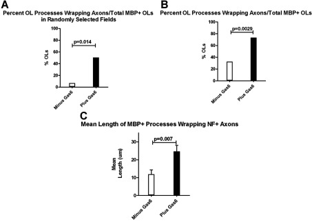
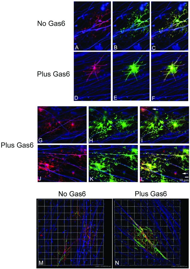
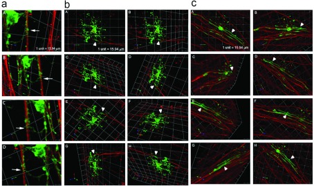
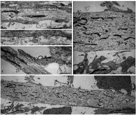
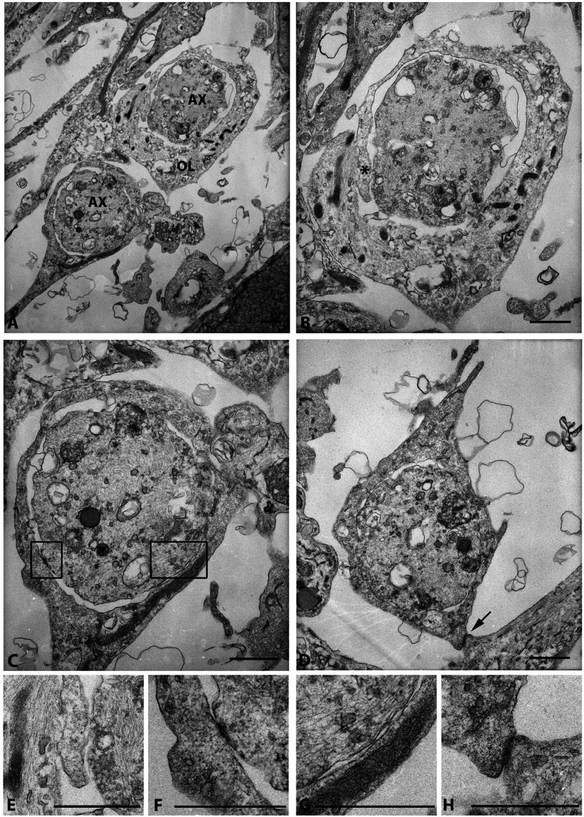
The molecular requirements for human myelination are incompletely defined, and further study is needed to fully understand the cellular mechanisms involved during development and in demyelinating diseases. We have established a human co-culture model to study myelination. Our earlier observations showed that addition of human γ-carboxylated growth-arrest-specific protein 6 (Gas6) to human oligodendrocyte progenitor cell (OPC) cultures enhanced their survival and maturation. Therefore, we explored the effect of Gas6 in co-cultures of enriched OPCs plated on axons of human fetal dorsal root ganglia explant. Gas6 significantly enhanced the number of myelin basic protein-positive (MBP+) oligodendrocytes with membranous processes parallel with and ensheathing axons relative to co-cultures maintained in defined medium only for 14 days. Gas6 did not increase the overall number of MBP+ oligodendrocytes/culture; however, it significantly increased the length of MBP+ oligodendrocyte processes in contact with and wrapping axons. Multiple oligodendrocytes were in contact with a single axon, and several processes from one oligodendrocyte made contact with one or multiple axons. Electron microscopy supported confocal Z-series microscopy demonstrating axonal ensheathment by MBP+ oligodendrocyte membranous processes in Gas6-treated co-cultures. Contacts between the axonal and oligodendrocyte membranes were evident and multiple wraps of oligodendrocyte membrane around the axon were visible supporting a model system in which to study events in human myelination and aspects of non-compact myelin formation.


Figures

Figure 1
Figure 1. Generation of human fetal DRG explants (A and B) with parallel arrays of NF+ axons, and enriched OPCs from mixed glial cultures (C and D)
(A) Axons radiating from a DRG explant maintained in culture for 3 weeks. (B) NF+-stained axons radiating from a DRG explant. (C and D) Enriched OPCs from human fetal brain maintained in the presence (C) and absence (D) of Gas6 are βIV tubulin+ (green) and express Olig1 (red) in the nucleus following 20 days in culture. Blue is Hoechst stain. An Olympus digital microscope was used.
Figure 2
Figure 2. Human OPC/DRG co-cultures in medium containing PDGF+Gas6 express MBP+ processes that align and elongate along NF+ axons, while OLs in co-cultures maintained in BDNF+IGF1+Gas6 have more mature MBP+ processes that ensheath axons
Immunofluorescent staining and confocal microscopy show MBP+ OLs (green) and NF+ axons (red; 1:2000; chick IgY-cy3) captured on an Olympus Digital microscope after 14 days in co-culture; scale bar for all images is 50 μm. (A) Co-cultures maintained in PDGF medium minus Gas6. (B) Co-cultures maintained in PDGF medium plus Gas6 have MBP+ OL processes aligned with axons. (C and E) Co-cultures maintained in IGF1+BDNF-defined medium minus Gas6. (D and F) Co-cultures maintained in IGF1+BDNF-defined medium plus Gas6. Scale bar in (A) designates magnification for all panels.
Figure 3
Figure 3. Gas6 enhances OL wrapping and mean process length in human co-cultures
For experiments 1 and 2, co-cultures were maintained in defined medium as defined in the DRG/co-culture section of the Materials and methods (DMEM supplemented with 2% B27, 1% N2, 20 ng/ml rhBDNF, 20 ng/ml IGF1, 40 ng/ml T3, 40 ng/ml T4, 5ng/ml NT3, antibiotic/antimycotic, plus and minus Gas6). (A) Experiment 1, multiple, randomly selected MBP+ fields were photographed. From each random field, the percentage of MBP+ oligodendrocytes with processes wrapping axons relative to the total number of MBP+ oligodendrocytes/field was calculated. (B) Experiment 2, MBP+ OLs with obvious processes aligned with an axon were photographed, and the percentage of MBP+ OLs with processes wrapping axons relative to the total number of MBP+ OLs/field was calculated. (C) Histogram depicts the mean length (μm) of MBP+ processes wrapping NF+ axons in Gas6 and Gas6+ co-cultures; P=0.007; Mann–Whitney test.
Figure 4
Figure 4. Multiple MAG+MBP+ OL processes from a single OL can ensheath the same axon and multiple axons
Co-cultures were maintained in defined medium plus and minus Gas6 for 14 days (see the Materials and methods section). (AL) Immunofluorescent staining and light microscopy. MAG+ (red), MBP+ (green) OLs, NF+ (blue) axons; scale bar=10 μm. (M and N) Z-series and confocal microscopy shows wrapping of MAG- and MBP-positive processes. Maximal projection of MBP+ processes was obtained in Volocity. Linear line of a box corresponds to 1 unit=24 μm. Backgrounds were adjusted to the negative primary antibody isotype controls. For example, the MAG primary antibody (IgG1) was substituted with primary antibody MOPC-31C purified clone for mouse IgG1 (BD Pharmingen; 557273).
Figure 5
Figure 5. MBP+ OL processes (green) spiral and ensheath multiple NF+ axons (red) in co-cultures maintained for 14 days in defined medium minus (a and b) and plus (c) Gas6
Identification of multiple Z-series images captured by confocal microscopy (Leica) and analyzed in Volocity. The arrow in the image a shows an MBP+ OL process spirally wrapping the axon. Images in (a and b) show MBP+ OLs wrapping NF+ axons in co-cultures maintained in defined medium only. (c) A representative image from a Gas6-treated co-culture. Arrowheads in (b and c) indicate areas where MBP+ (green) membranes totally ensheath the axon (red).
Figure 6
Figure 6. Multiple OL/axonal contacts are observed in Gas6-treated co-cultures
A co-culture maintained for 14 days in defined medium plus Gas6 was embedded in Epon, and uranyl-stained and 1-μm sections were examined by EM. Examination of grids found fields containing axons with multiple oligodendrocytes. Scale bars in (AD) are 1 μm; the scale bar in (E) is 0.5 μm.
Figure 7
Figure 7. Visualization of wrapping of axons in human co-cultures in the presence of Gas6 (2.6 nM)
(A) Two axons (AX) wrapped in myelin membrane. (B) Upper axon from (A) shows wrapping of one myelin membrane encircling the other (*); higher magnification * is shown in (E). (C) Lower axon from (A) shows OL membrane wrapping and multiple contacts/adhesions between the axonal membrane and the OL membrane; higher magnifications of the contacts are shown in (F) and (G). (D) OL membrane wrapping an axon, and contacting an additional membrane; higher magnification is shown in (H). Scale bars are 1 μm.

References

    1. Aggarwal S, Yurlova L, Snaidero N, Reetz C, Frey S, Zimmermann J, Pahler G, Janshoff A, Friedrichs J, Muller DJ, Goebel C, Simons M. A size barrier limits protein diffusion at the cell surface to generate lipid-rich myelin-membrane sheets. Dev Cell. 2011;21:445–456. - PubMed
    1. Allen MP, Zeng C, Schneider K, Xiong X, Meintzer MK, Bellosta P, Basilico C, Varnum B, Heidenreich KA, Wierman ME. Growth arrest-specific gene 6 (Gas6)/adhesion related kinase (Ark) signaling promotes gonadotropin-releasing hormone neuronal survival via extracellular signal-regulated kinase (ERK) and Akt. Mol Endocrinol. 1999;13:191–201. - PubMed
    1. Arnett HA, Fancy SP, Alberta JA, Zhao C, Plant SR, Kaing S, Raine CS, Rowitch DH, Franklin RJ, Stiles CD. bHLH transcription factor Olig1 is required to repair demyelinated lesions in the CNS. Science. 2004;306:2111–2115. - PubMed
    1. Avanzi GC, Gallicchio M, Bottarel F, Gammaitoni L, Cavalloni G, Buonfiglio D, Bragardo M, Bellomo G, Albano E, Fantozzi R, Garbarino G, Varnum B, Aglietta M, Saglio G, Dianzani U, Dianzani C. GAS6 inhibits granulocyte adhesion to endothelial cells. Blood. 1998;91:2334–2340. - PubMed
    1. Bakhti M, Snaidero N, Schneider D, Aggarwal S, Mobius W, Janshoff A, Eckhardt M, Nave KA, Simons M. Loss of electrostatic cell-surface repulsion mediates myelin membrane adhesion and compaction in the central nervous system. Proc Natl Acad Sci USA. 2013;110:3143–3148. - PMC - PubMed

Publication types

MeSH terms

Substances