

Review
doi: 10.1056/NEJMra1310050.
Necroptosis
Affiliations
- PMID: 24476434
- PMCID: PMC4035222
- DOI: 10.1056/NEJMra1310050
Item in Clipboard
Review
Necroptosis
N Engl J Med.
.
No abstract available
Figures
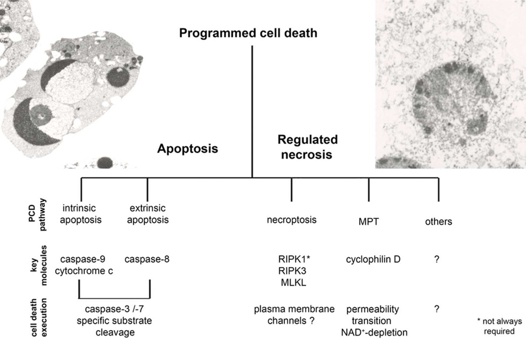
The term programmed cell death was widely used synonymously with apoptosis until necrotic cell death was demonstrated to depend on genetically defined signaling pathways. Whereas the role of caspase-mediated apoptosis in diseases has been revealed in detail over the last three decades, the contribution of regulated necrosis to the pathophysiology of diseases was investigated only recently. Pathways of regulated necrosis include necroptosis, dependent on RIPK3, and regulated necrosis mediated by mitochondrial permeability transition (MPT) which involves cyclophilin D-dependent opening of the mitochondrial permeability transition pore. Whereas MPT and necroptosis have been demonstrated to represent two distinct pathways, other emerging signaling cascades of regulated necrosis have been described, but it remains currently unclear to what extent such pathways may have overlapping mechanisms.
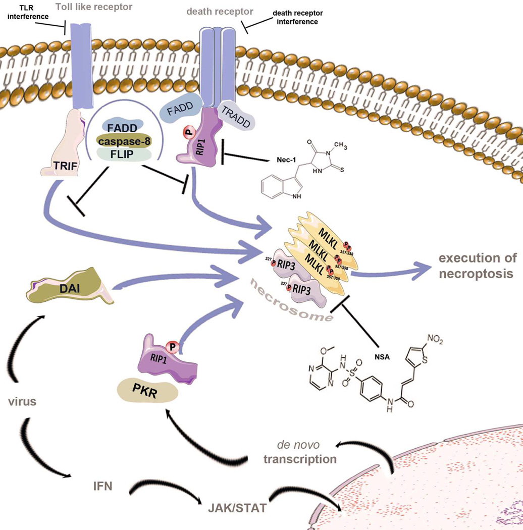
Various stimuli lead to the activation of the supramolecular necroptosis-inducing complex, referred to as the necrosome. Initially, studies of necroptosis employed models of death receptor stimulation in the presence of caspase-inhibition (not shown). The intracellular adapter molecules FADD and TRADD recruit RIPK1, which subsequently undergoes an incompletely understood series of ubiquitylation, deubiquitinylation, and phosphorylation events before exposing its rip homotypic interaction motif (RHIM-domain) to recruit RIPK3. RIPK1, RIPK3 and MLKL are phosphorylated during the assembly of the necrosome. Within the human genome, RIPK1, RIPK3 and two other proteins exhibit RHIM-domains. One of these is TRIF, an intracellular signal transducer that is capable of activating the necrosome downstream of Toll-like receptors which are triggered by microbial molecules. The fourth RHIM-domain protein, DAI, integrates signals from viral RNA sensors into the necrosome. Finally, viral infection is accompanied by production of interferon, which triggers the JAK/STAT-dependent de novo synthesis of protein kinase R (PKR) which phosphorylates FADD to directly interact with RIPK1 and induce necrosome formation. Death receptor-mediated necroptosis involves deubiquitinylation of RIPK1, the kinase domain of which is targeted by necrostatin-1 (Nec-1). Second generation RIP1-kinase inhibitors, such as Nec-1s, a stable version of Nec-1 which is more potent at lower concentrations, might reduce observed side effects. Necrosulfonamide (NSA) inhibits MLKL and prevents the activity of the necrosome in human cells. In addition, RIPK3-inhibitors, death receptor antagonists or plasma membrane channel blockers might be attractive therapeutical targets.
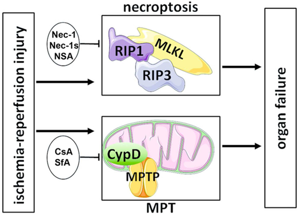
In ischemia-reperfusion injury, independent pathways of regulated necrosis contribute additively to overall organ damage. Whereas formation of the necroptotic pathway involves RIPK1-triggered assembly of the necrosome (MLKL-RIPK3), mitochondrial permeability transition (MPT) independently contributes in a cyclophilin D dependent manner. Necroptosis may be blocked by necrostatin-1 (Nec-1), second generation “stable” Nec-1 (Nec-1s) or the MLKL-inhibitor necrosulfonamide (NSA). MPT is inhibited by cyclosporine A (CsA) or sanglifehrin A (SfA). Combination therapy with Nec-1 and SfA exhibited significantly stronger protection compared to each monotherapy.
References
-
- He S, Wang L, Miao L, et al. Receptor interacting protein kinase-3 determines cellular necrotic response to TNF-alpha. Cell. 2009;137(6):1100–1111. - PubMed
-
- Vandenabeele P, Galluzzi L, Vanden Berghe T, Kroemer G. Molecular mechanisms of necroptosis: an ordered cellular explosion. Nat Rev Mol Cell Biol. 2010;11(10):700–714. - PubMed
-
- Zhang DW, Shao J, Lin J, et al. RIP3, an energy metabolism regulator that switches TNF-induced cell death from apoptosis to necrosis. Science. 2009;325(5938):332–336. - PubMed
Publication types
MeSH terms
Grants and funding
LinkOut - more resources
Full Text Sources
Other Literature Sources