. 2014 Apr;42(7):4414-26.
doi: 10.1093/nar/gku076. Epub 2014 Jan 29.

The α isoform of topoisomerase II is required for hypercompaction of mitotic chromosomes in human cells

Affiliations

The α isoform of topoisomerase II is required for hypercompaction of mitotic chromosomes in human cells

Christine J Farr et al. Nucleic Acids Res. 2014 Apr.

Abstract

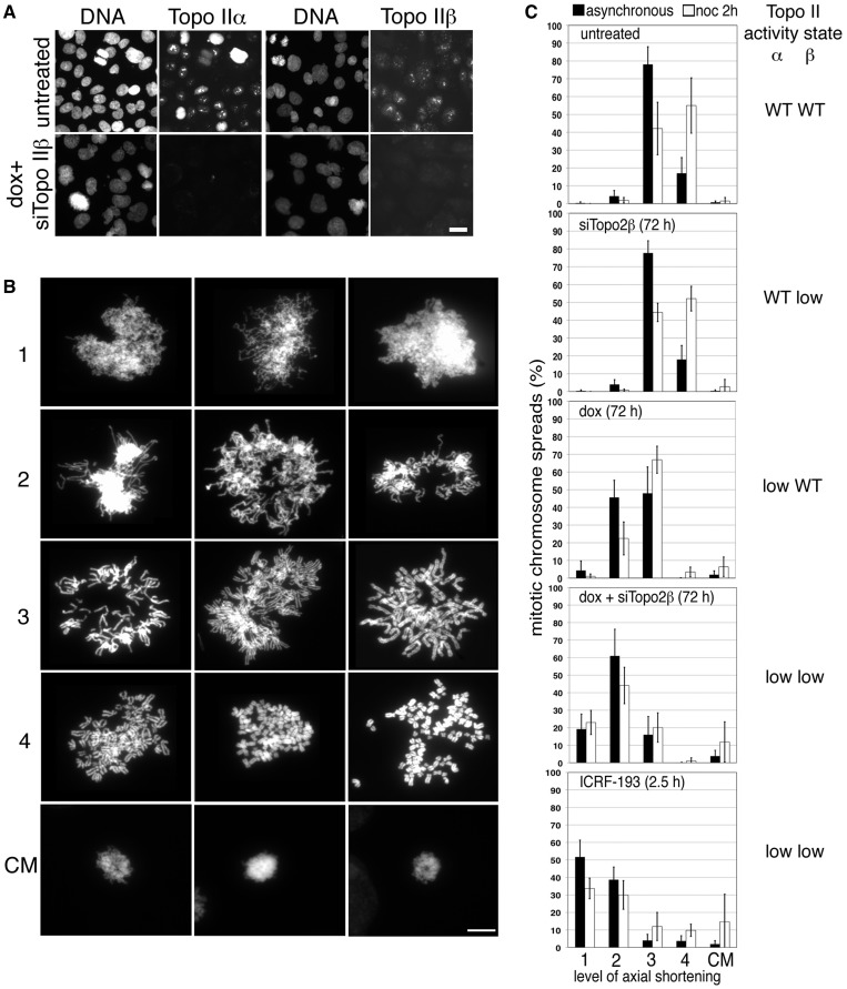
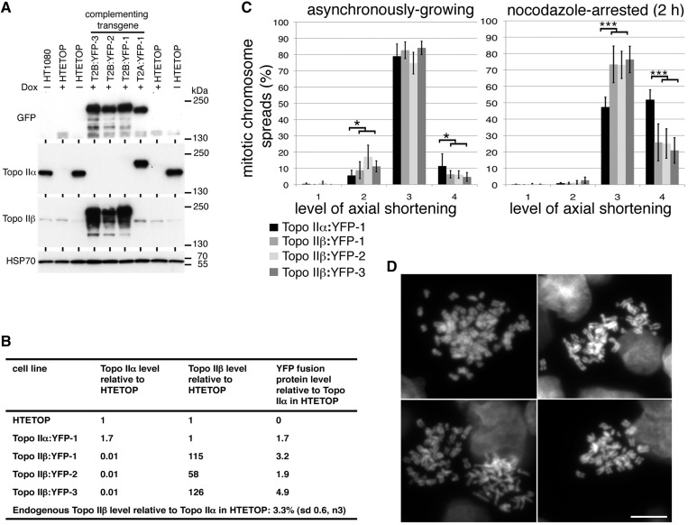
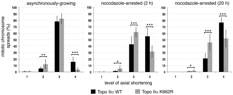
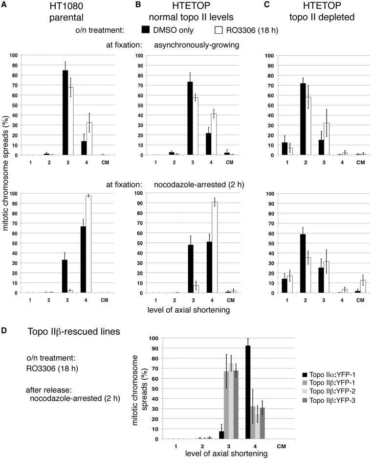
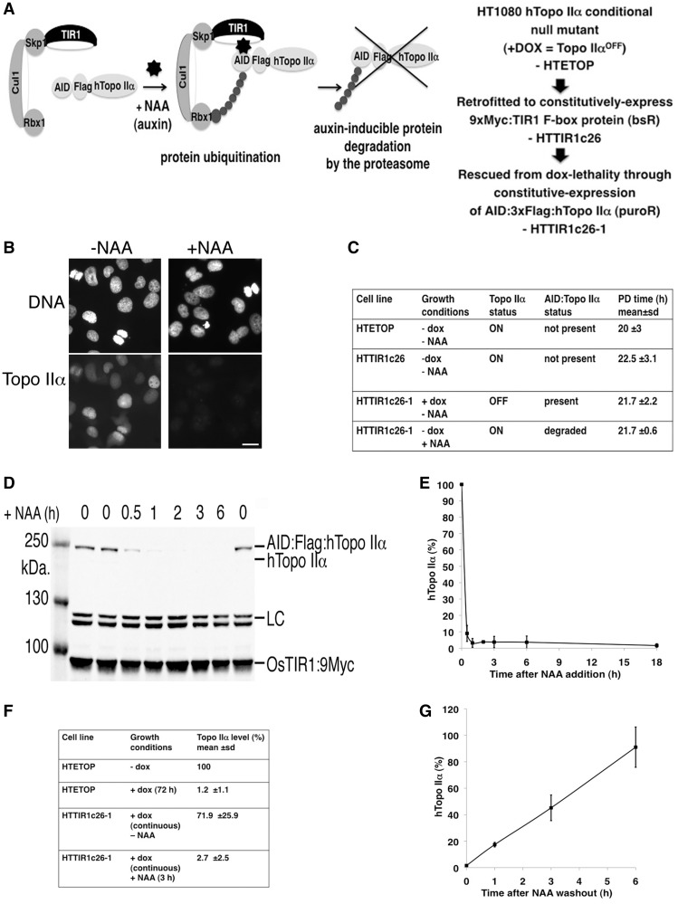
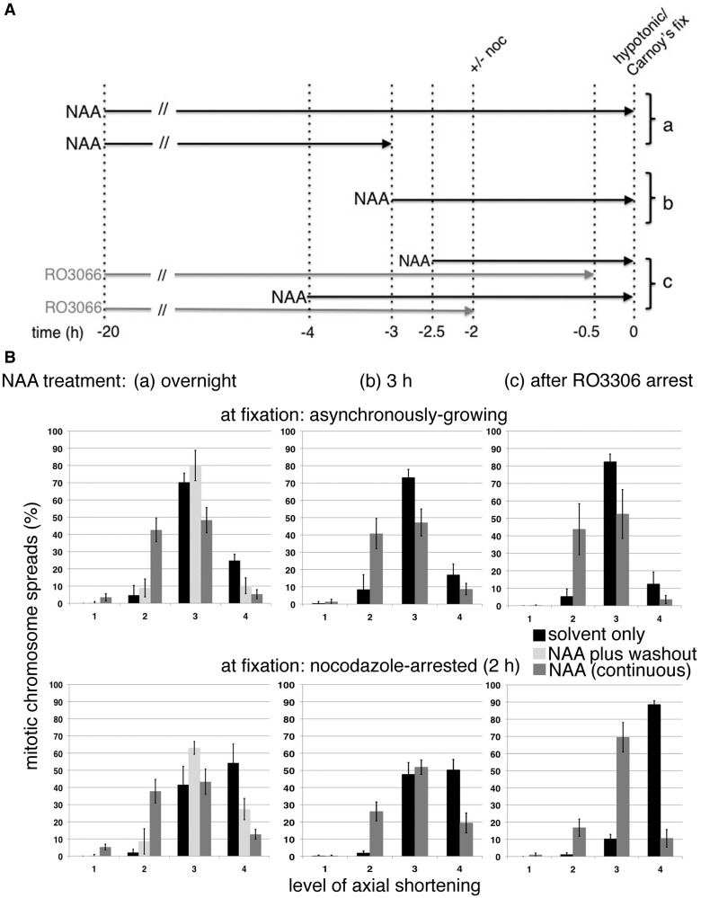
As proliferating cells transit from interphase into M-phase, chromatin undergoes extensive reorganization, and topoisomerase (topo) IIα, the major isoform of this enzyme present in cycling vertebrate cells, plays a key role in this process. In this study, a human cell line conditional null mutant for topo IIα and a derivative expressing an auxin-inducible degron (AID)-tagged version of the protein have been used to distinguish real mitotic chromosome functions of topo IIα from its more general role in DNA metabolism and to investigate whether topo IIβ makes any contribution to mitotic chromosome formation. We show that topo IIβ does contribute, with endogenous levels being sufficient for the initial stages of axial shortening. However, a significant effect of topo IIα depletion, seen with or without the co-depletion of topo IIβ, is the failure of chromosomes to hypercompact when delayed in M-phase. This requires much higher levels of topo II protein and is impaired by drugs or mutations that affect enzyme activity. A prolonged delay at the G2/M border results in hyperefficient axial shortening, a process that is topo IIα-dependent. Rapid depletion of topo IIα has allowed us to show that its function during late G2 and M-phase is truly required for shaping mitotic chromosomes.


Figures

Figure 1.
Figure 1.
The effect of depleting the two topo II isoforms on mitotic chromosome formation. (A) Indirect IF of topo IIα and topo IIβ in HTETOP cells either untreated or exposed to dox (topo IIα depletion) + siTopo IIβ for 3 days. Cells were fixed in situ using PTEMF. Topo II was detected using either anti-topo IIα or anti-topo IIβ antibody (FITC) and DNA counterstained using DAPI. Scale bar, 25 µm. (B) Representative images of DAPI-stained chromosome spreads assigned to various levels (1–4) of axial shortening are shown, together with examples of compact chromatin masses (CM). Scale bar, 10 µm. (C) Frequencies of the various levels of axial shortening observed in mitotic HTETOP cells expressing normal levels of both topo II isoforms compared with cells depleted of either topo IIα or IIβ, or both, over 72 h, or cells in which both isoforms have been chemically inhibited for 2.5 h (ICRF-193). Cells were grown on slides overnight, treated with hypotonic (75 mM KCl, 10 min) before fixation in ice-cold methanol: acetic acid and examined after DAPI staining of the DNA. Data were collected both from asynchronously growing populations and from cells arrested in M-phase (nocodazole 2 h). Data points represent the mean (±standard deviation (sd)) based on ≥3 independent experiments, with ∼100 cells scored per experiment.
Figure 2.
Figure 2.
Topo IIβ and mitotic chromosome formation. (A) Chemiluminescent immunoblot of HTETOP clones rescued from dox lethality by expression of YFP-fused topo IIα or IIβ. Whole cell lysates of the untransfected HTETOP parental cell line (grown in the absence or presence of dox), and dox-resistant HTETOP transfectant clones expressing topo II:YFP fusion proteins (+ dox) were subjected to sodium dodecyl sulfate–polyacrylamide gel electrophoresis (5% gels) and immunoblotting. Blots were hybridized with antibodies against GFP/YFP, human topo IIα, human topo IIβ and HSP70 (loading control). (B) Topo IIα and IIβ levels estimated by fluorescence immunoblotting of the various cell lines. Antibodies against topo IIα and IIβ were used to estimate levels of the two isoforms in the various transfectants. YFP-immunoblotting was then used to compare levels of the fusion protein in transfectants and determine topo IIα and IIβ levels relative to the parental. (C) Assessment of mitotic chromosome formation in three independent HTETOP transfectant clones expressing topo IIβ:YFP compared with a reference clone expressing topo IIα:YFP. Data were collected both from asynchronously growing and nocodazole-arrested (2 h) cell populations. Data points for each cell line represent the mean (±sd) based on ≥3 independent experiments, with ∼100 cells scored per experiment. The frequency of spreads showing the compact mass (CM) phenotype were <1% in all cases. (D) Representative DAPI-stained chromosome spreads showing level 4 axial shortening from cells in which ∼99% of topo II is topo IIβ (YFP-tagged and the endogenous untagged isoform). Scale bar, 10 µm.
Figure 3.
Figure 3.
The effect of catalytically compromised topo IIα (K662R) on mitotic chromosome formation. Chromosome compaction status in independently derived HTETOP stable cell clones (n6) rescued from dox lethality by expression of topo IIα K662R (N-terminally Flag-tagged) was compared with stable cell clones expressing Flag:topo IIα WT (n3). The results from asynchronously growing cell populations are shown, together with those from cells held in M-phase by exposure to nocodazole for either 2 or 20 h. Each stable cell line was analyzed in ≥3 independent experiments, with ∼100 cells scored per experiment. The data sets for all clones expressing either K662R or WT topo IIα were combined. Data points represent the mean ±sd. The frequency of spreads showing the compact mass (CM) phenotype were <2% in all cases.
Figure 4.
Figure 4.
Enhanced hypercompaction following RO3306 arrest. Cells were delayed at the G2/M boundary by an overnight exposure to RO3306 (or treated with DMSO only). Following release back into the cell cycle, cells were treated with hypotonic and fixed in methanol: acetic acid, either when growing asynchronously (30 min post RO3306 washout) or after a nocodazole-induced delay (2 h) and mitotic chromosome formation assessed in (A) HT1080 cells (the parental cell line of HTETOP), (B) untreated HTETOP (normal levels of both topo II isoforms), (C) HTETOP cells depleted of both topo II isoforms (dox + siTopo IIβ, 72 h) and (D) HTETOP cells that have been rescued from dox lethality by expression of topo IIβ:YFP compared with those expressing topo IIα:YFP. All data points represent the mean (±sd) based on ≥3 independent experiments, with ∼100 cells scored per experiment.
Figure 5.
Figure 5.
Rapid depletion of topo IIα from HTETOP cells. (A) Strategy used to generate an HTETOP derivative from which topo IIα can be rapidly depleted. (B) Indirect IF of topo IIα in HTTIR1c26-1 cells grown continuously in the presence of dox (to deplete untagged topo IIα) and blasticidin (to maintain TIR1 expression), with or without 1 mM NAA for 3 h (to degrade AID-tagged topo IIα). Topo IIα was detected using anti-topo IIα antibody (FITC). DNA was counterstained using DAPI. Scale bar, 25 µm. (C) Comparison of the PD times of the parental HTETOP cell line (-dox) with HTTIR1c26 (a derivative retrofitted to constitutively express TIR1) and HTTIR1c26-1 (which expresses both TIR1 and AID degron:Flag:Topo IIα). The growth of the latter clone was examined under both +dox/−NAA conditions (where the AID:Flag:Topo IIα fusion protein is the main form of topo IIα present in the cells) and under +NAA/−dox selection (when the bulk of the topo IIα present is untagged). (D) Fluorescence immunoblotting of topo IIα, myc-tagged TIR1 and an unidentified protein detected by the anti-topo IIα antibody (LC). (E) Graph showing topo IIα levels at various time points after addition of 1 mM NAA. Shown are mean values. Error bars represent sd. Each data point is based on <3 independent experiments. (F) Topo IIα level in the parental HTETOP cell line (− and + dox) compared with that in HTTIR1c26-1 after exposure to 1 mM NAA (3 h). (G) Graph showing the recovery of topo IIα levels at various time points after removal of 1 mM NAA. Shown are mean values. Error bars represent sd. Each data point is based on three independent experiments.
Figure 6.
Figure 6.
(A) Schematic outlining the experimental strategy used to compare the impact on mitotic chromosome formation of depleting topo IIα from late G2 only versus its continuous depletion throughout the cell cycle. In all experiments, cells were treated with siRNA (72 h) to deplete topo IIβ. (a), (b) and (c) refer to sections in 6B. (B) Assessment of mitotic chromosome formation following various NAA (or DMSO only) treatment regimens: (a) overnight, with or without a 3 h recovery period post NAA washout, (b) for 3 h and (c) cells delayed at the G2/M border by overnight treatment with RO3306 were exposed to NAA for 2 h (RO3306 + NAA) before release back into the cell cycle in the continued presence of NAA. Cells were treated with hypotonic and fixed in methanol–acetic acid either when growing asynchronously (cells arrested by RO3306 were fixed 30 min post RO3306 washout) or after M-phase arrest (nocodazole 2 h). For all conditions, data points represent the mean (±sd) based on ≥3 independent experiments, with ∼100 cells scored per experiment. The frequency of spreads showing the compact mass (CM) phenotype was <2.4% in all cases.

References

    1. Chung TD, Drake FH, Tan KB, Per SR, Crooke ST, Mirabelli CK. Characterization and immunological identification of cDNA clones encoding two human DNA topoisomerase II isozymes. Proc. Natl Acad. Sci. USA. 1989;86:9431–9435. - PMC - PubMed
    1. Tan KB, Dorman TE, Falls KM, Chung TD, Mirabelli CK, Crooke ST, Mao J. Topoisomerase II alpha and topoisomerase II beta genes: characterization and mapping to human chromosomes 17 and 3, respectively. Cancer Res. 1992;52:231–234. - PubMed
    1. Niimi A, Suka N, Harata M, Kikuchi A, Mizuno S. Co-localization of chicken DNA topoisomerase IIalpha, but not beta, with sites of DNA replication and possible involvement of a C-terminal region of alpha through its binding to PCNA. Chromosoma. 2001;110:102–114. - PubMed
    1. Heck MM, Hittelman WN, Earnshaw WC. Differential expression of DNA topoisomerases I and II during the eukaryotic cell cycle. Proc. Natl Acad. Sci. USA. 1988;85:1086–1090. - PMC - PubMed
    1. Adachi N, Nomoto M, Kohno K, Koyama H. Cell-cycle regulation of the DNA topoisomerase IIalpha promoter is mediated by proximal CCAAT boxes: possible involvement of acetylation. Gene. 2000;245:49–57. - PubMed

Publication types