. 2014 Mar;3(3):323-33.
doi: 10.5966/sctm.2013-0112. Epub 2014 Jan 29.

Transplantation of photoreceptors derived from human Muller glia restore rod function in the P23H rat

Affiliations

Transplantation of photoreceptors derived from human Muller glia restore rod function in the P23H rat

Hari Jayaram et al. Stem Cells Transl Med. 2014 Mar.

Abstract

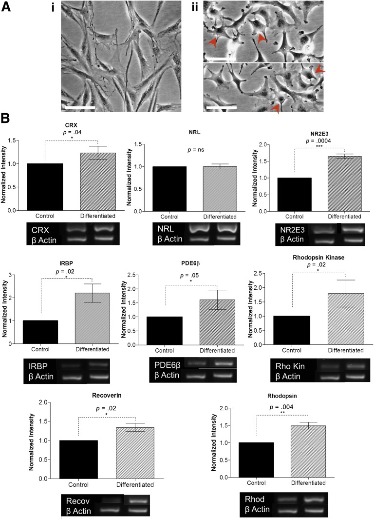
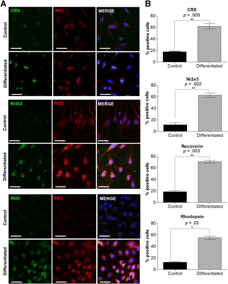
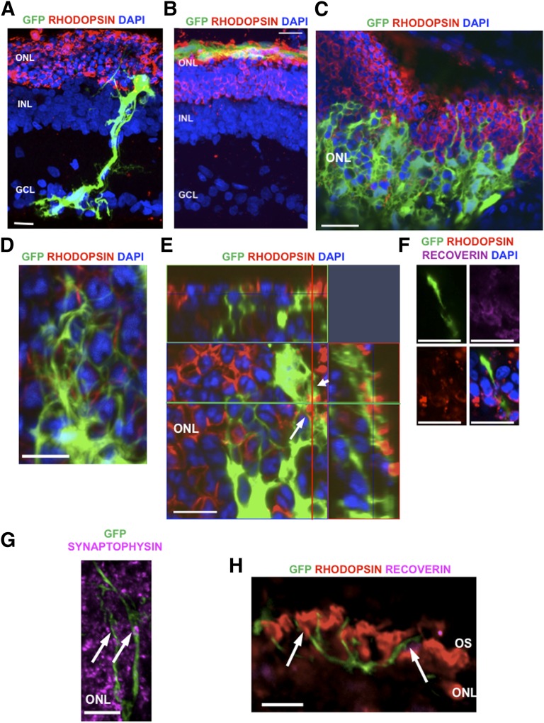
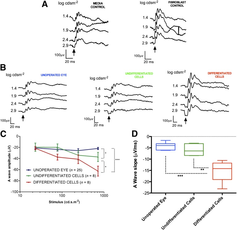
Müller glia possess stem cell characteristics that have been recognized to be responsible for the regeneration of injured retina in fish and amphibians. Although these cells are present in the adult human eye, they are not known to regenerate human retina in vivo. Human Müller glia with stem cell characteristics (hMSCs) can acquire phenotypic and genotypic characteristics of rod photoreceptors in vitro, suggesting that they may have potential for use in transplantation strategies to treat human photoreceptor degenerations. Much work has been undertaken in rodents using various sources of allogeneic stem cells to restore photoreceptor function, but the effect of human Müller glia-derived photoreceptors in the restoration of rod photoreceptor function has not been investigated. This study aimed to differentiate hMSCs into photoreceptor cells by stimulation with growth and differentiation factors in vitro to upregulate gene and protein expression of CRX, NR2E3, and rhodopsin and various phototransduction markers associated with rod photoreceptor development and function and to examine the effect of subretinal transplantation of these cells into the P23H rat, a model of primary photoreceptor degeneration. Following transplantation, hMSC-derived photoreceptor cells migrated and integrated into the outer nuclear layer of the degenerated retinas and led to significant improvement in rod photoreceptor function as shown by an increase in a-wave amplitude and slope using scotopic flash electroretinography. These observations suggest that hMSCs can be regarded as a cell source for development of cell-replacement therapies to treat human photoreceptor degenerations and may also offer potential for the development of autologous transplantation.

Keywords: Müller glia; Photoreceptors; Repair and regeneration; Retina; Stem cells; Transplantation.


Figures

Figure 1.
Figure 1.
In vitro differentiation with recombinant human basic fibroblast growth factor, taurine, retinoic acid, and insulin-like growth factor type 1 on basement membrane protein induces morphological changes and upregulates rod photoreceptor gene expression in human Müller glia with stem cell characteristics (hMSCs). (Ai): hMSCs cultured in the absence of growth or differentiation factors exhibit a typical elongated glial morphology. (Aii): hMSCs cultured for 5 days in the presence of fibroblast growth factor, taurine, retinoic acid, and insulin-like growth factor type 1 exhibited a marked change in cellular morphology, as seen with phase microscopy, with condensed cell bodies, phase bright nuclei, prominent nuclear chromatin, and several short neurite-like processes, similar to previous published reports of photoreceptors in culture [–38] (red arrowheads). Scale bars = 100 μm. (B): Analysis of reverse transcription-polymerase chain reaction band densities showed that differentiated cells exhibited a significant increase in mRNA expression of CRX (30%), Nr2e3 (60%), rhodopsin (50%), recoverin (40%), rhodopsin kinase (80%), PDE6β (60%), and IRBP (110%) when compared with untreated cells cultured in the absence of extracellular matrix (n = 5; error bars represent the SEM). Abbreviations: IRBP, interphotoreceptor retinoid-binding protein; ns, not significant; PDE6β, phosphodiesterase type 6β Recov, recoverin; Rhod, rhodopsin.
Figure 2.
Figure 2.
In vitro differentiation of human Müller glia with stem cell characteristics with fibroblast growth factor, taurine, retinoic acid, and insulin-like growth factor type 1 upregulates the expression of rod photoreceptor markers. (A): Differentiation of human Müller glia with stem cell characteristics was performed on basement membrane protein-coated glass slides in the presence of fibroblast growth factor, taurine, retinoic acid, and insulin-like growth factor type 1 and on uncoated plastic chamberwell slides as a control. Differentiating conditions led to an observed increase in staining for Nr2e3, CRX, rhodopsin, and recoverin. The images shown illustrate areas showing larger numbers of positive cells for each marker. Scale bars = 100 μm. (B): The histograms show the increase in the percentage of cells within the population staining for each marker. A significant increase in expression of CRX (p = .005), rhodopsin (p = .03), NR2E3 (p = .005), and recoverin (p = .003) was seen in differentiated cells when compared with control cells. These results are the average of four different fields in three separate experiments. Abbreviations: REC, recoverin; RHO, rhodopsin.
Figure 3.
Figure 3.
Migration of human Müller glia with stem cell characteristics-derived photoreceptor cells into the retina of P23H-1 transgenic rats. (A): Undifferentiated human Müller glia with stem cell characteristics migrated longitudinally across the host retina without localization to a specific retinal lamina adopting a typical Müller glial morphology. (B): Low-magnification confocal microscopy image of the whole retina showing localization of differentiated donor GFP+ cells at the outer retina following subretinal transplantation. (C): At 3 weeks after transplantation, differentiated donor GFP+ cells had migrated into the ONL of the host retina. (D): Differentiated donor GFP+ cells appeared to be well integrated into the ONL, as judged by their interwoven cellular projections observed around the host cells. (E): An example of differentiated donor GFP+ cells demonstrating colocalization with rhodopsin (arrow) within the ONL of the host retina. (F): Differentiated donor GFP+ cells were seen to localize within the host outer nuclear layer extending toward the location of the remaining host outer segments with a spherule-like projection remaining in the outer nuclear layer. (G): Synaptic connectivity between the differentiated donor GFP+ cells and host ONL was suggested by colocalization of synaptophysin (purple) with GFP+ cells (arrows). (H): Although classic outer segments were not seen in the differentiated donor GFP+ cells, neural-like projections of GFP+ cells were observed surrounding the host OS (arrows). Scale bars = 50 μm. Abbreviations: DAPI, 4′,6-diamidino-2-phenylindole; GCL, ganglion cell layer; GFP, green fluorescent protein; INL, inner nuclear layer; ONL, outer nuclear layer; OS, outer segments.
Figure 4.
Figure 4.
Scotopic flash electroretinography performed 3 weeks following subretinal transplantation in the P23H-1 transgenic rat. (A): Representative waveforms in controls that were transplanted with sham injections of tissue culture media alone or human Tenon’s fibroblasts, with the a-wave indicated by the arrow. (B): Representative waveforms from unoperated eyes and those transplanted with undifferentiated human Müller glia with stem cell characteristics. The a-wave (arrows) in the eyes transplanted with the differentiated cells showed significantly greater amplitude than the other experimental groups at the higher intensity flash stimuli. (C): Eyes transplanted with differentiated cells showed significantly greater a-wave amplitude than those transplanted with undifferentiated cells (p < .01) and unoperated eyes (p < .001). A difference in a-wave amplitudes was also observed between unoperated eyes and those transplanted with undifferentiated Müller stem cells (p < .03) (error bars represent the SEM). (D): Analysis of slope of the leading edge of the a-wave through quantification of the gradient of the first 4 ms of the leading edge of the waveform, a more specific measure of rod photoreceptor function before other light-induced currents commence, showed a significantly greater a-wave slope in eyes transplanted with differentiated cells when compared with unoperated eyes (p < .0001) or eyes transplanted with undifferentiated cells (p < .001).

References

    1. Meyer RL. Evidence from thymidine labeling for continuing growth of retina and tectum in juvenile goldfish. Exp Neurol. 1978;59:99–111. - PubMed
    1. Turner DL, Cepko CL. A common progenitor for neurons and glia persists in rat retina late in development. Nature. 1987;328:131–136. - PubMed
    1. Das AV, Mallya KB, Zhao X, et al. Neural stem cell properties of Müller glia in the mammalian retina: Regulation by Notch and Wnt signaling. Dev Biol. 2006;299:283–302. - PubMed
    1. Fischer AJ, Reh TA. Müller glia are a potential source of neural regeneration in the postnatal chicken retina. Nat Neurosci. 2001;4:247–252. - PubMed
    1. Bhatia B, Singhal S, Lawrence JM, et al. Distribution of Müller stem cells within the neural retina: Evidence for the existence of a ciliary margin-like zone in the adult human eye. Exp Eye Res. 2009;89:373–382. - PubMed

Publication types

MeSH terms