Review
. 2014 Jan 28;15(2):2024-52.
doi: 10.3390/ijms15022024.

Adenosine receptors: expression, function and regulation

Affiliations
Review

Adenosine receptors: expression, function and regulation

Sandeep Sheth et al. Int J Mol Sci. .

Abstract

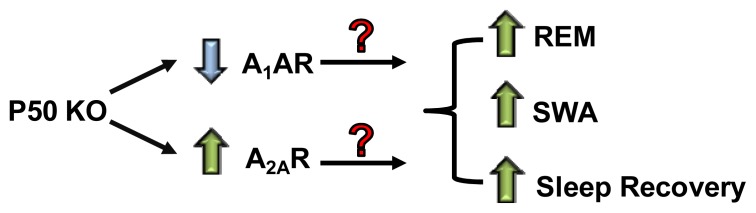
Adenosine receptors (ARs) comprise a group of G protein-coupled receptors (GPCR) which mediate the physiological actions of adenosine. To date, four AR subtypes have been cloned and identified in different tissues. These receptors have distinct localization, signal transduction pathways and different means of regulation upon exposure to agonists. This review will describe the biochemical characteristics and signaling cascade associated with each receptor and provide insight into how these receptors are regulated in response to agonists. A key property of some of these receptors is their ability to serve as sensors of cellular oxidative stress, which is transmitted by transcription factors, such as nuclear factor (NF)-κB, to regulate the expression of ARs. Recent observations of oligomerization of these receptors into homo- and heterodimers will be discussed. In addition, the importance of these receptors in the regulation of normal and pathological processes such as sleep, the development of cancers and in protection against hearing loss will be examined.


Figures

Figure 1.
Figure 1.
Proposed model of regulation of sleep by adenosine receptors in p50 KO mice. These mice express lower levels of A1AR in the cortex and striatum but higher expression of the A2AAR. P50 KO mice also demonstrate increased REM and SWA sleep and increased rate of sleep recovery following sleep deprivation. (?) indicates question as to which receptors to attribute the differences in sleep pattern to.
Figure 2.
Figure 2.
Protection of rat cochlear outer hair cells from cisplatin-induced damage by A1AR agonist, R-PIA. Rats were pre-treated with R-PIA (10 μL of a 10 μM solution added to the round window). The remaining liquid was removed after 1 h and rats were administered cisplatin by intraperitoneal injections (16 mg/kg). Panels AC are electron micrographs of the organ of Corti from rats treated with cisplatin and represent the hook, basal turn and the middle turn, respectively; Panels DF shows the effect of pre-treatment with R-PIA for the respective regions; Panels GI show the effect of R-PIA in presence of DPCPX, an A1AR antagonist, on cisplatin-induced damage. The inner hair cells are the three rows of cells with the “V” shaped stereociliary bundles. The inner hair cells are barely visible on the top of each micrograph. The data which was confirmed from different cochlear preparations support a protective role of the A1AR against cisplatin ototoxicity. (Reprinted with permission from [134], Copyright 2004 Elsevier). Scale bar = 5 μm.
Figure 3.
Figure 3.
Activation of A3AR suppresses ROS generation in AT6.1 prostate cancer cells. Cells were pretreated with the A3AR agonist, IB-MECA, the antagonist, MRS1523, or a combination of IB-MECA + MRS1523. ROS was determined by H2DCFDA fluorescence. IB-MECA significantly reduced fluorescence in these cells (A,B) which was reversed by MRS1523, indicating a role of the A3AR in this process. Similar effects were obtained with DPI and AEBSF, known inhibitors of NADPH oxidase (C,D). Experiments were replicated four times. Histograms represent the mean ± SEM. Asterisks (*) and (**) indicates statistically significant difference (p < 0.05) from vehicle and IB-MECA treatments, respectively. (Reprinted with permission from [169], Copyright 2009 Neoplasia Press, Inc.).

References

    1. Sebastiao A.M., Ribeiro J.A. Fine-tuning neuromodulation by adenosine. Trends Pharmacol. Sci. 2000;21:341–346. - PubMed
    1. de Mendoncca A., Ribeiro J.A. Adenosine and synaptic plasticity. Drug Dev. Res. 2001;52:283–290.
    1. Cunha R.A. Adenosine as a neuromodulator and as a homeostatic regulator in the nervous system: Different roles, different sources and different receptors. Neurochem. Int. 2001;38:107–125. - PubMed
    1. Ferreira J.M., Paes-de-Carvalho R. Long-term activation of adenosine A(2a) receptors blocks glutamate excitotoxicity in cultures of avian retinal neurons. Brain Res. 2001;900:169–176. - PubMed
    1. Fredholm B.B., AP I.J., Jacobson K.A., Linden J., Muller C.E. International Union of Basic and Clinical Pharmacology. LXXXI. Nomenclature and classification of adenosine receptors—An update. Pharmacol. Rev. 2011;63:1–34. - PMC - PubMed

Publication types