. 2014 Oct;229(10):1359-68.
doi: 10.1002/jcp.24569.

Phosphorylation-regulated degradation of the tumor-suppressor form of PED by chaperone-mediated autophagy in lung cancer cells

Affiliations

Phosphorylation-regulated degradation of the tumor-suppressor form of PED by chaperone-mediated autophagy in lung cancer cells

Cristina Quintavalle et al. J Cell Physiol. 2014 Oct.

Abstract

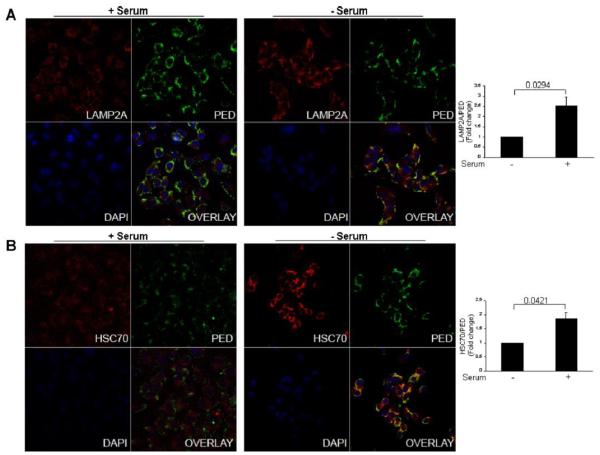
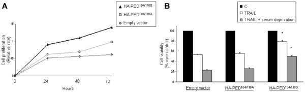
PED/PEA-15 is a death effector domain (DED) family member with a variety of effects on cell growth and metabolism. To get further insight into the role of PED in cancer, we aimed to find new PED interactors. Using tandem affinity purification, we identified HSC70 (Heat Shock Cognate Protein of 70 kDa)-which, among other processes, is involved in chaperone-mediated autophagy (CMA)-as a PED-interacting protein. We found that PED has two CMA-like motifs (i.e., KFERQ), one of which is located within a phosphorylation site, and demonstrate that PED is a bona fide CMA substrate and the first example in which phosphorylation modifies the ability of HSC70 to access KFERQ-like motifs and target the protein for lysosomal degradation. Phosphorylation of PED switches its function from tumor suppression to tumor promotion, and we show that HSC70 preferentially targets the unphosphorylated form of PED to CMA. Therefore, we propose that the up-regulated CMA activity characteristic of most types of cancer cell enhances oncogenesis by shifting the balance of PED function toward tumor promotion. This mechanism is consistent with the notion of a therapeutic potential for targeting CMA in cancer, as inhibition of this autophagic pathway may help restore a physiological ratio of PED forms.


Figures

Figure 1
Figure 1. Validation of the interaction between PED and Hsc-70. In vitro immunoprecipitation
HEK-293 were transfected with PED-Myc cDNA. After 48 hrs, 1 mg of protein extracts were immunoprecipitated with anti-PED (A) or anti-Hsc70 antibody (B) or with control IgG, as indicated. Samples were then resolved with SDS-PAGE and western blot with anti-Hsc70 or anti-Myc antibodies. 10 μg of total cellular extracts were loaded as control. (C)- GST pull down. A549 cellular extracts were incubated with Glutathione-Sepharose 4B beads pre-bound to GST-PED fusion protein. Negative control was obtained by incubating cellular extracts to Glutathione-Sepharose 4B beads not pre-bound to GST-PED. Positive control was obtained loading the pre-bound GST PED fusion beads (10 μg). 50 μg of total cellular extract was loaded. (D) Confocal experiments. PED and Hsc70 distribution in A549 cells was determined by immunofluorescence analysis using confocal microscopy. A549 cells were fixed with 4% PFA, permeabilized in blocking solution (0.1% Saponin, 10% FBS in PBS), incubated with PED or Hsc70 antibodies and then with secondary antibodies conjugated respectively with a green (anti-rabbit) or red (anti-mouse) fluorochrome. The intensity of the yellow signals represents the degree of co-localization of the two proteins.
Figure 2
Figure 2. PED localizes to CMA-positive lysosomes
(A) Cellular localization of PED and LAMP-2A protein in serum deprived A549 cells. Upon serum starvation, PED-LAMP-2A co-localization was evident (yellow signal) indicating the co-localization of PED with CMA-positive lysosomes. (B) Cellular localization of PED and Hsc70 protein in serum deprived A549 cells. After starvation, in A459 a yellow signalling was evident, indicating PED- Hsc70 co-localization.
Figure 3
Figure 3. Lysosomal uptake of PED by chaperone-mediated autophagy
(A) Intact rat liver lysosomes pretreated or not with protease inhibitors (PI) were incubated with purified PED (0.1mg) and at the end of the incubation collected by centrifugation and subjected to SDS-PAGE and immunoblot for PED. Left: representative immunoblot (input: 1ng). Right: binding, association and uptake of PED by lysosomes calculated as described under material and methods. Values are expressed as percentage of the protein added and are mean + S.E. n=4. (B) Lysosomes were incubated with increased concentrations of PED and processed as in A. Left: representative immunoblot. Right: Quantification of the percentage of PED bound to lysosomes. (C) Lysosomes were incubated with PED in the presence of ovalbumin or GAPDH, a non-CMA and a CMA substrate respectively and processed as in A. Left: representative immunoblot. Right: PED uptake in lysosomes incubated with PED alone or in the presence of the indicated proteins. Values are expressed as percentage of the protein added and are mean+ S.E. n=4. Percentage of inhibition is indicated. D-E. Lysosomes were incubated with a fix concentration of PED and increasing concentrations of Rnase A (D) or with RNase A and increasing concentrations of PED (E). Samples were subjected to immunoblot for both proteins.
Figure 4
Figure 4. PED expression is affected by CMA
(A) CMA activation. PED protein level analyzed in A549 cells by western blot upon A549 serum deprivation for 24 and 48 hr or (B) in A549 cells transfected for 24 hr with LAMP-2A cDNA. (C) CMA inhibition. PED protein level analyzed by western blot upon A549 treatment with different doses of cloroquine or (D) in A549 transfected for 24 hr with ShRNA-LAMP-2A or (E) with siRNA Hsc70 for 48hr. (F) A549 transfected for 24 hr with PED-Myc cDNA and then serum deprived or treated with 10μM of chloroquine for 24 hrs or 15mM NH4CL for 4 hrs. PED Myc levels were analyzed by Western blot.
Figure 5
Figure 5. CMA regulates the unphosphorylated form of PED
(A) Schematic representation of PED phosphorylation sites and putative KFEQR sites. (B) HEK-293 were transfected with PED-Myc cDNA, HA-PED-S104/116A and HA-PED-S104/116D. After 48 hrs, 1 mg of protein extracts were immunoprecipitated with anti-PED antibody or with control IgG. Samples were then resolved with SDS-PAGE and western blot with anti-Hsc70 antibodies. 10 μg of total cellular extract was loaded as control. (C) A549 cells transfected with PED-Myc, HA-PED-S104/116A and HA-PED-S104/116D were serum deprived for 24 hrs. Protein levels of PED wt and phosphorylation mutant were analyzed by Western blot (D) Western blot analysis of PED-Myc and HA-PED-S104/116A, HA-PED-S104/116D after A549 co-transfection with LAMP2A cDNA upon 24 hr. (E) Western blot analysis of PED-Myc and HA-PED-S104/116A, HA-PED-S104/116D upon A549 treatment with 15 mMol of NH4Cl for 4 hr. (F) Western blot analysis of PED-Myc and HA-PED-S104/116A, HA-PED-S104/116D upon A549 treatment with 10 μMol of chloroquine for 24 hr.
Figure 6
Figure 6. Role of PED phosphorylation on cell growth and apoptosis
(A) A549 were transfected with different PED mutant as indicated. After 24 hrs, cells were serum starved for different times point and cell viability was assessed with an MTT assay. (B) A549 were transfected with different PED mutant as indicated. After 24 hrs, cells were treated with SuperKiller TRAIL (50ng/ml) alone o associated with serum deprivation for 24hrs. Cell viability was assessed with an MTT assay.

References

    1. Condorelli G, Vigliotta G, Cafieri A, Trencia A, Andalo P, et al. PED/PEA-15: an anti-apoptotic molecule that regulates FAS/TNFR1-induced apoptosis. Oncogene. 1999;18:4409–4415. - PubMed
    1. Renault F, Formstecher E, Callebaut I, Junier MP, Chneiweiss H. The multifunctional protein PEA-15 is involved in the control of apoptosis and cell cycle in astrocytes. Biochem Pharmacol. 2003;66:1581–1588. - PubMed
    1. Zanca C, Garofalo M, Quintavalle C, Romano G, Acunzo M, et al. PED is overexpressed and mediates TRAIL resistance in human non-small cell lung cancer. J Cell Mol Med. 2008;225:63–72. - PMC - PubMed
    1. Incoronato M, Garofalo M, Urso L, Romano G, Quintavalle C, et al. miR-212 increases tumor necrosis factor-related apoptosis-inducing ligand sensitivity in non-small cell lung cancer by targeting the antiapoptotic protein PED. Cancer Res. 2010;70:3638–3646. - PubMed
    1. Stassi G, Garofalo M, Zerilli M, Ricci-Vitiani L, Zanca C, et al. PED mediates AKT-dependent chemoresistance in human breast cancer cells. Cancer Res. 2005;65:6668–6675. - PubMed

Publication types

MeSH terms

Substances

LinkOut - more resources