. 2014 Feb 11;111(6):2170-5.
doi: 10.1073/pnas.1316001111. Epub 2014 Jan 29.

Myosin-binding protein C displaces tropomyosin to activate cardiac thin filaments and governs their speed by an independent mechanism

Affiliations

Myosin-binding protein C displaces tropomyosin to activate cardiac thin filaments and governs their speed by an independent mechanism

Ji Young Mun et al. Proc Natl Acad Sci U S A. .

Abstract

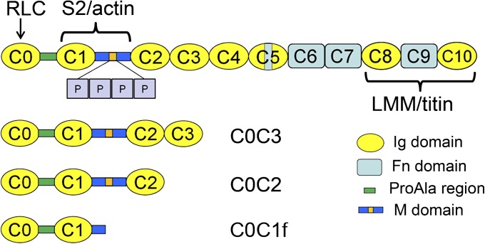
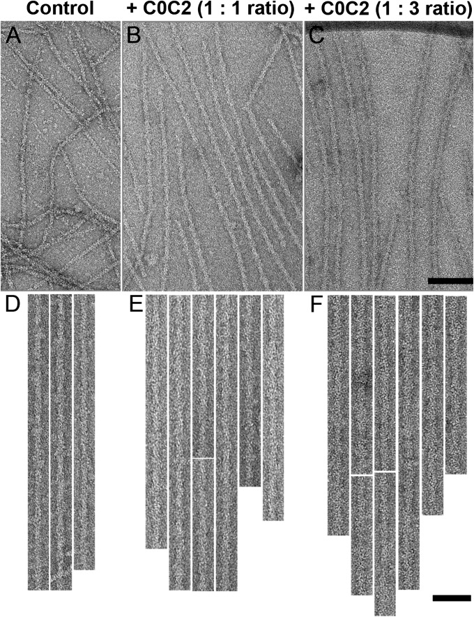
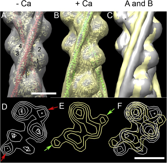
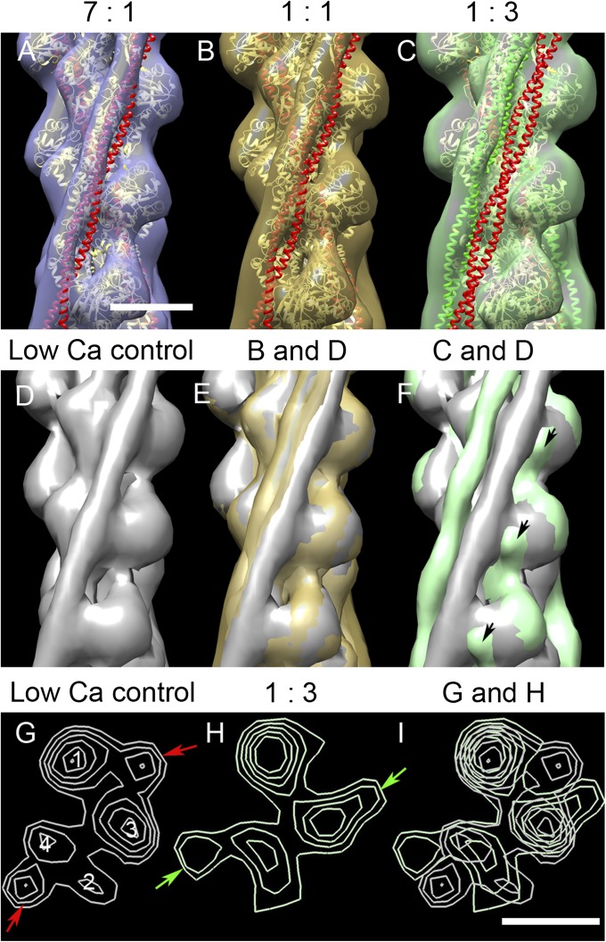
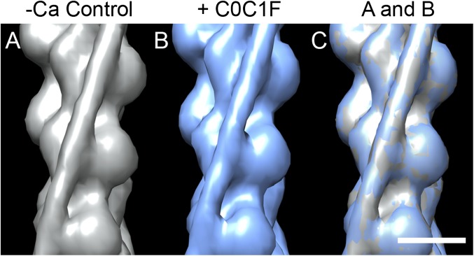
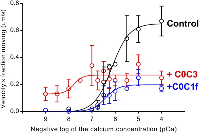
Myosin-binding protein C (MyBP-C) is an accessory protein of striated muscle thick filaments and a modulator of cardiac muscle contraction. Defects in the cardiac isoform, cMyBP-C, cause heart disease. cMyBP-C includes 11 Ig- and fibronectin-like domains and a cMyBP-C-specific motif. In vitro studies show that in addition to binding to the thick filament via its C-terminal region, cMyBP-C can also interact with actin via its N-terminal domains, modulating thin filament motility. Structural observations of F-actin decorated with N-terminal fragments of cMyBP-C suggest that cMyBP-C binds to actin close to the low Ca(2+) binding site of tropomyosin. This suggests that cMyBP-C might modulate thin filament activity by interfering with tropomyosin regulatory movements on actin. To determine directly whether cMyBP-C binding affects tropomyosin position, we have used electron microscopy and in vitro motility assays to study the structural and functional effects of N-terminal fragments binding to thin filaments. 3D reconstructions suggest that under low Ca(2+) conditions, cMyBP-C displaces tropomyosin toward its high Ca(2+) position, and that this movement corresponds to thin filament activation in the motility assay. At high Ca(2+), cMyBP-C had little effect on tropomyosin position and caused slowing of thin filament sliding. Unexpectedly, a shorter N-terminal fragment did not displace tropomyosin or activate the thin filament at low Ca(2+) but slowed thin filament sliding as much as the larger fragments. These results suggest that cMyBP-C may both modulate thin filament activity, by physically displacing tropomyosin from its low Ca(2+) position on actin, and govern contractile speed by an independent molecular mechanism.

Keywords: muscle activation; muscle regulation.


Conflict of interest statement

The authors declare no conflict of interest.

Figures

Fig. 1.
Fig. 1.
Schematic of cMyBP-C and the expressed N-terminal fragments C0C3, C0C2, and C0C1f used in this study. cMyBP-C consists of 8 Ig and 3 Fn domains together with a cMyBP-C-specific M-domain, containing a phosphorylation region (orange) with 4 phosphorylatable serines (P), a ProAla-rich domain, and a cardiac-specific insert (blue) in the C5 domain.
Fig. 2.
Fig. 2.
Decoration of native thin filaments with C0C2 under low Ca2+ conditions. (A, D) Undecorated control. (B, C, E, F) Filaments decorated with C0C2 at A:C0C2 molar ratios of 1:1 (B, E) and 1:3 (C, F). Filaments in DF have been computationally straightened. [Scale bar (AC) = 100 nm; (DF) = 50 nm.)
Fig. 3.
Fig. 3.
3D reconstructions of native control thin filaments under low and high Ca2+ conditions. (A) Low Ca2+ filament (gray surface rendering) fitted with ribbon depiction of low-Ca2+ A.Tm atomic model (39) (actin monomers, yellow; Tm, red). (B) High-Ca2+ reconstruction (yellow surface rendering), fitted with high-Ca2+ A.Tm atomic model (actin monomers, yellow; Tm, green). (C) Superposition of A and B demonstrating Tm shift on to inner domain of actin at high Ca2+ (note: slight variations in actin contours in A and B cause either gray or yellow to appear on the actin surface in C). (D and E) Transverse sections of low and high Ca2+ reconstructions, respectively, showing positioning of Tm (arrows) near the junction of actin SD1 and SD3 in low Ca2+, and on SD3 in high Ca2+. (F) Superposition of D and E, demonstrating the shift of Tm. Filaments in AC oriented with pointed end at top; actin subdomains are marked in A and D. (Scale bar = 5 nm.)
Fig. 4.
Fig. 4.
3D reconstructions of native thin filaments decorated with C0C2 under low Ca2+ conditions. (AC) Reconstructions with the indicated ratios of A:C0C2 fitted with A.Tm atomic models (39), as in Fig. 3, with Tm in blocked (red) or closed (green) position. With low levels of C0C2, there was a small movement of Tm from the blocked position (A and B), whereas with the highest level, Tm shifted to approximately the closed position (C). (E and F) show superposition of B and C, respectively, on the low Ca2+ control (D), demonstrating the smaller and larger shifts; black arrows indicate protrusion on SD1 surface, close to Tm, which we attribute to proximal region of C0C2. (G and H) Transverse sections of D and C, respectively, showing the shift of Tm from the blocked position in control (red arrows) to the closed position in C0C2-decorated filament (green arrows). (I) Superposition of G and H. Filaments in AF oriented with the pointed end up. (Scale bar = 5 nm.)
Fig. 5.
Fig. 5.
3D reconstructions of native thin filaments decorated with C0C1f under low Ca2+ conditions. (A) Low Ca2+ control filament (gray surface rendering). (B) C0C1f-decorated filament (blue). (C) Superposition of low Ca2+ control (A) and C0C1f-decorated filament (B) showing no Tm shift. (Scale bar = 5 nm.)
Fig. 6.
Fig. 6.
Effect of N-terminal cMyBP-C fragments on native thin filament sliding in in vitro motility assays. The graph shows “effective activation” (thin filament velocity × fraction of filaments moving) vs. pCa. The black line shows native thin filaments demonstrating a sigmoidal response to Ca2+. The red line shows C0C3 activated the thin filaments at low Ca2+, increased Ca2+ sensitivity, and inhibited maximal velocity. The blue line shows C0C1f had no effect on activation or Ca2+ sensitivity but inhibited maximal velocity. See Fig. S8 for individual velocity and fraction-moving data.

References

    1. Offer G, Moos C, Starr R. A new protein of the thick filaments of vertebrate skeletal myofibrils. Extraction, purification and characterization. J Mol Biol. 1973;74(4):653–676. - PubMed
    1. Winegrad S. Cardiac myosin binding protein C. Circ Res. 1999;84(10):1117–1126. - PubMed
    1. Barefield D, Sadayappan S. Phosphorylation and function of cardiac myosin binding protein-C in health and disease. J Mol Cell Cardiol. 2010;48(5):866–875. - PMC - PubMed
    1. Craig R, Offer G. The location of C-protein in rabbit skeletal muscle. Proc R Soc Lond B Biol Sci. 1976;192(1109):451–461. - PubMed
    1. Flashman E, Redwood C, Moolman-Smook J, Watkins H. Cardiac myosin binding protein C: Its role in physiology and disease. Circ Res. 2004;94(10):1279–1289. - PubMed

Publication types

LinkOut - more resources