. 2014 Mar 1;192(5):2432-41.
doi: 10.4049/jimmunol.1300247. Epub 2014 Jan 29.

B-1a cell diversity: nontemplated addition in B-1a cell Ig is determined by progenitor population and developmental location

Affiliations

B-1a cell diversity: nontemplated addition in B-1a cell Ig is determined by progenitor population and developmental location

Nichol E Holodick et al. J Immunol. .

Abstract

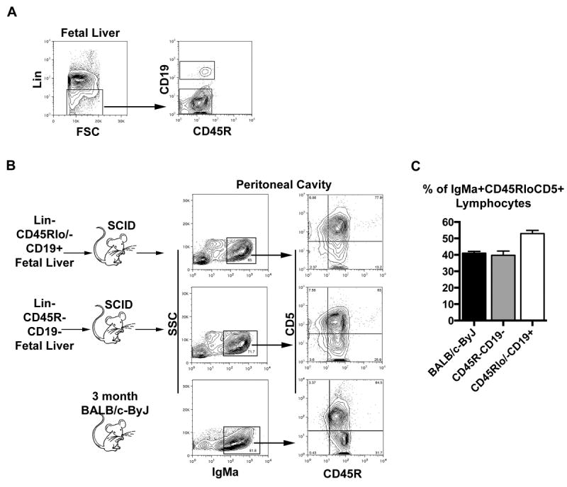
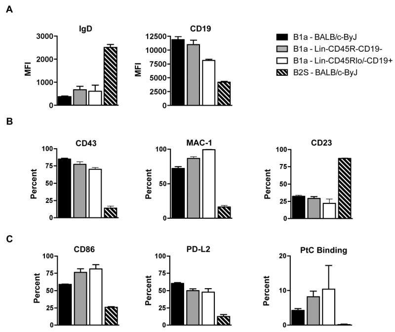
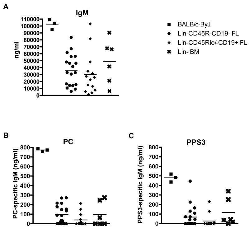
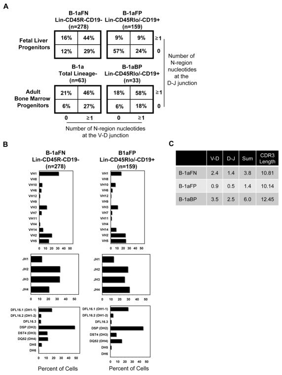
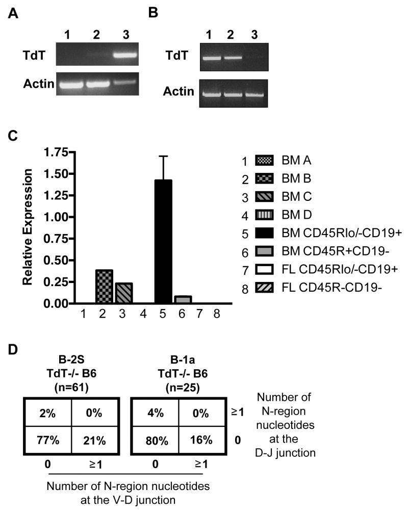
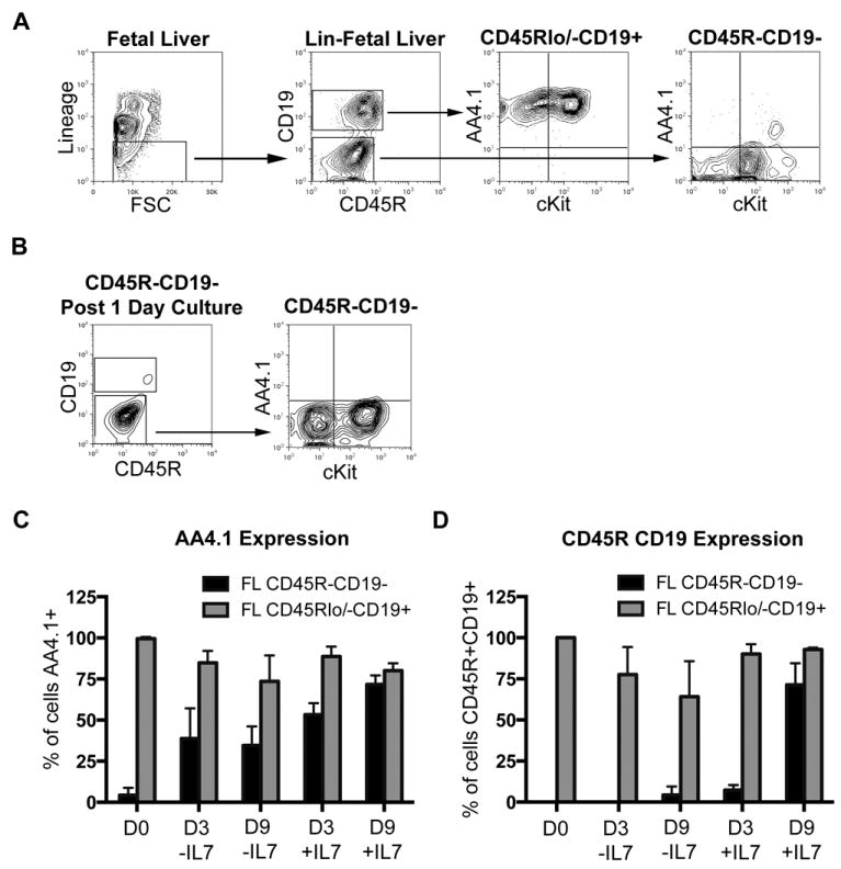
Natural Abs produced by B-1a cells are required for immediate protection against infection. The protective capacity of natural Abs is attributed to germline-like structure, which includes the relative absence of N-region addition. Previous studies have shown B-1a cell Ig from aged mice contains abundant nontemplated (N)-additions. B-1a cells have been shown to derive from a specific lineage-negative (Lin(-))CD45R(low/-)CD19(+) progenitor found both in fetal liver and adult bone marrow. In this study, we report identification of a fetal liver population characterized phenotypically as Lin(-)CD45R(-)CD19(-), which gives rise to IgM(+)IgD(low)CD45R(low)CD5(+)Mac-1(+)CD19(high)CD43(+)CD23(low) B-1a cells upon adoptive transfer to SCID recipients. These B-1a cells derived from the Lin(-)CD45R(-)CD19(-) fetal liver population produce natural Ab that binds pneumococcal Ags, but this Ig contains substantial N-addition despite initial absence of TdT. Furthermore, we show extensive N-addition is also present in B-1a cells derived from the Lin(-)CD45R(low/-)CD19(+) B-1 progenitor found in the bone marrow. Together these results demonstrate B-1a cell N-addition depends on the type of progenitor and the location of the progenitor during its development. These findings have implications for how regulation of different progenitors from fetal liver and bone marrow may play a role in the age-related increase in N-region addition by B-1a cells in normal animals.


Figures

Figure 1
Figure 1. Lin-CD45R-CD19- fetal liver cells reconstitute peritoneal IgMa+CD45RloCD5+ cells
Fetal liver cells collected from day 14, 15, or 18 embryos were stained with lineage markers, CD19, and CD45R. Lin-CD45R-CD19- and Lin-CD45Rlo/-CD19+ cells were sorted for transplantation. (A) A representative sorting scheme is displayed for day 15 fetal liver. (B) CB17 SCID mice were injected intravenously with 1-2 × 10⁵ Lin-CD45R-CD19- or 1-2 × 10⁵ Lin-CD45Rlo/-CD19+ fetal liver cells. Four to five weeks post transplantation the mice were euthanized and evaluated for reconstitution of lymphocytes in the spleen and peritoneal cavities. Results for a wild type 3-month old BALB/c-ByJ mouse are shown for comparison. (C) The percent of live lymphocytes in the peritoneal cavities of recipient SCID mice which are IgMa+ (fetal liver derived) CD45RloCD5+ (B-1a cells), as compared to the fraction of B-1a cells found in the peritoneal cavities of wild type 3-month old BALB/c-ByJ mice is displayed. The data shown are an average of six independent experiments with 3-5 mice per group in each experiment. Embryos were obtained from timed pregnant BALB/c-ByJ mice for three experiments (Jackson Labs) and Swiss Webster (Taconic) mice for three experiments.
Figure 2
Figure 2. Peritoneal B cells derived from Lin-CD45R-CD19- fetal liver cells phenotype as B-1a cells
Peritoneal IgMa+CD45RloCD5+ cells derived from Lin-CD45R-CD19- fetal liver cells were analyzed for surface markers typically found on peritoneal B-1a cells. These cells were compared to peritoneal B-1a and splenic B-2 cells from 3-month old BALB/c-ByJ mice in addition to peritoneal IgMa+CD45RloCD5+ cells derived from Lin-CD45Rlo/-CD19+ fetal liver cells. (A) MFI values of IgD and CD19 are shown. (B) Percent of peritoneal IgMa+CD45RloCD5+ cells or splenic IgMa+CD45RhighCD5- cells expressing CD43, Mac-1 (CD11b), or CD23 are shown. (C) Percent of peritoneal IgMa+CD45RloCD5+ cells or splenic IgMa+CD45RhighCD5- cells expressing CD86, PD-L2, or PtC liposome binding are shown. The data shown are an average of six independent experiments with 3-5 mice per group in each experiment.
Figure 3
Figure 3. Lin-CD45R-CD19- fetal liver derived B-1a cells produce natural IgM antibody that binds pneumococcal antigens
Serum was obtained from CB17 SCID mice transplanted with Lin-CD45R-CD19- fetal liver cells, Lin-CD45Rlo/-CD19+ fetal liver cells, or total Lin- bone marrow cells four to five weeks post transfer. The serum was evaluated for (A) total IgM, (B) PC-specific IgM, and (C) PPS3-specific IgM levels by ELISA. As a reference, serum from 3-month old BALB/c-ByJ mice was also evaluated at the same time. The results represent an average of serum samples taken from individual mice, which demonstrated reconstitution by flow cytometry analysis. The number of individual serum samples analyzed per group is as follows: 19 Lin-CD45R-CD19- fetal liver cell chimera mice, 14 Lin-CD45Rlo/-CD19+ fetal liver cell chimera mice, and 6 total Lin- bone marrow chimera mice.
Figure 4
Figure 4. Immunoglobulin heavy chain sequence analysis of Lin-CD45R-CD19- fetal liver and Lin-CD45Rlo/-CD19+ bone marrow derived B-1a cells show abundant N-addition
(A) N-region addition analysis at the D-J and V-D junctions in B-1a cells derived from Lin-CD45R-CD19- and Lin-CD45Rlo/-CD19+ fetal liver cells, Lin-CD45Rlo/-CD19+ bone marrow cells, and total Lin- bone marrow is shown. (B) V, D, and J gene segment usage in B-1a cells derived from Lin-CD45R-CD19- or Lin-CD45Rlo/-CD19+ fetal liver cells is displayed. (C) Average number of N-additions at the V-D, D-J, or sum of the two junctions is shown along with CDR3 length. Results are based on 3-5 independent experiments with sequences combined from each independent experiment.
Figure 5
Figure 5. The Lin-CD45R-CD19- fetal liver progenitor population does not express TdT
(A) cDNA was obtained from sort purified populations: 1) Lin-CD45R-CD19- fetal liver cells, 2) Lin-CD45Rlo/-CD19+ fetal liver cells, and 3) Lin-CD45Rlo/-CD19+ bone marrow cells, and assessed for TdT expression by PCR. (B) cDNA was obtained from sort purified bone marrow cell populations: 1) Lin-CD45Rlo/-CD19+, 2) Lin-CD45R+CD19-, or 3) total bone marrow cells, and assessed for TdT expression by PCR. (C) TdT expression in sort purified bone marrow and fetal liver populations was analyzed using Taqman PCR. The results shown are an average of 5 independent experiments. (D) N-region addition analysis at the D-J and V-D junctions in B-1a and B-2 cells obtained from CD57/B6 TdT-/- mice is shown.
Figure 6
Figure 6. Lin-CD45R-CD19- fetal liver cells express little to no AA4.1 and are responsive to IL-7
(A) Lin-CD45R-CD19- and Lin-CD45Rlo/-CD19+ fetal liver cells were assessed for AA4.1 and cKit expression at day 18 by immunofluorescent staining. (B) Lin-CD45R-CD19- fetal liver cells were sorted from day 18 embryos. The cells were placed in culture overnight with SCF, TPO, and Flt-3L. Expression of AA4.1 and cKit was determined following the 24-hour culture. The results shown are representative of 3 independent experiments. (C, D) Lin-CD45R-CD19- (black bars) and Lin-CD45Rlo/-CD19+ (grey bars) fetal liver cells were sorted from day 18 embryos. The cells were placed in culture with a cytokine mix containing IL-7 (SCF, Flt3-L, TSLP, IL-7) or lacking IL-7 (SCF, Flt3-L, TSLP, IL-6, IL-3). After 3 and 9 days expression of AA4.1 (C) or CD45R and CD19 (D) expression was determined. The results shown are an average of 3 independent experiments.

References

    1. Hardy RR, Hayakawa K. B cell development pathways. Annu Rev Immunol. 2001;19:595–621. - PubMed
    1. Rothstein TL. Cutting edge commentary: two B-1 or not to be one. J Immunol. 2002;168:4257–4261. - PubMed
    1. Wong SC, Chew WK, Tan JE, Melendez AJ, Francis F, Lam KP. Peritoneal CD5+ B-1 cells have signaling properties similar to tolerant B cells. J Biol Chem. 2002;277:30707–30715. - PubMed
    1. Holodick NE, Tumang JR, Rothstein TL. Continual signaling is responsible for constitutive ERK phosphorylation in B-1a cells. Mol Immunol. 2009;46:3029–3036. - PMC - PubMed
    1. Zhong X, Gao W, Degauque N, Bai C, Lu Y, Kenny J, Oukka M, Strom TB, Rothstein TL. Reciprocal generation of Th1/Th17 and T(reg) cells by B1 and B2 B cells. Eur J Immunol. 2007;37:2400–2404. - PubMed

Publication types

Substances