

Distribution of lymphocyte subpopulations in thyroid glands of human autoimmune thyroid disease
- PMID: 24478186
- PMCID: PMC6807618
- DOI: 10.1002/jcla.21674
Distribution of lymphocyte subpopulations in thyroid glands of human autoimmune thyroid disease
Abstract
Background: The autoimmune thyroid disease (AITD) is an organ-specific autoimmune disease characterized by the breakdown of self-tolerance to thyroid antigens. Some lymphocytes have been identified to be related notably to the pathogenesis of AITD. This article evaluated the distribution of the lymphocytic subpopulation in thyroid glands in order to develop the immunospecific forms of therapy for AITD.
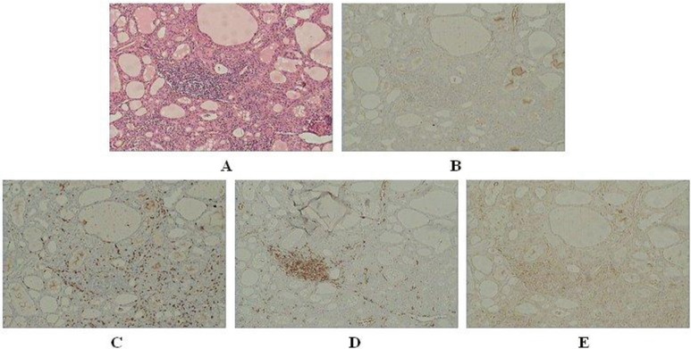
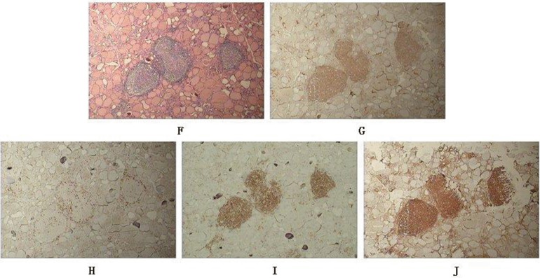
Methods: Damaged thyroid specimens were obtained from 18 Graves' disease (GD) and 17 Hashimoto's thyroiditis (HT) patients. Normal thyroid specimens were obtained from unaffected glands of 17 patients who underwent parathyroidectomy. We evaluated the distribution of lymphocytic subpopulation by analyzing the expression difference and correlationship among CD4+ T lymphocyte, CD8+ T lymphocyte, CD20+ B lymphocyte as well as regulatory T cells(Tregs)' marker FoxP3 in the thyroid tissues via immunohistochemistry.
Results: Our research uncovered that no distinct lymphocyte infiltrated in the normal thyroid specimens. Scarcely any lymphocyte infiltration could be found in half of the totally 18 GD thyroid specimens. For the rest 9 GD specimens, CD8+ T cells and CD20+ B cells were expressed more or less in all of them, FoxP3+ Tregs were detected in 7 of them and CD4+ T cells were weakly expressed in only 2 of them. For the 17 HT thyroid specimens, CD20+ B cells were stained strongly in all of them, CD4+, CD8+ T cells were expressed more or less in most of them and FoxP3+ Tregs could be detected in 9 of them.
Conclusion: Based on CD20+ B cells predominantly infiltrating in all HT thyroid tissues we suggested CD20 antibody might be of help for HT treatment. Furthermore based on FoxP3+ Tregs abundantly infiltrating in some of the AITD thyroid specimens, we considered that activating the Tregs' function in comparison to increasing the Tregs' number only, may be a more effective approach to the treatment of AITD in some cases.
Keywords: CD20; CD4; CD8; FoxP3; Graves’ disease; Hashimoto's thyroiditis; regulatory T cells.
© 2014 Wiley Periodicals, Inc.
Figures
References
-
- Weetman AP. Autoimmune thyroid disease: Propagation and progression. Eur J Endocrinol 2003;148:1–9. - PubMed
-
- Sepulveda H, Cerwenka A, Morgan T, Dutton RW. CD28, IL‐2‐independent costimulatory pathways for CD8 T lymphocyte activation. J Immunol 1999;163:1133–1142. - PubMed
-
- Miyara M, Sakaguchi S. Natural regulatory T cells: Mechanisms of suppression. Trends Mol Med 2007;13:108–116. - PubMed
-
- Baecher‐Allan C, Viglietta V, Hafler DA. Human CD4+CD25+ regulatory T cells. Semin Immunol 2004;16:89–98. - PubMed
-
- Banham AH, Powrie FM, Suri‐Payer E. FOXP3+ regulatory T cells: Current controversies and future perspectives. Eur J Immunol 2006;36:2832–2836. - PubMed
Publication types
MeSH terms
LinkOut - more resources
Full Text Sources
Other Literature Sources
Research Materials
