. 2014 Mar 14;289(11):7547-57.
doi: 10.1074/jbc.M113.545699. Epub 2014 Jan 29.

Sterol regulatory element-binding protein (SREBP) cleavage regulates Golgi-to-endoplasmic reticulum recycling of SREBP cleavage-activating protein (SCAP)

Affiliations

Sterol regulatory element-binding protein (SREBP) cleavage regulates Golgi-to-endoplasmic reticulum recycling of SREBP cleavage-activating protein (SCAP)

Wei Shao et al. J Biol Chem. .

Abstract

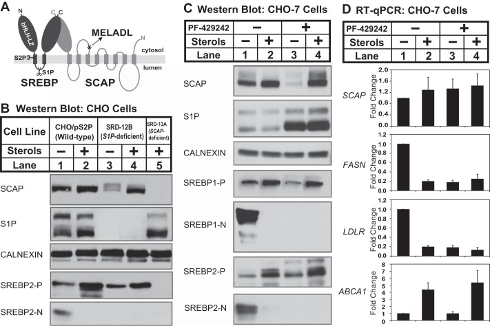
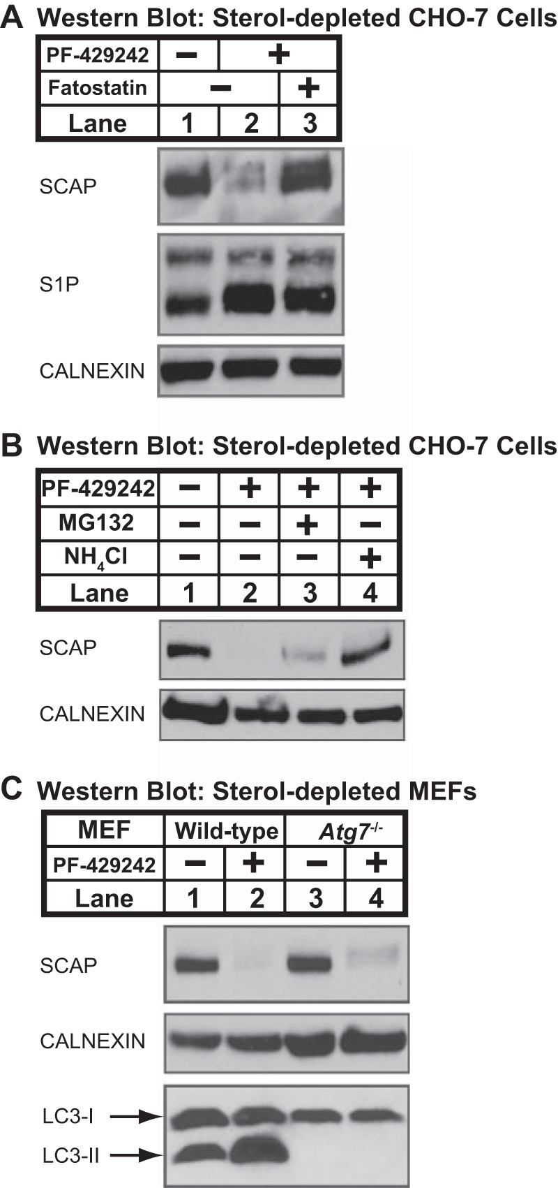
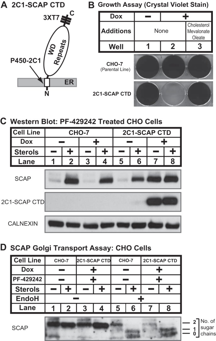
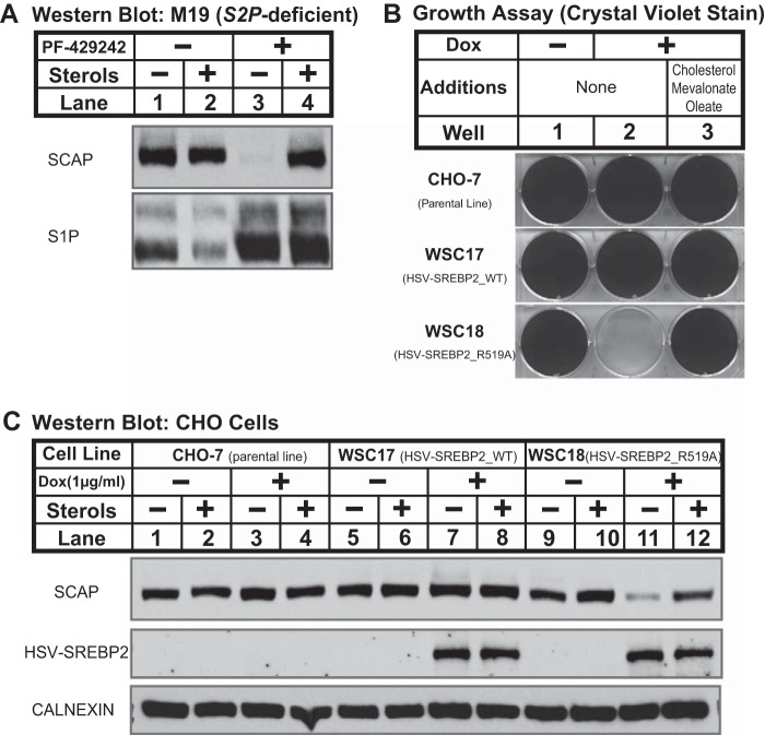
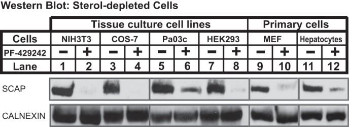
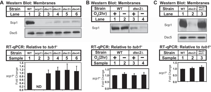
Sterol regulatory element-binding protein (SREBP) transcription factors are central regulators of cellular lipogenesis. Release of membrane-bound SREBP requires SREBP cleavage-activating protein (SCAP) to escort SREBP from the endoplasmic reticulum (ER) to the Golgi for cleavage by site-1 and site-2 proteases. SCAP then recycles to the ER for additional rounds of SREBP binding and transport. Mechanisms regulating ER-to-Golgi transport of SCAP-SREBP are understood in molecular detail, but little is known about SCAP recycling. Here, we have demonstrated that SCAP Golgi-to-ER transport requires cleavage of SREBP at site-1. Reductions in SREBP cleavage lead to SCAP degradation in lysosomes, providing additional negative feedback control to the SREBP pathway. Current models suggest that SREBP plays a passive role prior to cleavage. However, we show that SREBP actively prevents premature recycling of SCAP-SREBP until initiation of SREBP cleavage. SREBP regulates SCAP in human cells and yeast, indicating that this is an ancient regulatory mechanism.

Keywords: Cholesterol; Lipids; Lysosomes; Protein Degradation; Recycling; SCAP; SREBP; Transcription Factors.


Figures

FIGURE 1.
FIGURE 1.
Sterols regulate SCAP post-transcriptionally in cells lacking S1P. A, diagram of SREBP-SCAP complex showing sites of S1P and S2P cleavage in SREBP and the MELADL ER exit sequence in SCAP. B, wild-type CHO cells (CHO/pS2P), S1P-deficient CHO cells (SRD-12B), and SCAP-deficient cells (SRD-13A) were set up on day 0 at 1.5 × 10⁶ cells/100-mm dish in medium A supplemented with 5% (v/v) FCS (for CHO/pS2P) or medium B (for SRD-12B and SRD-13A). On day 1, cells were refed medium C in the absence or presence of sterols (1 μg/ml 25-HC, 10 μg/ml cholesterol). After 16 h, ALLN was added to a final concentration of 25 μg/ml, and cells were harvested 1 h later. C, CHO-7 cells were set up on day 0 at 1.5 × 10⁶ cells/100-mm dish in medium A supplemented with 5% (v/v) FCS. On day 1, the cells were refed medium C with the addition of sterols (1 μg/ml 25-HC, 10 μg/ml cholesterol) and S1P inhibitor PF-429242 (50 μm) as indicated. After 16 h, ALLN was added to a final concentration of 25 μg/ml, and cells were harvested 1 h later. For B and C, aliquots of membrane (25 μg protein/lane for SCAP, S1P, calnexin, and SREBP1/2-P) and nuclear extract (40 μg protein/lane for SREBP1/2-N) fractions were immunoblotted as indicated. D, CHO-7 cells were set up on day 0 at 2 × 10⁵ cells/well (6-well plate) in medium A supplemented with 5% (v/v) FCS. Cells were treated as in described in C. After 16 h, cells were harvested and subjected to RNA preparation and RT-qPCR analysis as described under “Experimental Procedures.” Error bars represent the standard deviation of fold changes from three biological replicates (mean ± S.D.). FASN and LDLR are targets of SREBP1 and SREBP2, respectively. ABCA1 is the target of nuclear receptor LXR.
FIGURE 2.
FIGURE 2.
Loss of SCAP requires ER exit and is blocked by ammonium chloride. A, CHO-7 cells were set up on day 0 at 1.5 × 10⁶ cells/100-mm dish in medium A supplemented with 5% (v/v) FCS. On day 1, cells were refed medium C containing PF-429242 (50 μm) and fatostatin (10 μm) as indicated. B, CHO-7 cells were set up on day 0 at 1.5 × 10⁶ cells/100-mm dish in medium A supplemented with 5% (v/v) FCS. On day 1, cells were refed medium C containing vehicle, PF-429242 (50 μm), MG132 (50 μm), and NH4Cl (50 mm) as indicated. C, MEFs (wild-type control and Atg7−/−) were set up for experiments on day 0 at 1.5 × 10⁶ cells/100-mm dish in medium D supplemented with 10% (v/v) FCS. On day 1, the cells were refed medium E in the absence or presence of PF-429242 (50 μm). A–C, after 16 h, the cells were fractionated, and membrane fractions (25 μg protein/lane) were subjected to immunoblot analysis using the indicated antibodies.
FIGURE 3.
FIGURE 3.
SCAP degradation requires binding to SREBP. A, diagram shows structure of 2C1-SCAP CTD fusion protein in which the N-terminal 1–29 amino acids of cytochrome P450–2C1 are fused to the C-terminal domain of hamster SCAP (amino acids 731–1276) with three repeats of T7 epitope. B, CHO-7 and 2C1-SCAP CTD cells were seeded on day 0 at a density of 3 × 10⁴ cells/well (6-well plate) in medium A supplemented with 5% (v/v) FCS. On day 1, cells were refed with medium A supplemented with 5% (v/v) LPDS in the absence or presence of doxycycline (Dox) (1 μg/ml) or medium B plus doxycycline (1 μg/ml). Cells were refed every 2 days. On day 14, cells were fixed in cold methanol and stained with 0.05% crystal violet. C, CHO-7 and 2C1-SCAP CTD cells were set up on day 0 at 1.5 × 10⁶ cells/100-mm dish in medium A supplemented with 5% (v/v) FCS. On day 1, cells were refed medium C in the absence or presence of sterols (1 μg/ml 25-HC, 10 μg/ml cholesterol), PF-429242 (50 μm), or doxycycline (1 μg/ml) as indicated. After 16 h, cells were fractionated, and membrane fractions (25 μg protein/lane) were subjected to immunoblot analysis with the indicated antibodies and anti-T7 to detect 2C1-SCAP CTD. D, CHO-7 and 2C1-SCAP CTD cells were set up on day 0 at 1.5 × 10⁶ cells/100-mm dish in medium A supplemented with 5% FCS plus 25-hydroxycholesterol (1 μg/ml). On day 1, cells were refed medium C in the absence or presence of sterols (1 μg/ml 25-HC, 10 μg/ml cholesterol), PF-429242 (50 μm), or doxycycline (1 μg/ml) as indicated. After 16 h, cells were harvested (4 dishes/condition) and subjected to cell fractionation. Aliquots of membrane fractions (50 μg) were analyzed using the SCAP endoglycosidase H (EndoH) assay described under “Experimental Procedures” and then subjected to immunoblot analysis with 10 μg/ml anti-SCAP mouse monoclonal 9D5. Filters were exposed to film at room temperature for 60 s.
FIGURE 4.
FIGURE 4.
SCAP recycling requires SREBP cleavage at site-1. A, S2P-deficient (M19) cells were set up on day 0 at 1.5 × 10⁶ cells/100-mm dish in medium B. On day 1, the cells were refed medium C in the absence or presence of sterols (1 μg/ml 25-HC, 10 μg/ml cholesterol) and PF-429242 (50 μm). After 16 h, cells were fractionated, and membrane fractions (25 μg protein/lane) were subjected to immunoblot analysis as indicated. B, CHO-7, WSC17 (HSV-SREBP2_WT), and WSC18 (HSV-SREBP2_R519A) cells were set up on day 0 at 3 × 10⁴ cells/well (6-well plate) in medium A supplemented with 5% (v/v) FCS. On day 1, cells were refed with medium A supplemented with 5% (v/v) LPDS in the absence or presence of doxycycline (Dox) (1 μg/ml) or medium B plus doxycycline (1 μg/ml). Cells were refed every 2 days. On day 14, cells were fixed in cold methanol and stained with 0.05% crystal violet. C, CHO-7 cells, WSC17 (HSV-SREBP2_WT), and WSC18 (HSV-SREBP2_R519A) cells were set up on day 0 at 1.5 × 10⁶ cells/100-mm dish in medium A supplemented with 5% (v/v) FCS. On day 1, the cells were refed medium C in the absence or presence of sterols (1 μg/ml 25-HC, 10 μg/ml cholesterol) and doxycycline (1 μg/ml) as indicated. After 16 h, cells were fractionated, and membrane fractions (25 μg protein/lane) were subjected to immunoblot analysis as noted.
FIGURE 5.
FIGURE 5.
Mammalian SREBP regulates SCAP recycling through a common mechanism. NIH3T3, COS-7, Pa03c, HEK293, and primary MEFs (MEF-P3, isolated from CF1 strain, passage 3) were set on day 0 at 1 × 10⁶ cells/100-mm dish in medium D supplemented with 10% (v/v) FCS. On day 1, the cells were refed medium E in the absence or presence of PF-429242 (50 μm). Primary mouse hepatocytes were isolated from C57BL/6J mice and set up for experiments on day 0 at 3 × 10⁶ cells/100-mm dish (BD BioCoat collagen-coated plates) in medium D supplemented with 10% (v/v) FCS. On day 1, the cells were refed medium E in the absence or presence of PF-429242 (50 μm). After 16 h, cells were fractionated, and membrane fractions (25 μg protein/lane) were subjected to immunoblot analysis using the indicated antibodies.
FIGURE 6.
FIGURE 6.
Yeast SREBP regulates SCAP recycling. A, Western blot of membrane fractions from wild-type and the indicated mutant strains probed with anti-Scp1 IgGs or anti-Dsc5 serum. Expression of scp1+ was analyzed by RT-qPCR. B, Western blot of membrane fractions from wild-type and dsc2Δ mutant cells grown for 2 h in the presence or absence of oxygen probed with anti-Scp1 IgGs. Expression of scp1+ was analyzed by RT-qPCR. C, Western blot of membrane fractions from wild-type, dsc2Δ, and dsc2Δsre1Δ mutant strains probed with anti-Scp1 IgGs and anti-Dsc5 serum. Expression of scp1+ was analyzed by RT-qPCR. Error bars represented the standard deviation of fold changes from three biological replicates (mean ± S.D.). ND, not detected.

References

    1. Cohen J. C., Horton J. D., Hobbs H. H. (2011) Human fatty liver disease: old questions and new insights. Science 332, 1519–1523 - PMC - PubMed
    1. Goldstein J. L., Hazzard W. R., Schrott H. G., Bierman E. L., Motulsky A. G. (1973) Hyperlipidemia in coronary heart disease. I. Lipid levels in 500 survivors of myocardial infarction. J. Clin. Invest. 52, 1533–1543 - PMC - PubMed
    1. Hachinski V., Graffagnino C., Beaudry M., Bernier G., Buck C., Donner A., Spence J. D., Doig G., Wolfe B. M. (1996) Lipids and stroke: a paradox resolved. Arch. Neurol. 53, 303–308 - PubMed
    1. Brown M. S., Goldstein J. L. (2009) Cholesterol feedback: from Schoenheimer's bottle to Scap's MELADL. J. Lipid Res. 50, (suppl.) S15–S27 - PMC - PubMed
    1. Rawson R. B., DeBose-Boyd R., Goldstein J. L., Brown M. S. (1999) Failure to cleave sterol regulatory element-binding proteins (SREBPs) causes cholesterol auxotrophy in Chinese hamster ovary cells with genetic absence of SREBP cleavage-activating protein. J. Biol. Chem. 274, 28549–28556 - PubMed

Publication types

MeSH terms

Substances

LinkOut - more resources