. 2014 Jan 29;34(5):1903-15.
doi: 10.1523/JNEUROSCI.4043-13.2014.

Omega-3 fatty acids protect the brain against ischemic injury by activating Nrf2 and upregulating heme oxygenase 1

Affiliations

Omega-3 fatty acids protect the brain against ischemic injury by activating Nrf2 and upregulating heme oxygenase 1

Meijuan Zhang et al. J Neurosci. .

Abstract

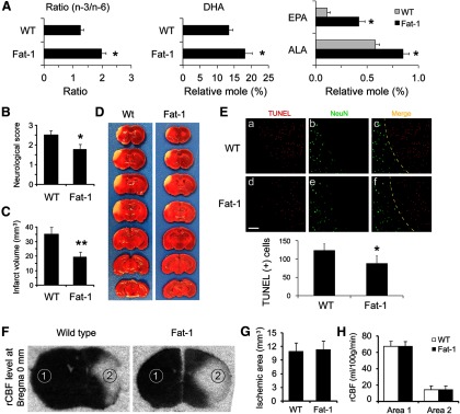
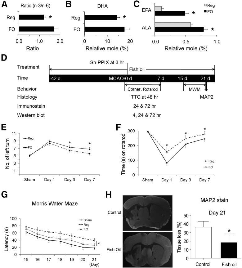
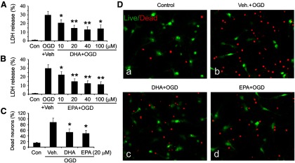
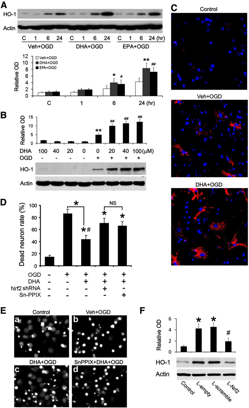
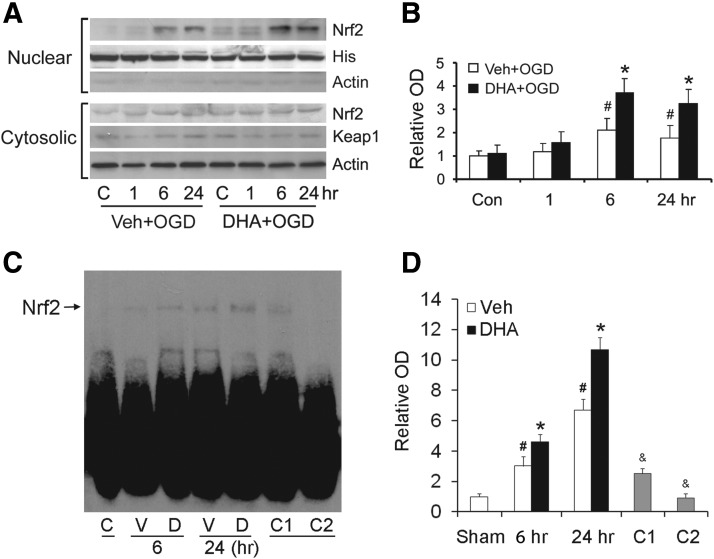
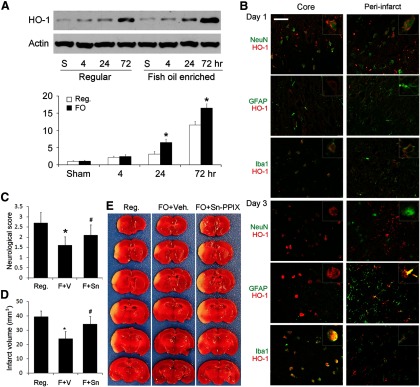
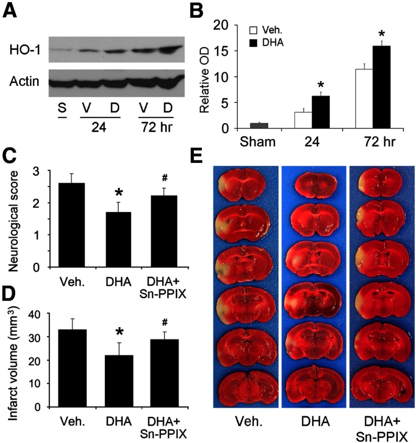
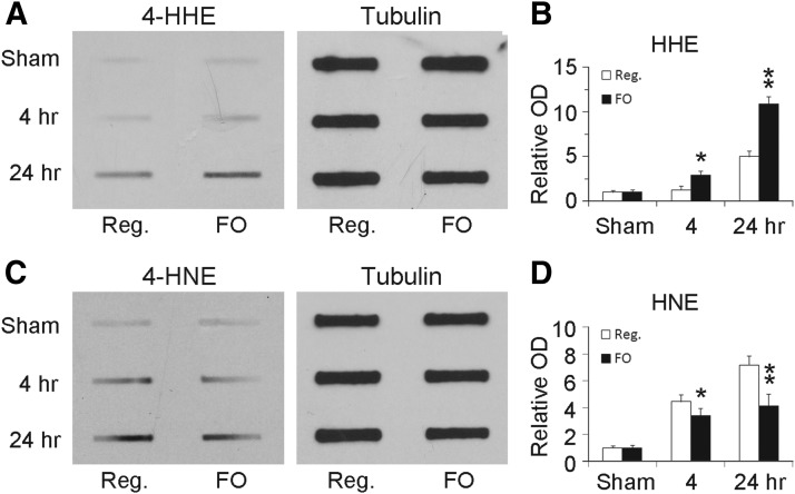
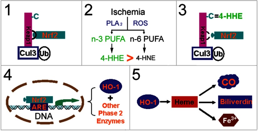
Ischemic stroke is a debilitating clinical disorder that affects millions of people, yet lacks effective neuroprotective treatments. Fish oil is known to exert beneficial effects against cerebral ischemia. However, the underlying protective mechanisms are not fully understood. The present study tests the hypothesis that omega-3 polyunsaturated fatty acids (n-3 PUFAs) attenuate ischemic neuronal injury by activating nuclear factor E2-related factor 2 (Nrf2) and upregulating heme oxygenase-1 (HO-1) in both in vitro and in vivo models. We observed that pretreatment of rat primary neurons with docosahexaenoic acid (DHA) significantly reduced neuronal death following oxygen-glucose deprivation. This protection was associated with increased Nrf2 activation and HO-1 upregulation. Inhibition of HO-1 activity with tin protoporphyrin IX attenuated the protective effects of DHA. Further studies showed that 4-hydroxy-2E-hexenal (4-HHE), an end-product of peroxidation of n-3 PUFAs, was a more potent Nrf2 inducer than 4-hydroxy-2E-nonenal derived from n-6 PUFAs. In an in vivo setting, transgenic mice overexpressing fatty acid metabolism-1, an enzyme that converts n-6 PUFAs to n-3 PUFAs, were remarkably resistant to focal cerebral ischemia compared with their wild-type littermates. Regular mice fed with a fish oil-enhanced diet also demonstrated significant resistance to ischemia compared with mice fed with a regular diet. As expected, the protection was associated with HO-1 upregulation, Nrf2 activation, and 4-HHE generation. Together, our data demonstrate that n-3 PUFAs are highly effective in protecting the brain, and that the protective mechanisms involve Nrf2 activation and HO-1 upregulation by 4-HHE. Further investigation of n-3 PUFA neuroprotective mechanisms may accelerate the development of stroke therapies.


Figures

Figure 1.
Figure 1.
n-3 PUFA protects the brain against transient focal ischemia in mice. A, Fatty acid analysis of forebrains from wild-type (WT) and fat-1 transgenic mice. Compared with WT mice, fat-1 mice showed an increased total n-3/n-6 ratio (left), increased levels of DHA (middle), and increased levels of ALA and EPA. Data are presented as mean ± SD (n = 6; *p ≤ 0.01 vs WT). Focal brain ischemia was then induced by 60 min MCAO followed by 48 h reperfusion. Neurological function was examined at 48 h after ischemia. B, Fat-1 mice showed improved neurological function recovery after ischemia. C, Fat-1 mice had smaller infarct volumes compared with WT mice. Data are presented as mean ± SD (n = 9 per group, *p ≤ 0.05 and **p ≤ 0.01 vs WT by t test). D, Representative photographs of TTC-stained brain sections showing reduced infarct sizes in fat-1 mice. E, Representative microphotographs and counts of TUNEL-positive cells in the infarct areas 48 h after MCAO in WT and fat-1 mice, expressed as the number of cells per field of view (200×) in randomly selected regions. Scale bar, 100 μm. Yellow dotted lines in C and F indicate the boundary between infarct (upper right) and peri-infarct (lower left) zones (n = 6 per group; *p ≤ 0.05 vs WT littermates). F, Representative autoradiographs of C14-IAP at bregma 0 mm, showing rCBF changes in mouse brains during MCAO. Dark region represents normal blood flow, while light area represents reduced blood flow. Area 1, contralateral hemisphere; area 2, ischemic inner boundary; area 3, ischemic core. G, WT and fat-1 mice demonstrated similar ischemic areas during MCAO. Data are presented as mean ± SD, analyzed with t test (n = 6 per group). H, WT and fat-1 mice demonstrated similar levels of CBF reduction during MCAO. Data are presented as mean ± SD, and analyzed with ANOVA and post hoc tests.
Figure 2.
Figure 2.
FO treatment confers long-term neuroprotection against stroke in mice. Mice were fed with either a regular diet or an FO-enriched diet for 6 weeks. Their brains were collected and the lipid profiles of forebrains were analyzed. A–C, The overall n-3/n-6 ratio (A), DHA content (B), and ALA and EPA contents (C) were calculated, showing increased n-3 PUFA contents in the brains (n = 3, *p ≤ 0.05 vs regular diet mice). D, A diagram showing the timeline of FO feeding and the time points for MCAO and various assessments. E, F, The corner test (E) and rotarod test (F) show that FO-fed mice recovered sensorimotor function better than control mice (n = 6–8, *p ≤ 0.05 vs regular diet mice). G, Cognitive function in the Morris water maze during the third week after MCAO is presented as latency to escape the water bath. FO-fed mice recovered better than control mice (n = 5–6, *p ≤ 0.05 vs regular diet-fed mice). H, Representative photographs of MAP2-stained coronal sections and quantitative analyses of tissue loss at day 21 after MCAO (n = 6; data are presented as mean ± SD, *p ≤ 0.05 vs control).
Figure 3.
Figure 3.
n-3 PUFA pretreatment reduces neuronal death after in vitro ischemia. A, B, DHA-treated (A) or EPA-treated (B) neuronal cultures were challenged with 60 min OGD; 24 h later, LDH release was measured. Data are presented as mean ± SE from three independent experiments, analyzed with ANOVA and post hoc tests (*p ≤ 0.05 and **p ≤ 0.01 vs OGD group). C, Neuronal cultures were treated with DHA or EPA, followed by OGD. Cultures were stained with a live/dead viability/toxicity kit 24 h later, and live and dead neurons were counted. Data are presented as the percentage of dead neurons as a function of total neuron number, and analyzed by χ2 tests (*p ≤ 0.05 vs OGD group). D, Representative micrographs of primary neuronal cultures 24 h after n-3 PUFA treatment and OGD. The cultures were stained with a live/dead kit; healthy neurons appear green and the condensed nuclei of dead neurons appear red.
Figure 4.
Figure 4.
HO-1 mediates the neuroprotective effects of n-3 PUFAs in vitro. Primary neuronal cultures were treated with n-3 PUFAs and harvested for Western blotting or fixed for Hoechst staining. A, Time course of HO-1 expression. Representative Western blots and semiquantitative analyses of HO-1 levels after n-3 PUFA and OGD treatments (*p ≤ 0.05 and **p ≤ 0.01 vs control and vehicle-treated OGD groups at the same time points; #p ≤ 0.05 and ##p ≤ 0.01 vs control group. B, Dose–response data related to HO-1 expression. Representative Western blots and semiquantitative analyses of HO-1 levels after n-3 PUFA treatment, with or without OGD (**p ≤ 0.01 vs control, ##p ≤ 0.01 vs control and OGD groups). C, Representative micrographs of HO-1 immunofluorescence in neuronal cultures. HO-1 was immunostained red with Cy3 and nuclei were stained blue with Hoechst. D, E, Quantitative analysis (D) and representative micrographs (E) showed that knockdown of Nrf2 or inhibition of HO-1 activity with 10 μm Sn-PPIX diminishes the neuroprotective effect of n-3 PUFAs (*p ≤ 0.05 vs control group, #p ≤ 0.05 vs OGD group). F, Nrf2 knockdown reduces neuronal HO-1 levels. Primary neurons were infected with Lenti-scramble or Lenti-Nrf2 shRNAs for 3 d, followed by DHA and OGD treatments. HO-1 levels were detected 6 h after OGD using Western blot (p ≤ 0.05 vs control group, and #p ≤ 0.05 vs lentiviral groups).
Figure 5.
Figure 5.
DHA enhances the nuclear translocation and DNA binding activity of Nrf2 in neurons. Primary neuronal cultures were treated with vehicle or DHA followed by OGD. A, B, Representative Western blots (A) and semiquantitative analyses (B) of Nrf2 levels, showing increased nuclear Nrf2 in DHA-treated groups (*p ≤ 0.05 vs control and vehicle-treated OGD groups at the same time points; #p ≤ 0.05 vs control groups, n = 3 per condition). C, D, Representative electrophoretic mobility shift assay (C) and semiquantitative analyses (D) of DNA binding of Nrf2 (*p ≤ 0.05 vs control and vehicle-treated OGD groups at the same time points; #p ≤ 0.05 vs control groups; and &p ≤ 0.01 vs DHA-treated OGD at 24 h; n = 3 per condition). The specificity of DNA binding activity was determined by a competition assay, whereas threefold (C1) or 50-fold (C2) excess of cold probe was added as a specific competitor.
Figure 6.
Figure 6.
FO protects the brain against focal ischemia via HO-1. Mice were fed with a regular diet (Reg.) or FO-enhanced diet for 6 weeks followed by 60 min MCAO. A, Representative Western blots and semiquantitative analyses of HO-1 levels in mouse brain after ischemia, showing increased HO-1 levels in the FO group (n = 3, *p ≤ 0.05 vs sham and regular diet at the same time points). B, Representative microphotographs of HO-1 expression and cellular distribution in the ischemic core and surrounding regions at 1 or 3 d after ischemia. HO-1 was stained red and cellular markers were stained green. Scale bar, 50 μm. Insets show high-power images of representative cells. C–E, Neurological score (C), infarct volumes (D), and representative TTC staining (E) at 48 h after MCAO, showing that FO treatment reduced neurological dysfunction and infarct volumes and that protection was partially blocked by Sn-PPIX, a HO-1 activity inhibitor (n = 6, *p ≤ 0.05 vs Reg., #p ≤ 0.05 vs FO mice injected with vehicle).
Figure 7.
Figure 7.
Postischemic DHA treatment protects the brain against focal ischemia. MCAO was induced in C57BL/6 mice and DHA was subcutaneously injected at the onset of reperfusion. A, B, Representative Western blots (A) and semiquantitative analyses (B) of HO-1 levels in mouse brain after ischemia, showing increased HO-1 levels in the DNA group (n = 3, *p ≤ 0.05 vs sham and vehicle group at the same time points). S, Sham-operated; V, vehicle; D, DHA. C–E, Neurological score (C), infarct volumes (D), and representative TTC staining (E) at 48 h after MCAO, showing that DHA treatment reduced neurological dysfunction and infarct volumes and that the protection was partially blocked by Sn-PPIX (n = 8, *p ≤ 0.05 vs vehicle, #p ≤ 0.05 vs DHA group).
Figure 8.
Figure 8.
Four-HHE is a potent Nrf2 activator. Primary cultures were incubated with 4-HHE (n-3) or 4-HNE (n-6) at a concentration of 10 μm for the indicated duration. A, Representative Western blots of cytosolic HO-1 and nuclear Nrf2. B, C, Quantitative analyses of HO-1 (B) and Nrf2 (C) levels indicated that lipid electrophiles can directly induce Nrf2 nuclear translocation and HO-1 expression, and that 4-HHE (n-3) is more potent than 4-HNE (n-6) in inducing Nrf2 activation. Data are presented as mean ± SE, and analyzed with ANOVA and post hoc tests (*p ≤ 0.05 vs 4-HNE-treated group at the same time point). D, Representative microphotographs of Nrf2-EGFP transfected neuronal cultures, showing that 4-HHE was a stronger inducer of Nrf2 than 4-HNE. Primary neurons were infected with lenti-Nrf2-EGFP for 3 d followed by treatment with 4-HHE and 4-HNE (10 μm) for 1 h. Cells were then fixed and counterstained with DAPI (blue). E, F, Representative Hoechst staining (E) and dead cell counts (F), showing that 4-HHE pretreatment reduced neuronal death. Primary neurons were treated with vehicle, 4-HNE, and 4-HHE (10 μm) for 2 h. After overnight recovery, neurons were challenged with OGD for 60 min and then stained with Hoechst (p ≤ 0.05 vs control, vehicle-treated, and 4-HNE-treated OGD groups.
Figure 9.
Figure 9.
FO treatment increases 4-HHE levels in mouse brains after MCAO. Mice were fed with a regular diet or an FO-enhanced diet for 6 weeks followed by 60 min MCAO. At the indicated time points, brain tissues were harvested and subjected to Western slot blots. A, B, Representative slot blots (A) and semiquantitative analyses (B) of the levels of 4-HHE modified proteins, showing increased 4-HHE levels in FO-treated groups (n = 3; **p ≤ 0.01 and *p ≤ 0.05 vs control and regular diet groups at the same time points). C, D, Representative slot blots (C) and semiquantitative analyses (D) of the levels of 4-HHE modified proteins, showing decreased 4-HNE levels in FO-treated groups (n = 3; **p ≤ 0.01 and *p ≤ 0.05 vs control and regular diet groups at the same time points).
Figure 10.
Figure 10.
Potential mechanism of n-3 PUFA-mediated neuroprotection. 1, Under physiological conditions, Nrf2 is physically “locked” with Keap1, which leads to proteasomal degradation of Nrf2 via the Cul3 (E3)-dependent pathway. 2, Under oxidative stress conditions, such as brain ischemia and reperfusion, lipid oxidation produces α,β-unsaturated carbonyl electrophiles. n-3 PUFA-derived 4-HHE is more powerful than n-6 PUFA-derived 4-HNE in electrophilicity. 3, Four-HHE covalently reacts with cysteine residues of Keap1, which causes conformational changes in Keap1, setting Nrf2 free. 4, Nrf2 then accumulates, translocates into the nucleus, and binds the antioxidant response element (ARE) of phase 2 genes, leading to upregulation of HO-1 and other enzymes. 5, HO-1 protects the brain against ischemic damage by breaking down heme into biliverdin (antioxidative) and carbon monoxide (CO; anti-inflammatory), reducing calcium overload and positive feedback on HO-1 expression. C, Cysteines; Cul3, cullin3; PLA2, phospholipase A2; Ub, ubiquitin.

Similar articles

Cited by

References

    1. Akbar M, Calderon F, Wen Z, Kim HY. Docosahexaenoic acid: a positive modulator of Akt signaling in neuronal survival. Proc Natl Acad Sci U S A. 2005;102:10858–10863. doi: 10.1073/pnas.0502903102. - DOI - PMC - PubMed
    1. Bagga D, Wang L, Farias-Eisner R, Glaspy JA, Reddy ST. Differential effects of prostaglandin derived from omega-6 and omega-3 polyunsaturated fatty acids on COX-2 expression and IL-6 secretion. Proc Natl Acad Sci U S A. 2003;100:1751–1756. doi: 10.1073/pnas.0334211100. - DOI - PMC - PubMed
    1. Bazan NG. Neuroprotectin D1 (NPD1): a DHA-derived mediator that protects brain and retina against cell injury-induced oxidative stress. Brain Pathol. 2005;15:159–166. - PMC - PubMed
    1. Begum G, Kintner D, Liu Y, Cramer SW, Sun D. DHA inhibits ER Ca2+ release and ER stress in astrocytes following in vitro ischemia. J Neurochem. 2012;120:622–630. doi: 10.1111/j.1471-4159.2011.07606.x. - DOI - PMC - PubMed
    1. Belayev L, Marcheselli VL, Khoutorova L, Rodriguez de Turco EB, Busto R, Ginsberg MD, Bazan NG. Docosahexaenoic acid complexed to albumin elicits high-grade ischemic neuroprotection. Stroke. 2005;36:118–123. doi: 10.1161/01.STR.0000149620.74770.2e. - DOI - PubMed

Publication types

MeSH terms

LinkOut - more resources