. 2014 Jan 29;34(5):1916-23.
doi: 10.1523/JNEUROSCI.3753-13.2014.

Tissue plasminogen activator contributes to alterations of neuronal migration and activity-dependent responses in fragile X mice

Affiliations

Tissue plasminogen activator contributes to alterations of neuronal migration and activity-dependent responses in fragile X mice

V Swaroop Achuta et al. J Neurosci. .

Abstract

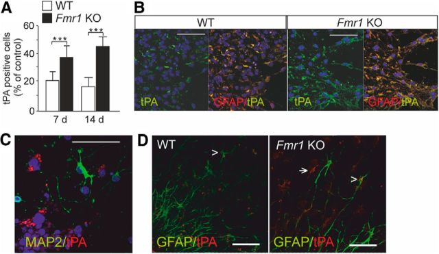
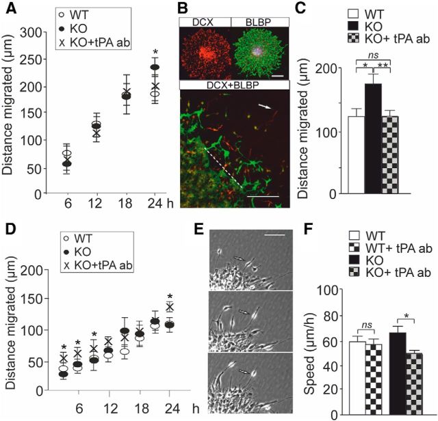
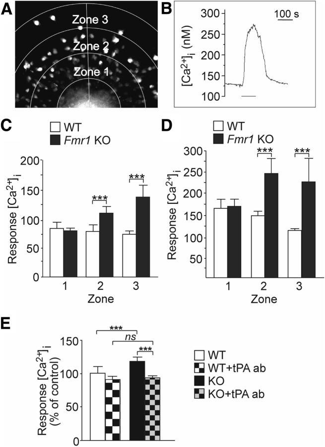
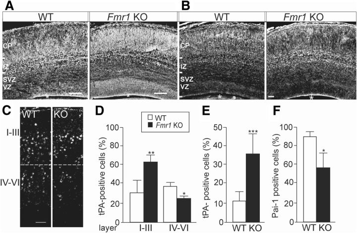
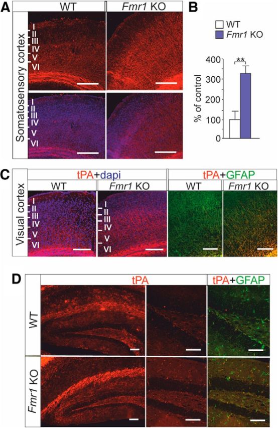
Fragile X syndrome (FXS) is the most common inherited neurodevelopmental disorder with intellectual disability. Here, we show that the expression of tissue plasminogen activator (tPA) is increased in glial cells differentiated from neural progenitors of Fmr1 knock-out mice, a mouse model for FXS, and that tPA is involved in the altered migration and differentiation of these progenitors lacking FMR1 protein (FMRP). When tPA function is blocked with an antibody, enhanced migration of doublecortin-immunoreactive neurons in 1 d differentiated FMRP-deficient neurospheres is normalized. In time-lapse imaging, blocking the tPA function promotes early glial differentiation and reduces the velocity of nuclear movement of FMRP-deficient radial glia. In addition, we show that enhanced intracellular Ca(2+) responses to depolarization with potassium are prevented by the treatment with the tPA-neutralizing antibody in FMRP-deficient cells during early neural progenitor differentiation. Alterations of the tPA expression in the embryonic, postnatal, and adult brain of Fmr1 knock-out mice suggest an important role for tPA in the abnormal neuronal differentiation and plasticity in FXS. Altogether, the results indicate that tPA may prove to be an interesting potential target for pharmacological intervention in FXS.

Keywords: FMRP; depolarization; differentiation; glia; neurons; stem cells.


Figures

Figure 1.
Figure 1.
Expression of tPA in cortical progenitors derived from Fmr1 KO mice. A, Number of tPA-immunoreactive cells was increased in differentiated neurospheres generated from Fmr1 KO mice compared with wild-type (WT) controls after differentiation for 7 and 14 d. Data are expressed as means ± SEM. *Significant difference from control (p < 0.001; n = 3–5/each group, Student's t test). B, tPA (green) immunoreactivity colocalized with GFAP (yellow) in 7 d differentiated neurospheres derived from wild-type and Fmr1 KO mouse brain. C, tPA immunoreactivity (red) was not seen in MAP2-positive neurons (green). DAPI staining show nuclei (blue). D, Glial tPA expression (red) in FMRP-deficient neurospheres after differentiation for 3 d. The arrow denotes radial glia; the arrowhead denotes colocalization of tPA with GFAP (green). Scale bars, 50 μm.
Figure 2.
Figure 2.
Effects of tPA on the migration of differentiating FMRP-deficient cells. A, In time-lapse imaging using the Cell-IQ system, the distance migrated by neurons from the edge of neurosphere cell cluster was significantly longer in neurospheres derived from Fmr1 KO mice than in wild-type (WT) controls after differentiation for 24 h (Student's t test, *p = 0.014) and the treatment with the neutralizing tPA antibody (KO + tPA ab) prevented the difference. The data are from 3–6 neurospheres in 3 experiments. B, Images of a representative neurosphere stained with DCX (red) and the radial glia marker BLBP (green) and the double staining (DCX + BLBP) used in the immunocytochemical analysis of neuronal migration in neurospheres differentiated for 24 h. The arrow denotes the outmost migrated DCX-immunopositive neuron and the dashed line the edge of the neurosphere. Scale bar, 50 μm. C, The average distance moved by the outmost migrated DCX-immunopositive neurons was significantly increased in FMRP-deficient neurospheres compared with wild-type controls and blockage of tPA with a neutralizing antibody normalized the migration defect (ANOVA, Bonferroni test, n = 15, *p = 0.013, **p = 0.008, ns = 0.955). D, The distance reached by radial glia in neurospheres derived from wild-type (WT) and Fmr1 KO mice did not differ during the differentiation for first 24 h. Treatment with the tPA antibody (KO + tPA ab) enhanced significantly the length of the processes after differentiation for 3, 6, 9, and 24 h compared with untreated controls in neurospheres derived from Fmr1 KO mice (Student's t test; *p < 0.05). Data are from wild-type neurospheres (n = 15) and neurospheres derived from Fmr1 KO mice (untreated n = 18 and treated with the tPA antibody n = 15) in three experiments. E, Interkinetic nuclear migration in radial glia was analyzed in time-lapse image sequences. F, Velocity of the interkinetic nuclear migration was reduced (Student's t test, *p = 0.041) in differentiated neurospheres derived from Fmr1 KO mice after treatment with the neutralizing tPA antibody (KO + tPA ab), but the velocity of the nuclear migration did not differ between nontreated wild-type (WT) neurospheres and neurospheres treated with the tPA antibody (WT + tPA ab). The data are from 3 experiments (n = 4–6 cells/experiment). Values are means ± SEM.
Figure 3.
Figure 3.
Effects of tPA on [Ca2+]i responses to depolarization with potassium activation in 1 d differentiated neurospheres derived from Fmr1 KO and wild-type mice. A, Fura-2 fluorescence image of the migration area that was categorized into 3 zones (zones 1–3). The cells in the migration area were divided to three groups based on their migration length, which correlated with the zones. B, Representative cell response after stimulation with [K+]e (75 mm) during the period indicated. C, D, Average calcium amplitude of the cells in each zone and its corresponding location in reference to the neurospheres derived from wild-type (WT) and Fmr1 KO male mice after stimulation with 17 mm (C) and with 75 mm [K+]e (D). WT(n) = 273 (n = 9) and KO(n) = 327 (n = 11) after exposure to 17 mm [K+]e; WT(n) = 332 (n = 9) and KO(n) = 349 (n = 11) after exposure to 75 mm [K+]e. E, Average calcium amplitude of [Ca2+]i) responses to 75 mm [K+]e in wild-type cells under basal condition (WT) and after treatment with the neutralizing tPA antibody (WT + tPA ab) compared with the amplitude of cells derived from Fmr1 KO mice under basal condition (KO) and after treatment with the neutralizing tPA antibody (KO + tPA ab). Data are from 4–5 neurospheres (n = 50–100 cells) in each group. Values are means ± SEM. *p < 0.05; **p < 0.01; ***p < 0.001.
Figure 4.
Figure 4.
Altered tPA expression in the developing brain of Fmr1 KO mouse. A, Pattern of anti-tPA immunostaining of a sagittal brain section from the neocortex of Fmr1 KO mouse differed from that of wild-type (WT) mouse at E17. B, As a control of the specificity of the tPA staining, immunostaining with anti-TrkB receptors that shows clearly different expression pattern than that of tPA. C, Anti-tPA immunostaining at P7. An asterisk denotes the lateral ventricle. D, Bar graph showing that, at P7, the proportion of tPA-expressing cells is increased in layers I-III and decreased in layers IV-V of Fmr1 KO mouse brain compared with wild-type controls (WT). E, F, Number of tPA-immunopositive cells was increased (E) and the number of PAI-1-immunopositive cells was significantly decreased (F) in the dentate gyrus of Fmr1 KO mouse hippocampus compared with controls at P7. n(WT) = 3–4; n(Fmr1 KO) = 4–5. CP, cortical plate; IZ, intermediate zone; SVZ/VZ, subventricular/ventricular zone. Scale bars, 100 μm in A and 50 μm in B. Values are means ± SEM. *p < 0.05; **p < 0.01; ***p < 0.001.
Figure 5.
Figure 5.
tPA expression in adult brain of Fmr1 KO mouse. A, Top, tPA immunoreactivity in red. Bottom, Merged images of tPA immunoreactivity in red and DAPI labeled nuclei in blue in the somatosensory cortex of adult wild-type (WT) and Fmr1 KO mouse. Scale bars, 200 μm. B, tPA immunoreactivity is significantly increased in the somatosensory cortex of Fmr1 KO mouse compared with that of WT controls. C, Merged images of anti-tPA immunostaining (red) combined with DAPI-labeled nuclei (blue) and anti-tPA immunostaining (red) combined with anti-GFAP (green) in sagittal brain sections from visual cortex of WT and Fmr1 KO mouse. D, tPA immunoreactivity (red) in the hippocampus. tPA expression (red) and its colocalization with GFAP (green) is shown in the dentate gyrus in a higher magnification. Scale bars, 200 μm. Data are expressed as means ± SEM. **p < 0.01, Student's t test.

References

    1. Bagni C, Greenough WT. From mRNP trafficking to spine dysmorphogenesis: the roots of fragile X syndrome. Nat Rev Neurosci. 2005;6:376–387. doi: 10.1038/nrn1667. - DOI - PubMed
    1. Bear MF, Huber KM, Warren ST. The mGluR theory of fragile X mental retardation. Trends Neurosci. 2004;27:370–377. doi: 10.1016/j.tins.2004.04.009. - DOI - PubMed
    1. Bhattacharyya A, McMillan E, Wallace K, Tubon TC, Jr, Capowski EE, Svendsen CN. Normal neurogenesis but abnormal gene expression in human fragile X cortical progenitor cells. Stem Cells Dev. 2008;17:107–117. doi: 10.1089/scd.2007.0073. - DOI - PubMed
    1. Brustein E, Côté S, Ghislain J, Drapeau P. Spontaneous glycine-induced calcium transients in spinal cord progenitors promote neurogenesis. Dev Neurobiol. 2013;73:168–175. doi: 10.1002/dneu.22050. - DOI - PubMed
    1. Callan MA, Zarnescu DC. Heads-up: New roles for the fragile X mental retardation protein in neural stem and progenitor cells. Genesis. 2011;49:424–440. doi: 10.1002/dvg.20745. - DOI - PubMed

Publication types

MeSH terms

LinkOut - more resources