Review
. 2014 Mar 1;6(3):a009142.
doi: 10.1101/cshperspect.a009142.

Tie2 and Eph receptor tyrosine kinase activation and signaling

Affiliations
Review

Tie2 and Eph receptor tyrosine kinase activation and signaling

William A Barton et al. Cold Spring Harb Perspect Biol. .

Abstract

The Eph and Tie cell surface receptors mediate a variety of signaling events during development and in the adult organism. As other receptor tyrosine kinases, they are activated on binding of extracellular ligands and their catalytic activity is tightly regulated on multiple levels. The Eph and Tie receptors display some unique characteristics, including the requirement of ligand-induced receptor clustering for efficient signaling. Interestingly, both Ephs and Ties can mediate different, even opposite, biological effects depending on the specific ligand eliciting the response and on the cellular context. Here we discuss the structural features of these receptors, their interactions with various ligands, as well as functional implications for downstream signaling initiation. The Eph/ephrin structures are already well reviewed and we only provide a brief overview on the initial binding events. We go into more detail discussing the Tie-angiopoietin structures and recognition.


Figures

Figure 1.
Figure 1.
Schematic representation of the Tie receptors and angiopoietin ligands. The Tie receptors are highly homologous endothelial-specific receptor tyrosine kinases. Each receptor consists of three Ig domains (shown in red, green, and blue), three EGF domains (yellow, magenta, orange), and three fibronectin type III repeats (gray) in the ectodomain, followed by a single-pass transmembrane domain, and a split tyrosine kinase domain in the cytoplasm. Tie2 interacts with all four of the structurally similar angiopoietin ligands (Ang1–4), although each ligand is functionally distinct. The angiopoietins contain an amino-terminal super-clustering domain (green), a coiled-coil domain, and a fibrinogen-like receptor-binding domain. Ang1 (blue) and Ang3 (purple) are agonists of Tie2 activation, Ang4 (orange) is an antagonist, and Ang2 (yellow) is a context-dependent antagonist as indicated. Despite the high level of sequence conservation between the two receptors, Tie1 is an orphan receptor, yet is able to heterodimerize with Tie2 on the cell surface.
Figure 2.
Figure 2.
Crystal structures of the angiopoietin receptor binding domains and the Tie2 ectodomain unbound and in complex with Ang1 and Ang2. (A) The Ang1 and Ang2 receptor-binding domains superimposed in blue and yellow, respectively. The calcium ion located within the P domain is displayed in space-filling representation in black. The surface loop, which mediates Ang1/Ang2 functional differences, is labeled. (B, C) The Ang1-Tie2 and Ang2-Tie2 crystal structures illustrated in two orientations. The Tie2 receptor is shown in green and its domains are labeled. The Ang1-Tie2 model contains an additional fibronectin type-III repeat not present in the Ang2-Tie2 structure.
Figure 3.
Figure 3.
The structure of the Tie2 TKD. Two views rotated 90° about the y-axis are shown (PBD 1FVR). The two conserved lobes, amino terminal and carboxy terminal, are colored blue and red, respectively. The three catalytic loops are labeled and colored yellow. The extended carboxy-terminal tail, containing the substrate tyrosines 1101, 1107, and 1112, is colored cyan.
Figure 4.
Figure 4.
Model of Tie-angiopoietin signaling. The angiopoietin growth factors initiate complex signaling pathways through the Tie receptor tyrosine kinases on the endothelial cell surface. Ang1 activation of the primary receptor, Tie2, leads to prosurvival signaling through pathways such as Akt and MAPK, and results in endothelial cell quiescence and recruitment of surrounding support cells. Disruption of Tie2 signaling gives increase to a leaky vessel wall as support cells detach and endothelial cells begin to migrate. The interaction of Tie2 with its coreceptor Tie1 leads to such a vessel branching phenotype. The context-dependent antagonist Ang2 inactivates the Tie2 receptor by facilitating the inhibitory Tie1/Tie2 interactions; however, if Tie1 is not present in the cell, Ang2 is capable of clustering and activating Tie2 in a manner similar to the agonist Ang1. Tie2 is in cyan, Tie1 is in yellow, and Ang1 and Ang2 are in magenta. Ang1 and Ang2 represent ligand dimers. Blue and red regions indicate electrostatically positive and negative surface regions, including phosphorylation of the Tie2 kinase domain.
Figure 5.
Figure 5.
Schematic representation of A and B types of ephrins and Eph receptors. Shown are the receptor-binding domain (RBD) of the ephrins, the Eph’s ligand-binding domain (LBD), the kinase domain (TK), the Cys-rich domain (CRD), and the fibronectin III domains (FN3). The A ephrin family are GPI-linked to the membrane, whereas the B ephrin family has a transmembrane domain and a short cytoplasmic tail.
Figure 6.
Figure 6.
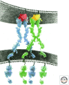
Schematic presentation of the Eph receptors bound to ephrin ligands at the cell–cell contact regions. Eph receptors are in blue and green, the ephrins are in red and yellow. The receptors use a “seeding” mechanism for creating signaling-competent assemblies where the ligand-binding domains first form receptor dimers (2:2 Eph/ephrin complexes), after which they bind other dimers via their cystein-rich domains, thus creating large clusters containing hundreds of molecules. Seemingly, the main role of the ligand is to increase the local receptor concentration so that full downstream signaling can be attained.

References

    1. Abengozar MA, de Frutos S, Ferreiro S, Soriano J, Perez-Martinez M, Olmeda D, Marenchino M, Canamero M, Ortega S, Megias D, et al. 2012. Blocking ephrinB2 with highly specific antibodies inhibits angiogenesis, lymphangiogenesis, and tumor growth. Blood 119: 4565–4576 - PubMed
    1. Adams RH, Alitalo K 2007. Molecular regulation of angiogenesis and lymphangiogenesis. Nat Rev 8: 464–478 - PubMed
    1. Artemenko EO, Egorova NS, Arseniev AS, Feofanov AV 2008. Transmembrane domain of EphA1 receptor forms dimers in membrane-like environment. Biochim Biophys Acta 1778: 2361–2367 - PubMed
    1. Barton WA, Tzvetkova D, Nikolov DB 2005. Structure of the angiopoietin-2 receptor binding domain and identification of surfaces involved in Tie2 recognition. Structure 13: 825–832 - PubMed
    1. Barton WA, Tzvetkova-Robev D, Miranda EP, Kolev MV, Rajashankar KR, Himanen JP, Nikolov DB 2006. Crystal structures of the Tie2 receptor ectodomain and the angiopoietin-2-Tie2 complex. Nat Struct Mol Biol 13: 524–532 - PubMed

Publication types

LinkOut - more resources