Reduced frontal brain volume in non-treatment-seeking cocaine-dependent individuals: exploring the role of impulsivity, depression, and smoking
- PMID: 24478673
- PMCID: PMC3894477
- DOI: 10.3389/fnhum.2014.00007
Reduced frontal brain volume in non-treatment-seeking cocaine-dependent individuals: exploring the role of impulsivity, depression, and smoking
Abstract
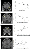
In cocaine-dependent patients, gray matter (GM) volume reductions have been observed in the frontal lobes that are associated with the duration of cocaine use. Studies are mostly restricted to treatment-seekers and studies in non-treatment-seeking cocaine abusers are sparse. Here, we assessed GM volume differences between 30 non-treatment-seeking cocaine-dependent individuals and 33 non-drug using controls using voxel-based morphometry. Additionally, within the group of non-treatment-seeking cocaine-dependent individuals, we explored the role of frequently co-occurring features such as trait impulsivity (Barratt Impulsivity Scale, BIS), smoking, and depressive symptoms (Beck Depression Inventory), as well as the role of cocaine use duration, on frontal GM volume. Smaller GM volumes in non-treatment-seeking cocaine-dependent individuals were observed in the left middle frontal gyrus. Moreover, within the group of cocaine users, trait impulsivity was associated with reduced GM volume in the right orbitofrontal cortex, the left precentral gyrus, and the right superior frontal gyrus, whereas no effect of smoking severity, depressive symptoms, or duration of cocaine use was observed on regional GM volumes. Our data show an important association between trait impulsivity and frontal GM volumes in cocaine-dependent individuals. In contrast to previous studies with treatment-seeking cocaine-dependent patients, no significant effects of smoking severity, depressive symptoms, or duration of cocaine use on frontal GM volume were observed. Reduced frontal GM volumes in non-treatment-seeking cocaine-dependent subjects are associated with trait impulsivity and are not associated with co-occurring nicotine dependence or depression.
Keywords: cocaine dependence; depression; drug abuse; frontal; nicotine; voxel-based morphometry.
Figures


Similar articles
-
Trait impulsivity and prefrontal gray matter reductions in cocaine dependent individuals.Drug Alcohol Depend. 2012 Oct 1;125(3):208-14. doi: 10.1016/j.drugalcdep.2012.02.012. Epub 2012 Mar 4. Drug Alcohol Depend. 2012. PMID: 22391134
-
A voxel-based morphometry study of frontal gray matter correlates of impulsivity.Hum Brain Mapp. 2009 Apr;30(4):1188-95. doi: 10.1002/hbm.20588. Hum Brain Mapp. 2009. PMID: 18465751 Free PMC article.
-
Gray matter volume changes following antipsychotic therapy in first-episode schizophrenia patients: A longitudinal voxel-based morphometric study.J Psychiatr Res. 2019 Sep;116:126-132. doi: 10.1016/j.jpsychires.2019.06.009. Epub 2019 Jun 15. J Psychiatr Res. 2019. PMID: 31233895
-
A comparison of impulsivity, depressive symptoms, lifetime stress and sensation seeking in healthy controls versus participants with cocaine or methamphetamine use disorders.J Psychopharmacol. 2015 Jan;29(1):50-6. doi: 10.1177/0269881114560182. Epub 2014 Nov 25. J Psychopharmacol. 2015. PMID: 25424624
-
Relationship between trait impulsivity and cortical volume, thickness and surface area in male cocaine users and non-drug using controls.Drug Alcohol Depend. 2014 Nov 1;144:210-7. doi: 10.1016/j.drugalcdep.2014.09.016. Epub 2014 Sep 22. Drug Alcohol Depend. 2014. PMID: 25278147
Cited by
-
Common abnormality of gray matter integrity in substance use disorder and obsessive-compulsive disorder: A comparative voxel-based meta-analysis.Hum Brain Mapp. 2021 Aug 15;42(12):3871-3886. doi: 10.1002/hbm.25471. Epub 2021 Jun 9. Hum Brain Mapp. 2021. PMID: 34105832 Free PMC article.
-
Discrimination of smoking status by MRI based on deep learning method.Quant Imaging Med Surg. 2018 Dec;8(11):1113-1120. doi: 10.21037/qims.2018.12.04. Quant Imaging Med Surg. 2018. PMID: 30701165 Free PMC article.
-
Self-reported impulsivity is negatively correlated with amygdalar volumes in cocaine dependence.Psychiatry Res. 2015 Aug 30;233(2):212-7. doi: 10.1016/j.pscychresns.2015.07.013. Epub 2015 Jul 8. Psychiatry Res. 2015. PMID: 26187551 Free PMC article.
-
Causal pathways between impulsiveness, cocaine use consequences, and depression.Addict Behav. 2015 Feb;41:1-6. doi: 10.1016/j.addbeh.2014.09.017. Epub 2014 Sep 22. Addict Behav. 2015. PMID: 25280245 Free PMC article.
-
A hypo-status in drug-dependent brain revealed by multi-modal MRI.Addict Biol. 2017 Nov;22(6):1622-1631. doi: 10.1111/adb.12459. Epub 2016 Sep 22. Addict Biol. 2017. PMID: 27654848 Free PMC article.
References
-
- Beck A. T., Steer R. A. (1987). Manual for Revised Beck Depression Inventory. San Antonio, TX: Psychological Corporation
LinkOut - more resources
Full Text Sources
Other Literature Sources
Miscellaneous

