Review
. 2014 Jan 20:4:409.
doi: 10.3389/fphys.2013.00409. eCollection 2013.

Possible contribution of chronobiology to cardiovascular health

Affiliations
Review

Possible contribution of chronobiology to cardiovascular health

Miho Sato et al. Front Physiol. .

Abstract

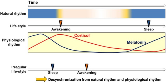
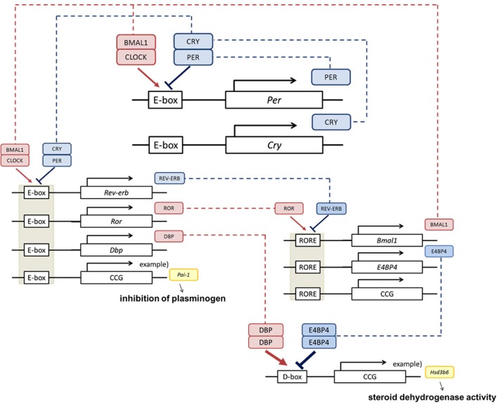
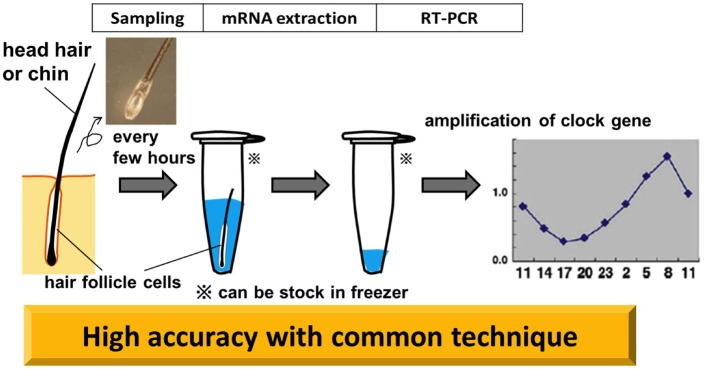
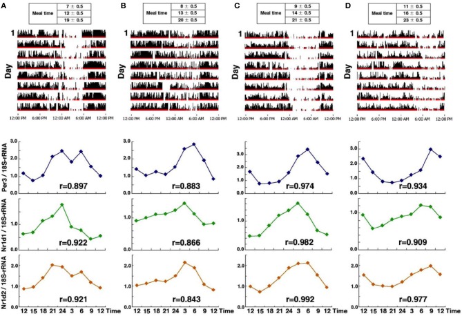
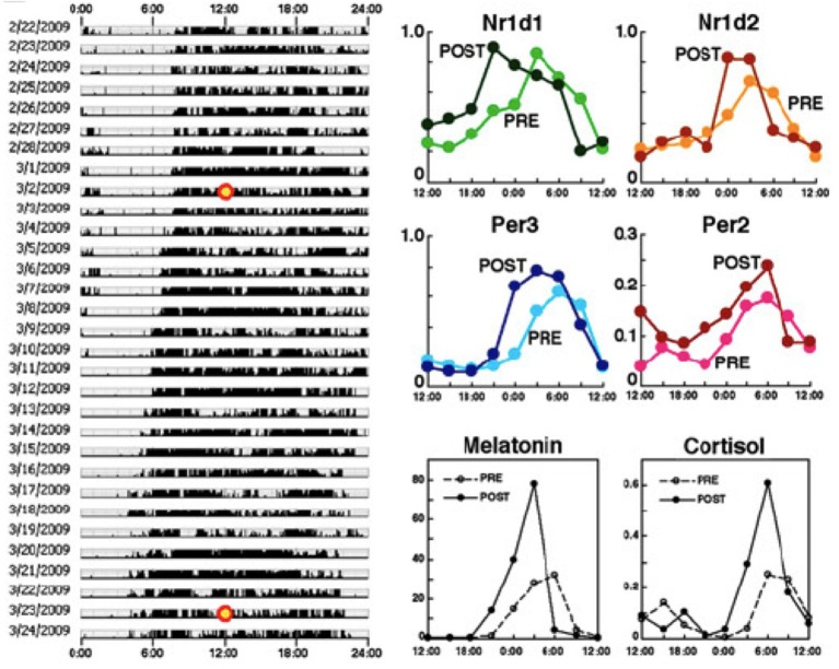
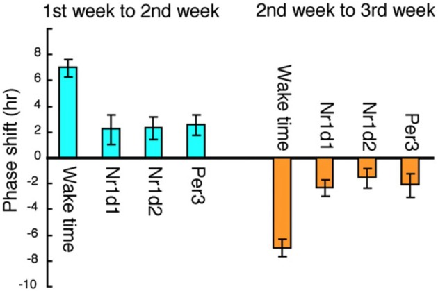
The daily variations found in many aspects of physiology are collectively known as circadian rhythm (from "circa" meaning "about" and "dien" meaning "day"). Circadian oscillation in clock gene expression can generate quantitative or functional variations of the molecules directly involved in many physiological functions. This paper reviews the molecular mechanisms of the circadian clock, the transmission of circadian effects to cardiovascular functions, and the effects of circadian dysfunction on cardiovascular diseases. An evaluation of the operation of the internal clock is needed in clinical settings and will be an effective tool in the diagnosis of circadian rhythm disorders. Toward this end, we introduce a novel non-invasive method for assessing circadian time-regulation in human beings through the utilization of hair follicle cells.

Keywords: cardiovascular diseases; circadian; clock gene; hair follicle; non-invasive method.


Figures

Figure 1
Figure 1
Circadian clock and life style. Circadian variation of physiological functions is set to synchronize with the natural environment. This enables the smooth operation of physiological functions in harmony with the basic rhythms (Weitzman et al., ; Lewy and Markey, ; Arendt, ; Haus, ; Benloucif et al., 2008) and cycles of nature (light-dark) and with the correlated but not always continuous rhythms and cycles of human life-style (waking-sleep). Irregular life-styles bring about the desynchronization of physiological rhythms from other sources of cyclicity.
Figure 2
Figure 2
Clock genes and physiological function. The core clock is composed by the Per, Cry, Bmal1, and Clock clock genes (Okamura et al., ; Takeda and Maemura, 2011). BMAL1 and CLOCK activate transcriptional levels through E-boxes, and CRY and PER suppress this activity. Cis elements such as RORE and D-box can be regulated by ROR, REV-ERB, DBP, and E4BP4, and multilayered rigid circadian rhythms are ticked down. This negative feedback loop produces the rhythm of transcription. These regulations are transmitted via transcriptional fluctuations of clock controlled genes (ccg) such as plasminogen activator inhibitor-1 (Pai-1) (Maemura et al., ; Haus, 2007), type VI 3 beta-hydroxyl-steroid dehydrogenase (Hsd3b6) (Bass, 2012).
Figure 3
Figure 3
Clock genes and diabetes. Glucose levels (left panel) and insulin levels (right panel) during glucose tolerance tests in pancreas-specific Bmal1-deficient mice (PdxCre Bmal1flx/flx). PdxCre and Bmal1flx/flx mice are negative controls for the experiments. Bmal1 knockout mice displayed decreased insulin secretion and higher glucose levels after glucose administration. The figures are adapted from Macmillan Publishers Ltd: Nature, Marcheva et al. (2010), copyright 2010. Data were analysed by one-way ANOVA and asterisks denote significance between Bmal1flx/flx and PdxCre Bmal1flx/flx, and plus symbols denote significance between PdxCre and PdxCre Bmal1flx/flx. *p = 0.05; ***p = 0.001.
Figure 4
Figure 4
Clock genes and hypertension. Fifteen-day mean arterial blood pressure recorded under high-salt diet conditions. Mice lacking the core clock components Cryptochrome1 (Cry1) and Cryptochrome2 (Cry2) (Cry1−/− Cry2−/−, upper panel) show salt-sensitive hypertension, while the wild type (WT) doesn't. This hypertension can be avoided with the aldosterone inhibitor, Eplerenone. The figures are adapted from Macmillan Publishers Ltd: Nature Medicine, Doi et al. (2010), copyright 2010.
Figure 5
Figure 5
Non-invasive circadian clock detection with hair follicle cells. Overview from sampling to quantification by realtime-PCR (RT-PCR). Plucking head hair sharply at the roots with non-slip tweezers in the direction of hair growth tends to extract a good deal of hair follicles. Pre-analysis, samples can be stocked in a deep freezer. The standard protocol for RT-PCR is adopted for the analysis. The graph in this diagram is adapted from Akashi et al. (2010).
Figure 6
Figure 6
Clock gene expression from four subjects who maintained a regular lifestyle with a specific phase. Activity data over an 8-day period (upper panel) and clock gene expression (Per3, Nr1d1, and Nr1d2) (lower panel) in four healthy individuals (A–D), from the earliest riser on the left (A), to the latest riser on the right (D). Interestingly, in all subjects, Per3 expression peaked just around waking time and the earliest phase of circadian gene expression was observed in subject (A). The r, correlation coefficient is calculated by cosine curve fitting. The figures are adapted from Akashi et al. (2010).
Figure 7
Figure 7
A 3-week adaptation period is not enough to advance the phase of the molecular clock by 4 h. Behavioral rhythms (left), rhythms in four clock genes from hair follicles (upper right) and melatonin and cortisol concentrations from saliva (lower right). Specimens were collected every 3 h from the time points marked with a yellow circle (3/2/2009: PRE, 3/23/2009: POST) and compared between PRE and POST. These results indicate that a 3-week adaptation period is not enough to advance the phase of the molecular clock by 4 h. PRE: data before the phase shift; POST: data after the 4-h phase advance of life style rhythms. The figures are adapted from Akashi et al. (2010).
Figure 8
Figure 8
Correlation between shift-work schedules and clock gene expression in six rotating shift workers. The average phase shift of waking-time and circadian gene expression was calculated among the six subjects. The phase delay from the 1st to the 2nd week and the phase advance from the 2nd to the 3rd week are shown. The life style was phase-delayed by about 7 h from the 1st to the 2nd week, whereas the phase of circadian gene expression was delayed only by about 2 h. The figures are adapted from Akashi et al. (2010).

References

    1. Akashi M., Soma H., Yamamoto T., Tsugitomi A., Yamashita S., Nishida E., et al. (2010). Noninvasive method for assessing the human circadian clock using hair follicle cells. Proc. Natl. Acad. Sci. U.S.A. 107, 15643–15648 10.1073/pnas.1003878107 - DOI - PMC - PubMed
    1. Anea C. B., Zhang M., Stepp D. W., Simkins G. B., Reed G., Fulton D. J., et al. (2009). Vascular disease in mice with a dysfunctional circadian clock. Circulation 119, 1510–1517 10.1161/CIRCULATIONAHA.108.827477 - DOI - PMC - PubMed
    1. Arendt J. (1998). Melatonin and the pineal gland: influence on mammalian seasonal and circadian physiology. Rev. Reprod. 3, 13–22 10.1530/ror.0.0030013 - DOI - PubMed
    1. Arendt J. (2005). Melatonin: characteristics, concerns, and prospects. J. Biol. Rhythms 20, 291–303 10.1177/0748730405277492 - DOI - PubMed
    1. Aschoff J. (1965). Circadian rhythms in man. Science 148, 1427–1432 10.1126/science.148.3676.1427 - DOI - PubMed

LinkOut - more resources