. 2014 Jan;184(1):159-70.
doi: 10.1016/j.ajpath.2013.09.015.

The level of hepatic ABCC6 expression determines the severity of calcification after cardiac injury

The level of hepatic ABCC6 expression determines the severity of calcification after cardiac injury

Christopher Brampton et al. Am J Pathol. 2014 Jan.

Erratum in

  • Am J Pathol. 2014 Mar;184(3):878. Erdfdi, Jeannette [corrected to Erdmann, Jeannette]

Abstract

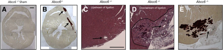
Because vascular or cardiac mineralization is inversely correlated with morbidity and long-term survival, we investigated the role of ABCC6 in the calcification response to cardiac injury in mice. By using two models of infarction, nonischemic cryoinjury and the pathologically relevant coronary artery ligation, we confirmed a large propensity to acute cardiac mineralization in Abcc6−/− mice. Furthermore, when the expression of ABCC6 was reduced to approximately 38% of wild-type levels in Abcc6+/− mice, no calcium deposits in injured cardiac tissue were observed. In addition, we used a gene therapy approach to deliver a functional human ABCC6 via hydrodynamic tail vein injection to approximately 13% of mouse hepatocytes, significantly reducing the calcification response to cardiac cryoinjury. We observed that the level and distribution of known regulators of mineralization, such as osteopontin and matrix Gla protein, but not osteocalcin, were concomitant to the level of hepatic expression of human and mouse ABCC6. We notably found that undercarboxylated matrix Gla protein precisely colocalized within areas of mineralization, whereas osteopontin was more diffusely distributed in the area of injury, suggesting a prominent association for matrix Gla protein and osteopontin in ABCC6-related dystrophic cardiac calcification. This study showed that the expression of ABCC6 in liver is an important determinant of calcification in cardiac tissues in response to injuries and is associated with changes in the expression patterns of regulators of mineralization.


Figures

Figure 1
Figure 1
Surface lesions and calcification of Abcc6−/− and control mice. Cardiac lesions were obtained through a single transdiaphragm cryoinjury. Mice were sacrificed 7 days after surgery, and hearts were exposed by opening the chest cavity and quickly imaged before excision. A: A representative image obtained with an Abcc6−/− mouse illustrates the macroscopic appearances of the surface lesions outlined with a dashed circle. The calcification is visible as white deposits. B and C: The lesions of a heterozygous Abcc6+/− mouse and a sham–operated on Abcc6−/− mouse devoid of any obvious white deposits indicating the lack of calcification. D: The discoloration of the myocardial tissue after ischemic injury (4 weeks, permanent occlusion) is delineated by the dashed circle. The arrowhead points to the suture ligating the left coronary artery.
Figure 2
Figure 2
Histological characterization of myocardium of Abcc6−/− mice and mice after cryoinjury or sham treatment. After excision, hearts were fixed in formalin and paraffin embedded. Sections were stained with H&E or Alizarin Red S and imaged. A and B: Images of heart cross sections of Abcc6−/− mice show the mineralization occurring after cyroinjury but not in the sham–operated mouse. C and D: The consequence of the ischemic injury after left coronary artery ligation (H&E staining). Arrows point to the left coronary artery upstream (C) and downstream of the ligation and the affected portion of the myocardium, which is outlined (D). E: An image of a serial section from D, stained with Alizarin Red S to reveal the calcification. Scale bar = 500 μm (A and C).
Figure 3
Figure 3
Quantification of the calcification after cardiac injury. A: The calcium content in the murine hearts after cryoinjury was expressed as a ratio between the injured ventral/uninjured dorsal sections. We also compared the cryoinjury calcification after the transient expression of the WT ABCC6 and the inactive mutant, V1298F, for various control mice and at 4 weeks after injury. B and C: Representative images of the surface lesions (dashed circles) and calcification in Abcc6−/− mice without and with the expression of the human ABCC6 cDNA. The calcified deposits (white) were visibly reduced after the hydrodynamic tail vein injection of ABCC6 (arrows). D: The level of calcification after ischemic injury was expressed as the calcium concentration derived from total heart because of the diffuse nature of this type of injury. The results for control mice are also shown. P < 0.05, ∗∗P < 0.01.
Figure 4
Figure 4
Verification of the efficiency of HTVI and the overall distribution of the transfected hepatocytes. The commercial pLIVE vector carrying liver-specific promoter, enhancer, and β-galactosidase was used. After excision, the whole liver and both kidneys were lightly fixed and prepared for X-gal staining to examine the distribution of expression. AC: The method revealed an extensive distribution throughout all lobes of the liver, although on close examination. D: The distribution of the individual cells was found to be heterogeneous. E and F: No visible expression was detected in either kidney, demonstrating the tissue-specific expression and the efficient delivery achieved by the HTVI expression of the pLIVE vector.
Figure 5
Figure 5
Evaluation of ABCC6 protein levels after HTVI. The level of expression of human ABCC6 in HTVI-treated Abcc6−/− mice was estimated and compared with WT levels using immunofluorescent images (A) and ImageJ software version 1.45k, by manually counting the number of cells with membrane expression of the protein and comparing with the number of DAPI-stained nuclei (B) and by morphometric analysis of pixel intensity (n = 5 HTVI-treated mice and n = 3 for WT mice with 10 random nonserial sections counted from each liver) (C). The mouse ABCC6 protein was detected on frozen sections by immunofluorescence with the S-20 polyclonal antibody (green). Human ABCC6 was revealed with the M6II-31 monoclonal antibody (red) showing sporadic staining, as expected. Liver tissue from an Abcc6−/− mouse was used as a negative control for the S-20 antibody. D: Representative Western blot images that include the clear detection of the human and mouse ABCC6 in the liver of Abcc6−/− mice are shown. E: The level of mouse ABCC6 expression in heterozygous mice was quantified by using Western blot analysis with the K-14 antibody. The detection of β-actin was used as a normalizing control. F: Representative Western blot images showing the absence of detection of the mouse ABCC6 in liver extracts of two Abcc6−/− mice with the K-14 antibody. The detection of β-actin was used as a loading control. Scale bars: 100 μm (A). P < 0.05, ∗∗P < 0.01.
Figure 6
Figure 6
ELISA quantification of calcification regulators in the myocardium of Abcc6−/− mice after cardiac injury. The injured cardiac regions were excised and total proteins were extracted. The levels of OC, OPN, and MGP were quantified with commercially available kits. The results are shown as pg/mL per mg of tissues. P < 0.05, ∗∗∗P < 0.001. Cryo, cryoinjured.
Figure 7
Figure 7
Immunofluorescent detection of calcification regulators in the myocardium of Abcc6−/− mice after ischemic injury and cryoinjury. Immunofluorescent detection of calcification regulators in experimental animals. Frozen serial sections from the heart of Abcc6−/− mice and Abcc6−/− mice expressing the human ABCC6 protein were used for both immunofluorescent and Alizarin Red S staining. We observed large positive staining of OC, OPN, and ucMGP (green) in the injured regions, which suggested a role for these regulators in the mineralization response to the cryoinjuries and ischemic injuries. Immunofluorescent detection of calcification regulators is shown for control animals. Frozen serial sections from the heart of heterozygous Abcc6+/− and WT mice were used. Little or no evidence of calcification (Alizarin Red S) or change in the immunofluorescent staining patterns was observed in these control tissues. Negative controls were performed by omitting the primary anti-mouse ucMGP antibody. Nuclei were stained with DAPI. The circles indicate the position of the ligating sutures placed to induce permanent cardiac ischemia. Scale bar = 100 μm.
Figure 8
Figure 8
Mineralization is not due to altered cell death. At 1, 3, and 7 days after cryoinjury, Abcc6−/− and WT hearts were harvested and stained with Alizarin Red S to reveal the progression of cardiac calcification (arrows). The level of apoptosis detected by TUNEL showed similar levels and patterns of apoptosis (green fluorescence) throughout the injured sections of the Abcc6−/− and WT tissues. Nuclei were stained with DAPI. The images were centered on the injured areas, which varied slightly for each mouse. Scale bars: 100 μm.

References

    1. Abedin M., Tintut Y., Demer L.L. Vascular calcification: mechanisms and clinical ramifications. Arterioscler Thromb Vasc Biol. 2004;24:1161–1170. - PubMed
    1. Atzeni F., Sarzi-Puttini P., Bevilacqua M. Calcium deposition and associated chronic diseases (atherosclerosis, diffuse idiopathic skeletal hyperostosis, and others) Rheum Dis Clin North Am. 2006;32:413–426. viii. - PubMed
    1. Brean H.P., Marks J.H. Massive calcification in infarcted myocardium. Radiology. 1950;54:33–42. illust. - PubMed
    1. El-Bialy A., Shenoda M., Saleh J., Tilkian A. Myocardial calcification as a rare cause of congestive heart failure: a case report. J Cardiovasc Pharmacol Ther. 2005;10:137–143. - PubMed
    1. Shackley B.S., Nguyen T.P., Shivkumar K., Finn P.J., Fishbein M.C. Idiopathic massive myocardial calcification: a case report and review of the literature. Cardiovasc Pathol. 2011;20:e79–e83. - PubMed

Publication types

MeSH terms

Substances