. 2014 Jan 1;385(1):41-51.
doi: 10.1016/j.ydbio.2013.10.016.

Dkk1 in the peri-cloaca mesenchyme regulates formation of anorectal and genitourinary tracts

Affiliations

Dkk1 in the peri-cloaca mesenchyme regulates formation of anorectal and genitourinary tracts

Chaoshe Guo et al. Dev Biol. .

Abstract

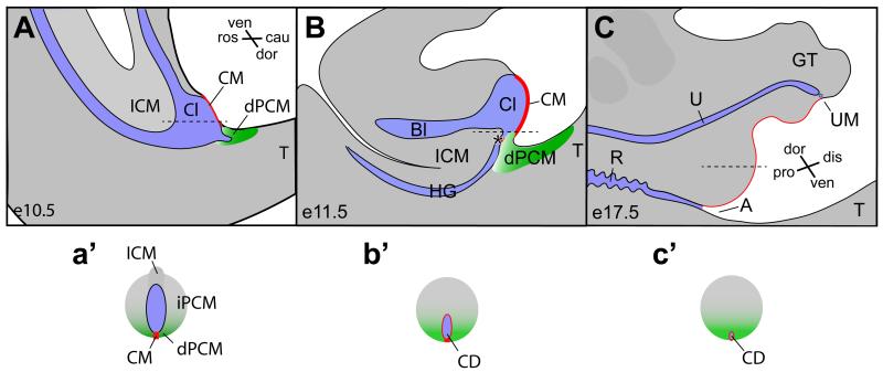
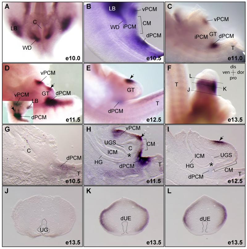
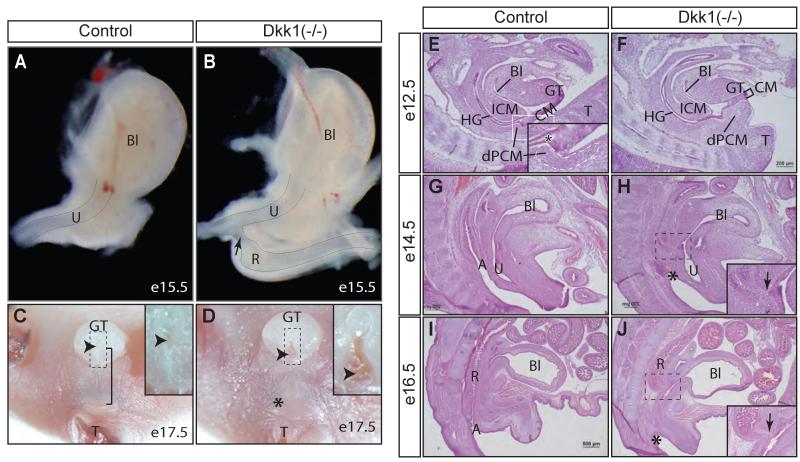
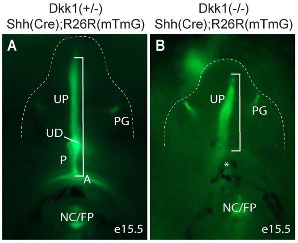
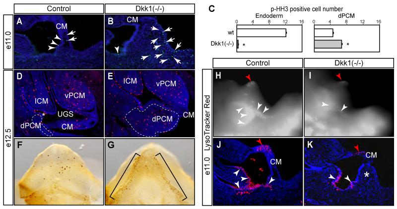
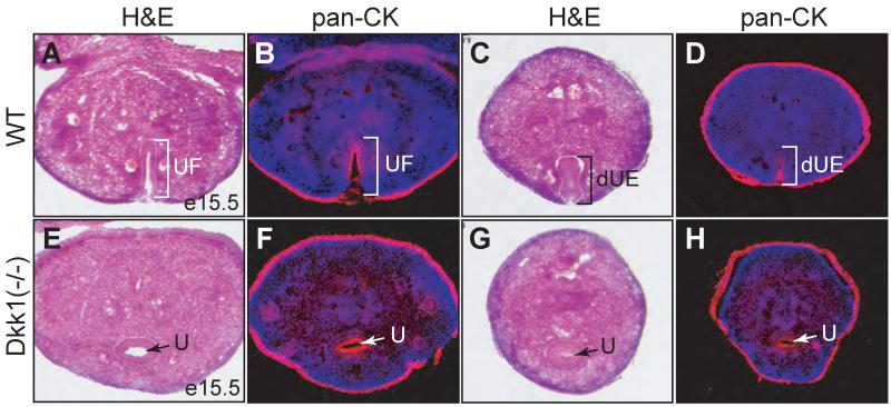
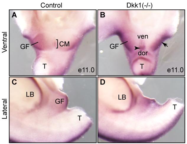
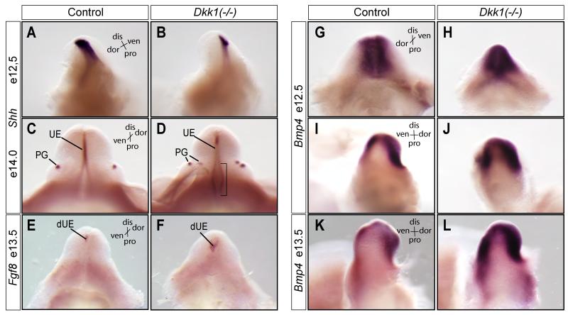
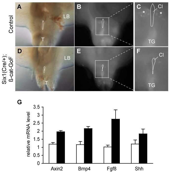
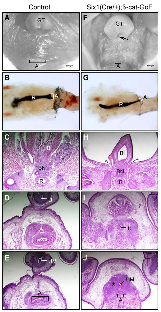
Anorectal malformation (ARM) is a common birth defect but the developmental history and the underlying molecular mechanism are poorly understood. Using murine genetic models, we report here that a signaling molecule Dickkopf-1 (Dkk1) is a critical regulator. The anorectal and genitourinary tracts are major derivatives of caudal hindgut, or the cloaca.Dkk1 is highly expressed in the dorsal peri-cloacal mesenchymal (dPCM) progenitors. We show that the deletion of Dkk1 causes the imperforate anus with rectourinary fistula. Mutant genital tubercles exhibit a preputial hypospadias phenotype and premature urethral canalization.Dkk1 mutants have an ectopic expansion of the dPCM tissue, which correlates with an aberrant increase of cell proliferation and survival. This ectopic tissue is detectable before the earliest sign of the anus formation, suggesting that it is most likely the primary or early cause of the defect. Deletion of Dkk1 results in an elevation of the Wnt/ß-catenin activity. Signaling molecules Shh, Fgf8 and Bmp4 are also upregulated. Furthermore, genetic hyperactivation of Wnt/ß-catenin signal pathway in the cloacal mesenchyme partially recapitulates Dkk1 mutant phenotypes. Together, these findings underscore the importance ofDKK1 in regulating behavior of dPCM progenitors, and suggest that formation of anus and urethral depends on Dkk1-mediated dynamic inhibition of the canonical Wnt/ß-catenin signal pathway.


Figures

Figure 1
Figure 1. The occlusion model of cloaca morphogenesis
The cloaca occlusion model suggests that the dorsal peri-cloacal mesenchyme (dPCM, green) and the cloacal membrane (CM, red) play prominent roles in morphogenesis of the cloaca (Cl). blue, endoderm cavity. The CM and the dPCM function as two pivot points through which asymmetric growth of mesenchymal cells surrounding cloacal leads to separation of the hindgut (HG) and developing bladder (Bl), as well as outgrowth of genital tubercle (GT). Both midline sagittal views (A-C) and horizontal views (a’-c’, indicated in A-C as dash lines) are shown. A, anus; Bl, bladder; Cl, cloaca; cau, caudal; dis, distal; dor, dorsal; ICM, intra-cloacal mesenchyme; pro, proximal; R, rectum; ros, rostra; T, tail; ven, ventral; asterisk, juxtaposition of the ICM, the dPCM and the CM.
Figure 2
Figure 2. Dynamic expression pattern of Dkk1 during cloaca morphogenesis
(A-F) Whole mount RNA in situ hybridization using a Dkk1 specific probe (n=2). All images are lateral views except A and an inset in D, which are ventral views. (G-I) Midline sections of Dkk1 whole mount stained embryos. (J-L) Cross sections of the genital tubercle of a e13.5 embryo shown in F. dUE, distal urethral epithelium; LB, limb bud; UG, urethral groove; vPCM, ventral peri-cloacal mesenchyme; WD, Wolffian ducts; asterisk, juxtaposition of the ICM, the dPCM and the CM. See Figure 1 for more abbreviations.
Figure 3
Figure 3. Dkk1 mutants exhibit an imperforate anus with rectourinary fistula
(A - D), whole mount views of lower urinary tract at e15.5 (A and B, lateral view, n=3) and anogenital structures at e17.5 (C and D, ventral view, n=5). Insets in C and D show the urethral meatus (arrowhead). Bracket, anogenital distance; Asterisk, missing anus. (E- J) Histological midline sagittal sections from staged embryos (n=2 from each stage). Insets in H and J indicate the rectourinary fistula (arrow). See Figures 1 and 2 for more abbreviations.
Figure 4
Figure 4. Genetic lineage mapping of Shh-expression cells
Ventral view eGFP-positive Shh lineages of e15.5 genital tubercle (outlined) and perineum. A, anus; NC/FP, notochord/floor plate; P, perineum; PG, preputial gland; UD, urethra duct; UF, urethral fold; asterisk, denote missing anus and perineum; bracket, ventral midline endoderm derived epithelium from anus to distal urethral plate.
Figure 5
Figure 5. Proliferation and apoptosis defects of Dkk1 mutants
(A-E) A mitotic marker phospho-histone H3 (p-HH3) staining of midline sagittal sections (A-E) and whole mount genital tubercle (F and G, ventral views). Results from e11.0 embryos are quantified in C. Total p-HH3-positive mesenchyme (arrow) and endoderm (arrowhead) cells from each section are counted separately. Asterisk, p<0.05, Student t test, n=3. (H-K) LysoTracker Red® staining of e11.0 wild type control (H and J) and Dkk1 mutant (I and K). Positive signals appeared to be bright white in whole mount images (H and I) and red in midline sagittal sections (J and K). Double asterisk, ectopic dPCM tissue. Red arrowhead, distal urethral plate epithelium.
Figure 6
Figure 6. Deletion of Dkk1 causes premature canalization of the genital urethra
Histological and pan-cytokeratin immunostaining of adjacent cross sections from e15.5 control (A-D) or Dkk1 mutant (E-H) genital tubercles. dUE, distal urethral epithelia; U, urethra; UF, urethral fold.
Figure 7
Figure 7. Axin2, an indicator of the Wnt/ß-catenin activity, is upregulated
Whole mount RNA in situ of e11.0 urogenital structures from control (A and C) and Dkk1 mutant (B and D) using Axin2 probe (n=3). Both ventral (A and B) and lateral (C and D) views were shown. CM, cloacal membrane; dor, dorsal; GF, genital fold; LB, limb bud; T, tail; ven, ventral; arrow, enhanced expression in the GF; arrowheaad, enhanced expression in the dorsal CM.
Figure 8
Figure 8. Aberrant expressions of Shh, Fgf8 and Bmp4 in the mutant genital tubercles
(A-D) Whole mount RNA in situ hybridizations using Shh (A-D), Fgf8 (E and F) and Bmp4 (G-L) probes (n=2). dis, distal; dor, dorsal; pro, proximal; ven, ventral; UE, erethral epithelia; PG, preputial gland; dUE, distal urethral epithelia; bracket, proximal urethral fold epithelia.
Figure 9
Figure 9. Constitutive activation of ß-catenin in the PCM
(A-F) Ventral views of LysoTracker stained e10.5 embryos from control (A-C) and ß-catenin GoF mutants (D-F). A and D, visual light images; B and E, fluorescent images of labeled cells; enlarged views are shown in C and F. Cl, cloaca; T, tail; TG, tail gut. (G) Significant increase of marker gene expression in ß-catenin GoF mutants (Student-t test, n=3). Real time quantitatively PCR analysis of expression of specific genes in e13.5 genital tubercle using 18S RNA as an internal control. Open column, littermate control; solid column, Six1Cre/+;ß-cateinn-GoF mutants.
Figure 10
Figure 10. ß-catenin GoF mutants partially recapitulate the urogenital phenotypes of Dkk1 mutants
(A and F) Whole mount ventral view of urogenital structures from e17.5 wild type control (A) and Six1Cre/+;ß-Cat-GoF compound mutants (F). (B and G) India Ink paint injection of anorectal lumen (n=5). (C-E and H-J) Serial histology of cross sections of the anal canal from e17.5 male embryos. A, anus; Bl, bladder; BN, bladder neck; GT, genital tubercle; R, rectum; U, genital urethra; UM, urethra meatus; asterisk, ectopic mesenchymal condensation (n=3).

References

    1. Blaschko SD, Cunha GR, Baskin LS. Molecular mechanisms of external genitalia development. Differentiation. 2012 - PMC - PubMed
    1. Cuschieri A. Anorectal anomalies associated with or as part of other anomalies. Am J Med Genet. 2002;110:122–130. - PubMed
    1. Glinka A, Wu W, Delius H, Monaghan AP, Blumenstock C, Niehrs C. Dickkopf-1 is a member of a new family of secreted proteins and functions in head induction. Nature. 1998;391:357–362. - PubMed
    1. Grotewold L, Ruther U. The Wnt antagonist Dickkopf-1 is regulated by Bmp signaling and c-Jun and modulates programmed cell death. Embo J. 2002;21:966–975. - PMC - PubMed
    1. Guo C, Sun Y, Zhou B, Adam RM, Li X, Pu WT, Morrow BE, Moon A, Li X. A Tbx1-Six1/Eya1-Fgf8 genetic pathway controls mammalian cardiovascular and craniofacial morphogenesis. J Clin Invest. 2011;121:1585–1595. - PMC - PubMed

Publication types

MeSH terms

LinkOut - more resources