. 2014 Jan 31;16(1):R38.
doi: 10.1186/ar4467.

Expression and function of visfatin (Nampt), an adipokine-enzyme involved in inflammatory pathways of osteoarthritis

Expression and function of visfatin (Nampt), an adipokine-enzyme involved in inflammatory pathways of osteoarthritis

Marie-Charlotte Laiguillon et al. Arthritis Res Ther. .

Abstract

Introduction: Visfatin is an adipokine that may be involved in intertissular joint communication in osteoarthritis (OA). With a homodimeric conformation, it exerts nicotinamide phosphoribosyltransferase (Nampt) enzymatic activity, essential for nicotinamide adenine dinucleotide biosynthesis. We examined the tissular origin and conformation of visfatin/Nampt in human OA joints and investigated the role of visfatin/Nampt in chondrocytes and osteoblasts by studying Nampt enzymatic activity.

Methods: Synovium, cartilage and subchondral bone from human OA joints were used for protein extraction or incubated for 24 hours in serum-free media (conditioned media), and synovial fluid was obtained from OA patients. Visfatin/Nampt expression in tissular extracts and conditioned media was evaluated by western blot and enzyme-linked immunosorbent assay (ELISA), respectively. Nampt activity was assessed in OA synovium by colorimetric assay. Primary cultures of murine chondrocytes and osteoblasts were stimulated with visfatin/Nampt and pretreated or not with APO866, a pharmacologic inhibitor of Nampt activity. The effect on cytokines, chemokines, growth factors and hypertrophic markers expression was examined by quantitative reverse transcriptase polymerase chain reaction and/or ELISA.

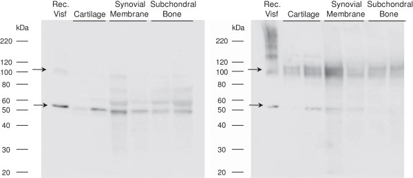
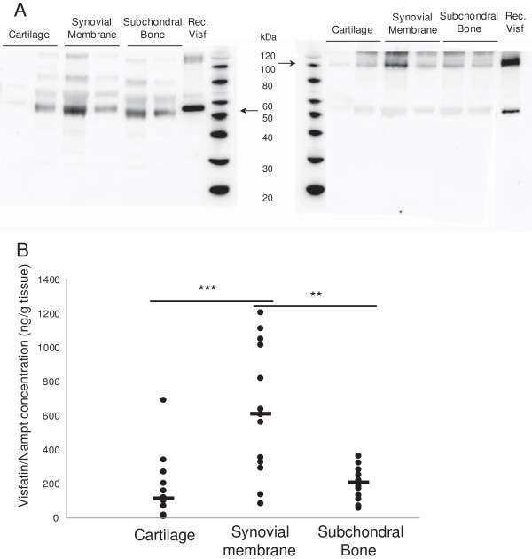
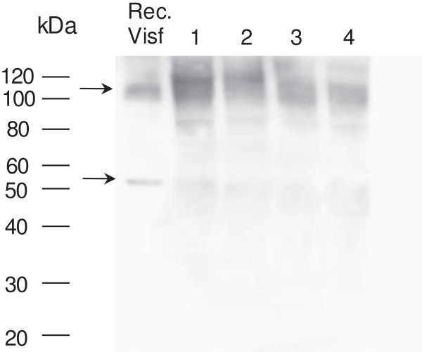
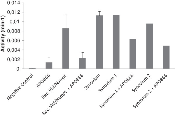
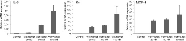
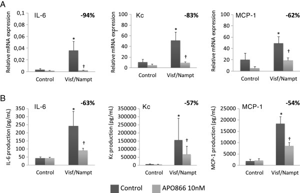
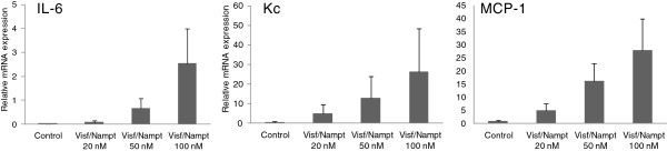
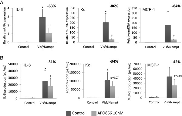
Results: In tissular explants, conditioned media and synovial fluid, visfatin/Nampt was found as a homodimer, corresponding to the enzymatically active conformation. All human OA joint tissues released visfatin/Nampt (synovium: 628 ± 106 ng/g tissue; subchondral bone: 195 ± 26 ng/g tissue; cartilage: 152 ± 46 ng/g tissue), with significantly higher level for synovium (P <0.0005). Nampt activity was identified ex vivo in synovium. In vitro, visfatin/Nampt significantly induced the expression of interleukin 6, keratinocyte chemoattractant and monocyte chemoattractant protein 1 in chondrocytes and osteoblasts. APO866 decreased the mRNA and protein levels of these pro-inflammatory cytokines in the two cell types (up to 94% and 63% inhibition, respectively). Levels of growth factors (vascular endothelial growth factor, transforming growth factor β) and hypertrophic genes were unchanged with treatment.

Conclusion: Visfatin/Nampt is released by all human OA tissues in a dimeric enzymatically active conformation and mostly by the synovium, which displays Nampt activity. The Nampt activity of visfatin is involved in chondrocyte and osteoblast activation, so targeting this enzymatic activity to disrupt joint tissue interactions may be novel in OA therapy.


Figures

Figure 1
Figure 1
Intratissular expression of visfatin/Nampt from osteoarthritic human joint tissues. Human osteoarthritic joint tissues were obtained after surgery, separated, frozen and ground to obtain protein extracts. Western blot analysis of the visfatin/nicotinamide phosphoribosyltransferase (Nampt) protein level in tissues. Left panel: denaturing conditions, showing a monomeric conformation. Right panel: nondenaturing conditions, showing polymeric conformation. Arrows show bands specific to visfatin/Nampt.
Figure 2
Figure 2
Production of visfatin/Nampt by human osteoarthritic joint tissues. Human osteoarthritic joint tissues were incubated for 24 hours in serum-free medium (6 ml/g tissue). Retrieved media were considered conditioned media. (A) Western blot analysis of visfatin/nicotinamide phosphoribosyltransferase (Nampt) protein level in conditioned media. Left panel: denaturing conditions, showing monomeric conformation. Right panel: nondenaturing conditions, showing dimeric conformation. Arrows show bands specific to visfatin/Nampt. (B) Enzyme-linked immunosorbent assay of visfatin/Nampt released by cartilage (n = 15), synovial membrane (n = 13) and subchondral bone (n = 13). **P < 0.001, ***P < 0.0005. Horizontal lines are medians. Each dot represents one sample.
Figure 3
Figure 3
Presence of visfatin/Nampt in synovial fluid. Synovial fluids were obtained from four osteoarthritis patients with joint effusion. Western blot analysis of visfatin/nicotinamide phosphoribosyltransferase (Nampt) protein level under nondenaturing conditions, showing polymeric conformation. Arrows show bands specific to visfatin/Nampt.
Figure 4
Figure 4
Enzymatic activity of visfatin/Nampt in synovial membrane of human osteoarthritis. Enzymatic activity of visfatin/nicotinamide phosphoribosyltransferase (Nampt) assayed in protein extracts of human osteoarthritis synovial membranes by colorimetric assay. Absorbance at 450 nm was measured every 5 minutes for 1 hour, representing the appearance of Nampt product, nicotinamide mononucleotide, over time. Negative control, water; positive control, recombinant visfatin/Nampt (50 μg/ml). Data are mean ± standard error of the mean of 3 samples. In parallel, recombinant visfatin/Nampt and two synovium samples were pretreated for 1 hour at 37°C with APO866 (10 nM). Histograms show the enzymatic Nampt activity displayed by the sample (per minute), calculated from the slope of the straight line of the curve, which is obtained by representing the absorbance (optical density) over time (per minute).
Figure 5
Figure 5
Dose–response effect of visfatin/Nampt on the mRNA expression of interleukin-6 (IL-6), keratinocyte chemoattractant (Kc) and monocyte chemoattractant protein 1 (MCP-1) by murine chondrocytes (from left to right). Murine chondrocytes were treated with recombinant visfatin/nicotinamide phosphoribosyltransferase (Nampt) at 20, 50 and 100 nM for 24 hours. Quantitative reverse transcriptase-polymerase chain reaction analysis of mRNA levels relative to that of murine hypoxanthine–guanine phosphoribosyltransferase. Data are mean ± standard error of the mean of three experiments.
Figure 6
Figure 6
Proinflammatory effect of visfatin/Nampt and effect of its enzymatic blockade by APO866 on murine chondrocytes. Murine chondrocytes were pretreated or not with 10 nM APO866 for 4 hours, and then with 100 nM recombinant visfatin/nicotinamide phosphoribosyltransferase (Nampt). (A) Quantitative reverse transcriptase-polymerase chain reaction analysis of mRNA levels of interleukin (IL)-6, keratinocyte chemoattractant (Kc) and monocyte chemoattractant protein 1 (MCP-1) relative to that of hypoxanthine–guanine phosphoribosyltransferase, n = 6. (B) Enzyme-linked immunosorbent assay of protein release of IL-6, Kc and MCP-1, n = 6. *P < 0.05 versus nonstimulated control; †P < 0.05 versus visfatin/Nampt alone. The percentage corresponds to the average decrease of mRNA or protein level with APO866 pretreatment. Data are mean ± standard error of the mean of six experiments.
Figure 7
Figure 7
Dose–response effect of visfatin/Nampt on the mRNA expression of interleukin-6, keratinocyte chemoattractant and monocyte chemoattractant protein 1 by murine osteoblasts. Murine osteoblasts were treated with recombinant visfatin/nicotinamide phosphoribosyltransferase (Nampt) at 20, 50 and 100 nM for 24 hours. Quantitative reverse transcriptase-polymerase chain reaction analysis of mRNA levels of (left to right) interleukin-6, keratinocyte chemoattractant and monocyte chemoattractant protein 1 relative to that of hypoxanthine–guanine phosphoribosyltransferase. Data are mean ± standard error of the mean of four experiments.
Figure 8
Figure 8
Proinflammatory effect of visfatin/Nampt and effect of its enzymatic blockade by APO866 on murine osteoblasts. Murine osteoblasts were pretreated or not with 10 nM APO866 for 4 hours, and then with 100 nM recombinant visfatin/nicotinamide phosphoribosyltransferase (Nampt). (A) Quantitative reverse transcriptase-polymerase chain reaction analysis of mRNA levels of interleukin (IL)-6, keratinocyte chemoattractant (Kc) and monocyte chemoattractant protein 1 (MCP-1) relative to that of hypoxanthine–guanine phosphoribosyltransferase, n = 6. (B) Enzyme-linked immunosorbent assay of protein release of IL-6, Kc and MCP-1, n = 6. *P < 0.05 versus nonstimulated control; †P < 0.05 versus visfatin/Nampt alone. The percentage corresponds to the average decrease of mRNA or protein level with APO866 pretreatment. Data are mean ± standard error of the mean of six experiments.

References

    1. Loeser RF, Goldring SR, Scanzello CR, Goldring MB. Osteoarthritis: a disease of the joint as an organ. Arthritis Rheum. 2012;16:1697–1707. doi: 10.1002/art.34453. - DOI - PMC - PubMed
    1. Sellam J, Berenbaum F. The role of synovitis in pathophysiology and clinical symptoms of osteoarthritis. Nat Rev Rheumatol. 2010;16:625–635. doi: 10.1038/nrrheum.2010.159. - DOI - PubMed
    1. Kapoor M, Martel-Pelletier J, Lajeunesse D, Pelletier JP, Fahmi H. Role of proinflammatory cytokines in the pathophysiology of osteoarthritis. Nat Rev Rheumatol. 2011;16:33–42. doi: 10.1038/nrrheum.2010.196. - DOI - PubMed
    1. Scanzello CR, Goldring SR. The role of synovitis in osteoarthritis pathogenesis. Bone. 2012;16:249–257. doi: 10.1016/j.bone.2012.02.012. - DOI - PMC - PubMed
    1. Mahjoub M, Berenbaum F, Houard X. Why subchondral bone in osteoarthritis? The importance of the cartilage bone interface in osteoarthritis. Osteoporos Int. 2012;16:S841–S846. - PubMed

Publication types

Substances