. 2014 Jan 30;11(1):8.
doi: 10.1186/1743-7075-11-8.

Beneficial effects of the nutritional supplements on the development of diabetic retinopathy

Affiliations

Beneficial effects of the nutritional supplements on the development of diabetic retinopathy

Renu A Kowluru et al. Nutr Metab (Lond). .

Erratum in

Abstract

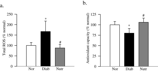
Purpose: Increased oxidative stress and inflammatory mediators are implicated in the development of diabetic retinopathy, and in rats, its development can be prevented by antioxidants. Carotenoids are some of the powerful antioxidants, and diabetes decreases lutein and zeaxanthin levels in the serum and retina. The aim of this study is to investigate the effect of carotenoid containing nutritional supplements (Nutr), which is in clinical trials for 'Diabetes Vision Function', on diabetic retinopathy.

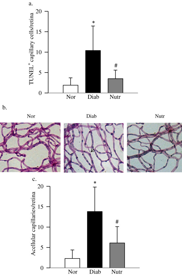
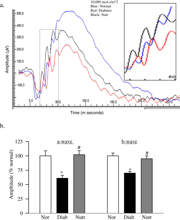
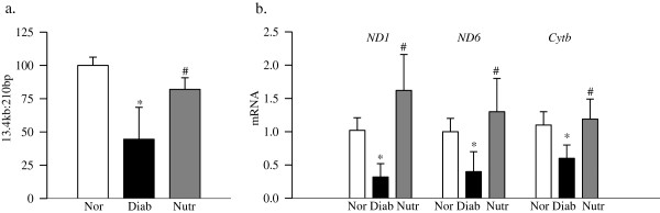
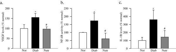
Methods: Streptozotocin-induced diabetic rats (Wistar, male) were fed Purina 5001 supplemented with nutritional supplements containing zeaxanthin, lutein, lipoic acid, omega-3 fatty acids and other nutrients, or without any supplementation. Retinal function was analyzed at ~4 months of diabetes by electroretinography. After 11 months of diabetes, capillary cell apoptosis (TUNEL-staining) and histopathology (degenerative capillaries) were quantified in trypsin-digested retinal vasculature. Retina was also analyzed for mitochondrial damage (by quantifying gene expressions of mtDNA-encoded proteins of the electron transport chain), VEGF and inflammatory mediators, interleukin-1β and NF-kB.

Results: Diabetes impaired retinal function decreasing the amplitudes of both a- and b-waves. In the same animals, retinal capillary cell apoptosis and degenerative capillaries were increased by 3-4 fold. Gene expressions of mtDNA encoded proteins were decreased, and VEGF, interleukin-1β and NF-kB levels were elevated. Supplementation with the nutrients prevented increased capillary cell apoptosis and vascular pathology, and ameliorated these diabetes-induced retinal abnormalities.

Conclusions: Nutritional supplementation prevents diabetic retinopathy, and also maintains normal retinal function, mitochondrial homeostasis and inflammatory mediators. Thus, this supplementation could represent an achievable and inexpensive adjunct therapy to also inhibit retinopathy, a slow progressing disease feared most by diabetic patients.


Figures

Figure 1
Figure 1
Nutrient administration inhibits retinal capillary cell apoptosis and degeneration in diabetic rats. Trypsin digested retinal microvasculature was (a) analyzed for capillary cell apoptosis by TUNEL staining. (b) After TUNEL staining, the microvessels were stained with periodic acid-Schiff-hematoxylin; the arrow indicates a capillary which has lost endothelial cell. (c) The number of acellular capillaries was counted in the entire retinal vasculature, and represented as number of acellular capillaries/retina. Results are expressed as mean ± SD of 7–8 rats each in normal (Nor), diabetic (Diab) and diabetic rats receiving the nutrients (Nutr) groups. *p < 0.05 compared to age-matched normal, and #p < 0.05 compared to diabetes.
Figure 2
Figure 2
Retinal dysfunction is ameliorated by the nutritional supplementation. ERG was performed in dark-adapted rats at ~4 months of diabetes using Ocuscience HMsERG system. A dark-adapted intensity-response series was recorded using a series of Ganzfeld flashes with intensities ranging from 100–25,000 mcd.s/m^2. (a) Representative plots of a- and b-wave amplitudes at 10,000 mcd.s/m^2 are shown. The boxed portion is enlarged to show the delayed ERG response. (b) a-wave and b-wave amplitudes, obtained from normal rats are considered as 100%. The results are representative of 5 or more rats in each of the three groups (normal, diabetes and diabetes + Nutr). Blue = normal rats, Red = diabetic rats and Black = diabetic rats receiving the nutrients. *p < 0.05 compared to age-matched normal, and #p < 0.05 compared to diabetes.
Figure 3
Figure 3
Oxidative stress is ameliorated in diabetic rats receiving the supplementation. (a) Total ROS levels were quantified in the retina (5-10 μg) using 2 μM of DCHFDA and the fluorescence emitted was measured at 485 nm and 530 nm. (b) Total antioxidant capacity of the retina was quantified by measuring the ability of the retina to inhibit oxidation of ABTS by metmyoglobin using 5-10 μg retina protein. Each sample was measured in duplicate, and the values are represented as mean ± SD of 6–8 rats in each group. *p < 0.05 and #p < 0.05 compared to normal and diabetes respectively.
Figure 4
Figure 4
The nutritional supplementation prevents diabetes-induced mitochondrial damage in the retina. (a) Damage of mtDNA was evaluated by amplifying long (13.4 kb) and short (210 bp) regions by extended length PCR and conventional PCR respectively. (b) Gene expressions of ND1, ND6 and Cytb were quantified by real time PCR using β-actin as a housekeeping gene. Measurements were made at least in duplicate, and each value represents the mean ± SD from 6–8 rats/group. Nor = normal, Diab = Diabetes and Nutr = diabetic rats receiving the nutrients supplementation.*p < 0.05 compared to normal and #p < 0.05 compared to diabetes.
Figure 5
Figure 5
Diabetes-induced increase in the retinal inflammatory cytokines is prevented in rats receiving nutritional supplementation. Levels of (a) VEGF, (b) NF-kB and (c) IL-1β were quantified in the retina by using their specific ELISA kits. Each measurement was made in duplicate using retina from 5–6 rats in each of the four groups, and the values are represented as mean ± SD. *p < 0.05 and #p < 0.05 compared to normal and diabetes respectively.

References

    1. Kowluru RA. Mitochondria damage in the pathogenesis of diabetic retinopathy and in the metabolic memory associated with its continued progression. Curr Med Chem. 2013;20:3226–3233. doi: 10.2174/09298673113209990029. - DOI - PMC - PubMed
    1. Kowluru RA, Abbas SN. Diabetes-induced mitochondrial dysfunction in the retina. Inves Ophthalmol Vis Sci. 2003;44:5327–5334. doi: 10.1167/iovs.03-0353. - DOI - PubMed
    1. Kowluru RA, Kennedy A. Therapeutic potential of anti-oxidants and diabetic retinopathy. Expert Opin Investig Drugs. 2001;10:1665–1676. doi: 10.1517/13543784.10.9.1665. - DOI - PubMed
    1. Kowluru RA, Tang J, Kern TS. Abnormalities of retinal metabolism in diabetes and experimental galactosemia. VII. Effect of long-term administration of antioxidants on the development of retinopathy. Diabetes. 2001;50:1938–1942. doi: 10.2337/diabetes.50.8.1938. - DOI - PubMed
    1. Madsen-Bouterse S, Zhong Q, Mohammad G, Ho YS, Kowluru RA. Oxidative damage of mitochondrial DNA in diabetes, and its protection by manganese superoxide dismutase. Free Rad Research. 2010;44:313–321. doi: 10.3109/10715760903494168. - DOI - PMC - PubMed

LinkOut - more resources