

Sequence of the Essex-Lopresti lesion--a high-speed video documentation and kinematic analysis
- PMID: 24479620
- PMCID: PMC3967261
- DOI: 10.3109/17453674.2014.887952
Sequence of the Essex-Lopresti lesion--a high-speed video documentation and kinematic analysis
Abstract
Background and purpose: The pathomechanics of the Essex-Lopresti lesion are not fully understood. We used human cadavers and documented the genesis of the injury with high-speed cameras.
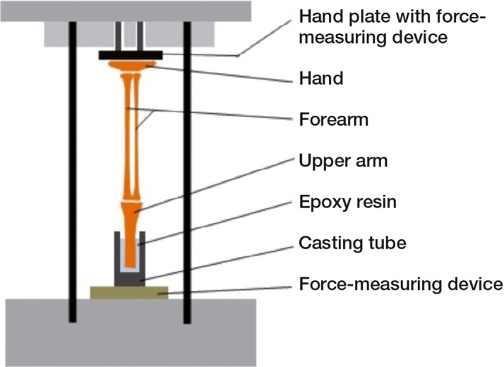
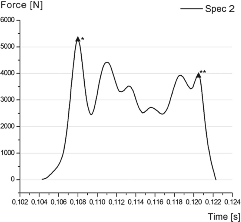
Methods: 4 formalin-fixed cadaveric specimens of human upper extremities were tested in a prototype, custom-made, drop-weight test bench. An axial high-energy impulse was applied and the development of the lesion was documented with 3 high-speed cameras.
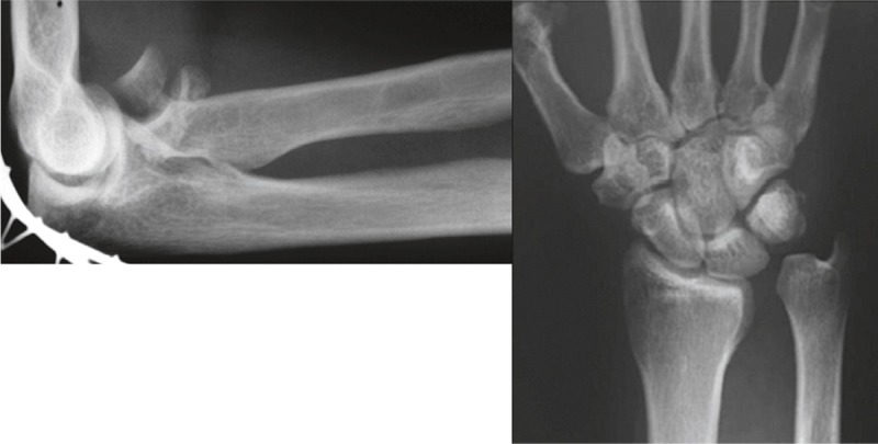
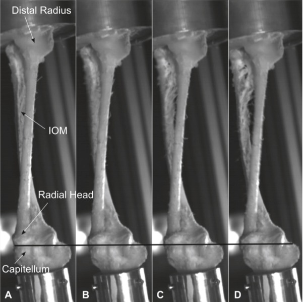
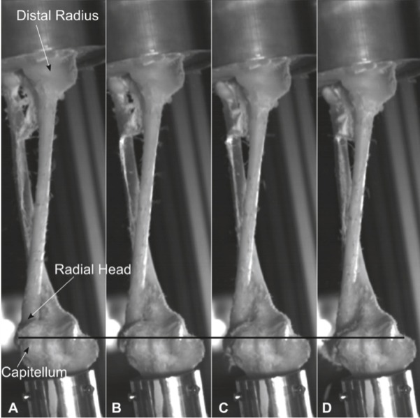
Results: The high-speed images showed a transversal movement of the radius and ulna, which moved away from each other in the transversal plane during the impact. This resulted into a transversal rupture of the interosseous membrane, starting in its central portion, and only then did the radius migrate proximally and fracture. The lesion proceeded to the dislocation of the distal radio-ulnar joint and then to a full-blown Essex-Lopresti lesion.
Interpretation: Our findings indicate that fracture of the radial head may be preceded by at least partial lesions of the interosseous membrane in the course of high-energy axial trauma.
Figures
Comment in
-
Sequence of the Essex-Lopresti lesion--a high-speed video documentation and kinematic analysis.Acta Orthop. 2014 Sep;85(5):545. doi: 10.3109/17453674.2014.949058. Epub 2014 Aug 20. Acta Orthop. 2014. PMID: 25140987 Free PMC article. No abstract available.
-
Author reply: To PMID 24479620.Acta Orthop. 2014 Sep;85(5):545-6. Acta Orthop. 2014. PMID: 25392871 No abstract available.
References
-
- Burkhart KJ, Nowak TE, Blum J, Kuhn S, Welker M, Sternstein W, et al. Influence of formalin fixation on the biomechanical properties of human ... . Biomed Tech (Berl) 2010;55:6, 361–5. - PubMed
-
- Davidson PA, Moseley JB, Jr, Tullos HS. Radial head fracture. A potentially complex injury . Clin Orthop. 1993;297:224–30. - PubMed
-
- Essex-Lopresti P. Fractures of the radial head with distal radio-ulnar dislocation; report... . J Bone Joint Surg (Br) 1951;33:2, 244–7. - PubMed
-
- Götz LP, Schulz R. Komplexe instabile Luxationsfrakturen des Ellenbogens – Klinische Ergebnisse nach Versorgung unter prothetischem Ersatz des Radiusköpfchens. Obere Extremität. 2013;8:1, 22–6.
-
- Green JB, Zelouf DS. Forearm instability. J Hand Surg Am. 2009;34:5, 953–61. - PubMed
MeSH terms
LinkOut - more resources
Full Text Sources
Other Literature Sources
Medical