Review
. 2014 Apr;25(100):57-66.
doi: 10.1016/j.sbi.2013.12.002. Epub 2014 Jan 28.

Structural studies of the spliceosome: zooming into the heart of the machine

Affiliations
Review

Structural studies of the spliceosome: zooming into the heart of the machine

Wojciech P Galej et al. Curr Opin Struct Biol. 2014 Apr.

Abstract

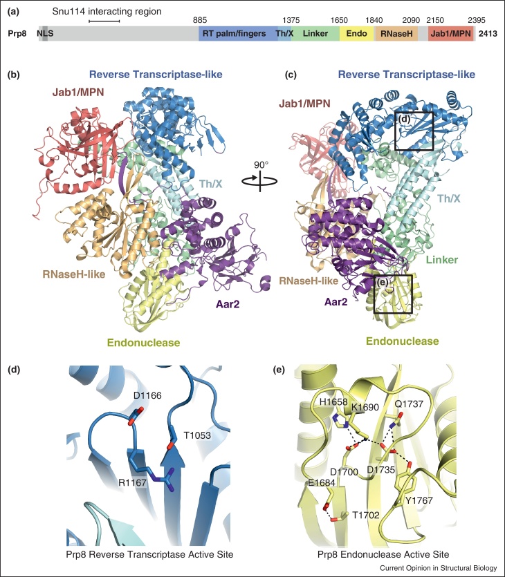
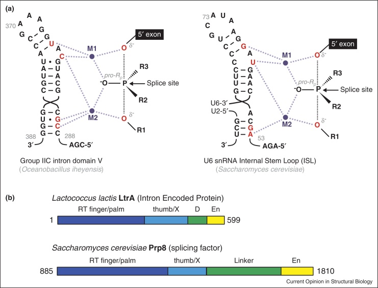
Spliceosomes are large, dynamic ribonucleoprotein complexes that catalyse the removal of introns from messenger RNA precursors via a two-step splicing reaction. The recent crystal structure of Prp8 has revealed Reverse Transcriptase-like, Linker and Endonuclease-like domains. The intron branch-point cross-link with the Linker domain of Prp8 in active spliceosomes and together with suppressors of 5' and 3' splice site mutations this unambiguously locates the active site cavity. Structural and mechanistic similarities with group II self-splicing introns have encouraged the notion that the spliceosome is at heart a ribozyme, and recently the ligands for two catalytic magnesium ions were identified within U6 snRNA. They position catalytic divalent metal ions in the same way as Domain V of group II intron RNA, suggesting that the spliceosome and group II intron use the same catalytic mechanisms.


Figures

Figure 1
Figure 1
Structure of the yeast Prp8-Aar2 complex [9••]. (a) Primary domain architecture of yeast Prp8. (b) Three dimensional arrangement of the six domains of Prp8 and their interactions with Aar2. (c) Orthogonal view of the complex. (d) Impaired active site of the RT domain of Prp8. (e) the active site of Prp8 Endonuclease domain, showing conserved catalytic residues interacting with a polypeptide loop positioned on top of the active site.
Figure 2
Figure 2
The active site cavity of the spliceosome [9••]. Splice site suppressor mutations (red spheres) co-localize mostly to the inner surface of a cavity formed by RNaseH-like and Large domains of Prp8. Branch-point crosslinks (BP + 2 XL) analysed in stalled and affinity purified, yeast step 2 spliceosomes map to a short sequence (1585–1598), part of a disordered loop between residues marked with blue spheres (Norman and Newman, unpublished data). A cross-link between 5′-splice site and the RNaseH-like domain in the human system is marked with green spheres [17]. Positions of suppressor mutations together with site-specific RNA-protein cross-linking sites unambiguously locate the active site cavity of the spliceosome.
Figure 3
Figure 3
Crystal structures of the Brr2 helicase [10,11••,12••]. (a) RNA interaction network in pre-catalytic and activated spliceosomes. Extensively based-paired U4/U6 snRNA duplex undergoes remodelling by the action of Brr2 helicase leading to the formation of the catalytically active secondary structures in the U6 snRNA. Adapted from [2]. (b) Schematic representation of the domain organizations of Brr2. Domains are colour coded: pink, N-terminal extension; light grey, RecA1; dark grey, RecA2; blue: winged-helix (WH); cyan, ratchet; yellow, helix-loop-helix (HLH) and orange, fibronectin3-like (FN3). (c) Overall structure of Brr2:Jab1/MPN complex. The structural domains are coloured as in (a) and labelled with N- and C- to indicate which helicase cassette they belong to.
Figure 4
Figure 4
U5 snRNP biogenesis. (a) Schematic representation of the key stages in yeast U5 snRNP biogenesis. The precursor particle, Aar2-U5 snRNP is transported from the cytoplasm to the nucleus, where after phosphorylation Aar2 is replaced by the helicase Brr2 [55]. (b) Superposition of Jab1/MPN domain of Prp8-Aar2 and Brr2-Jab1/MPN complexes [9••,11••]. (c) Intermolecular beta sheet formed between Aar2 and Jab1/MPN and RNaseH-like domains of Prp8. (d) A steric clash between Brr2 and the RNaseH-like domain of Prp8 reveals molecular basis of the competitive binding of Brr2 and Aar2 to Prp8.
Figure 5
Figure 5
Similarities between components of group II introns and the spliceosomes. (a) Secondary structures and catalytic metal interactions in the domain V of group IIC intron [42] and spliceosomal U6 snRNA Internal Stem Loop (ISL); catalytic magnesium ions M1 and M2 are coordinated by RNA nucleotides labeled in red. For the first step reaction, R1 represents 2′ hydroxyl of branch point adenosine (or equivalent water molecule in hydrolytic group IIC intron); R2, the intron; R3, the pro-Sp oxygen. For the second step exon ligation reaction, R1 represents 3′ oxygen leaving group; R2 the pro-Sp oxygen; R3 the 3′ exon. Adapted from [37••]. (b) Comparison of the domain architecture of Prp8 and Intron Encoded Protein reveals a previously unknown evolutionary link between the two systems. RT, reverse transcriptase domain; En, endonuclease domain; D, DNA binding domain.

References

    1. Wahl M.C., Will C.L., Lührmann R. The spliceosome: design principles of a dynamic RNP machine. Cell. 2009;136:701–718. - PubMed
    1. Will C.L., Lührmann R. Spliceosome structure and function. Cold Spring Harb Perspect Biol. 2011;3:1–23. - PMC - PubMed
    1. Cordin O., Hahn D., Beggs J.D. Structure, function and regulation of spliceosomal RNA helicases. Curr Opin Cell Biol. 2012;24:431–438. - PubMed
    1. Newman A.J., Nagai K. Structural studies of the spliceosome: blind men and an elephant. Curr Opin Struct Biol. 2010;20:82–89. - PubMed
    1. Lührmann R., Stark H. Structural mapping of spliceosomes by electron microscopy. Curr Opin Struct Biol. 2009;19:96–102. - PubMed

Publication types

MeSH terms

Substances