

Imaging of adipose tissue
- PMID: 24480341
- PMCID: PMC4272855
- DOI: 10.1016/B978-0-12-411619-1.00004-5
Imaging of adipose tissue
Abstract
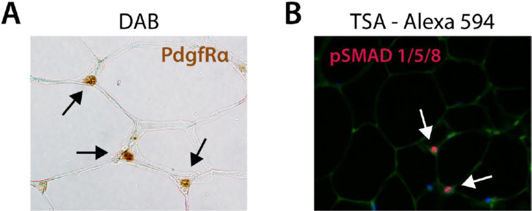
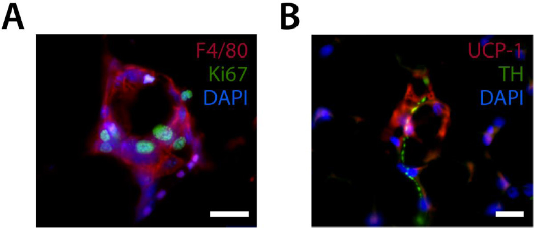
Adipose tissue is an endocrine organ that specializes in lipid metabolism and is distributed throughout the body in distinct white adipose tissue (WAT) and brown adipose tissue (BAT) depots. These tissues have opposing roles in lipid metabolism with WAT storing excessive caloric intake in the form of lipid, and BAT burning lipid through nonshivering thermogenesis. As accumulation of lipid in mature adipocytes of WAT leads to obesity and increased risk of comorbidity (Pi-Sunyer et al., 1998), detailed understanding of the mechanisms of BAT activation and WAT accumulation could produce therapeutic strategies for combatting metabolic pathologies. As morphological changes accompany alterations in adipose function, imaging of adipose tissue is one of the most important tools for understanding how adipose tissue mass fluctuates in response to various physiological contexts. Therefore, this chapter details several methods of processing and imaging adipose tissue, including bright-field colorimetric imaging of paraffin-sectioned adipose tissue with a detailed protocol for automated adipocyte size analysis; fluorescent imaging of paraffin and frozen-sectioned adipose tissue; and confocal fluorescent microscopy of whole mounted adipose tissue. We have also provided many example images showing results produced using each protocol, as well as commentary on the strengths and limitations of each approach.
Keywords: Adipose; Cell profiler; Confocal; Frozen; Lineage tracing; Paraffin; Whole mount.
© 2014 Elsevier Inc. All rights reserved.
Figures
Similar articles
-
Chronic l-menthol-induced browning of white adipose tissue hypothesis: A putative therapeutic regime for combating obesity and improving metabolic health.Med Hypotheses. 2016 Aug;93:21-6. doi: 10.1016/j.mehy.2016.05.006. Epub 2016 May 11. Med Hypotheses. 2016. PMID: 27372851
-
Overexpression of Adiponectin Receptor 1 Inhibits Brown and Beige Adipose Tissue Activity in Mice.Int J Mol Sci. 2021 Jan 18;22(2):906. doi: 10.3390/ijms22020906. Int J Mol Sci. 2021. PMID: 33477525 Free PMC article.
-
GQ-16, a TZD-Derived Partial PPARγ Agonist, Induces the Expression of Thermogenesis-Related Genes in Brown Fat and Visceral White Fat and Decreases Visceral Adiposity in Obese and Hyperglycemic Mice.PLoS One. 2016 May 3;11(5):e0154310. doi: 10.1371/journal.pone.0154310. eCollection 2016. PLoS One. 2016. PMID: 27138164 Free PMC article.
-
White, brown, beige/brite: different adipose cells for different functions?Endocrinology. 2013 Sep;154(9):2992-3000. doi: 10.1210/en.2013-1403. Epub 2013 Jun 19. Endocrinology. 2013. PMID: 23782940 Review.
-
Do estrogens enhance activation of brown and beiging of adipose tissues?Physiol Behav. 2018 Apr 1;187:24-31. doi: 10.1016/j.physbeh.2017.09.026. Epub 2017 Oct 6. Physiol Behav. 2018. PMID: 28988965 Review.
Cited by
-
Reflection mode polarimetry guides laser mass spectrometry to diagnostically important regions of human breast cancer tissue.Sci Rep. 2024 Oct 31;14(1):26230. doi: 10.1038/s41598-024-77963-w. Sci Rep. 2024. PMID: 39482347 Free PMC article.
-
Genetic Deletion of Syndecan-4 Alters Body Composition, Metabolic Phenotypes, and the Function of Metabolic Tissues in Female Mice Fed A High-Fat Diet.Nutrients. 2019 Nov 18;11(11):2810. doi: 10.3390/nu11112810. Nutrients. 2019. PMID: 31752080 Free PMC article.
-
Adipocyte PU.1 knockout promotes insulin sensitivity in HFD-fed obese mice.Sci Rep. 2019 Oct 14;9(1):14779. doi: 10.1038/s41598-019-51196-8. Sci Rep. 2019. PMID: 31611602 Free PMC article.
-
Mature white adipocyte plasticity during mammary gland remodelling and cancer.Cell Insight. 2023 Aug 20;2(5):100123. doi: 10.1016/j.cellin.2023.100123. eCollection 2023 Oct. Cell Insight. 2023. PMID: 37771567 Free PMC article. Review.
-
"The ubiquitin ligase SIAH2 is a female-specific regulator of circadian rhythms and metabolism".PLoS Genet. 2022 Jul 5;18(7):e1010305. doi: 10.1371/journal.pgen.1010305. eCollection 2022 Jul. PLoS Genet. 2022. PMID: 35789210 Free PMC article.
References
-
- CellProfiler cell image analysis software. 2013 from http://www.cellprofiler.org/
-
- Cinti S, Zingaretti MC, Cancello R, Ceresi E, Ferrara P. Morphologic techniques for the study of brown adipose tissue and white adipose tissue. Methods Mol Biol. 2001;155:21–51. - PubMed
Publication types
MeSH terms
Grants and funding
LinkOut - more resources
Full Text Sources
Other Literature Sources
Medical
Research Materials
Miscellaneous
