. 2014 Aug;35(8):1717-25.
doi: 10.1093/carcin/bgu025. Epub 2014 Jan 30.

SHP2E76K mutant promotes lung tumorigenesis in transgenic mice

Affiliations

SHP2E76K mutant promotes lung tumorigenesis in transgenic mice

Valentina E Schneeberger et al. Carcinogenesis. 2014 Aug.

Abstract

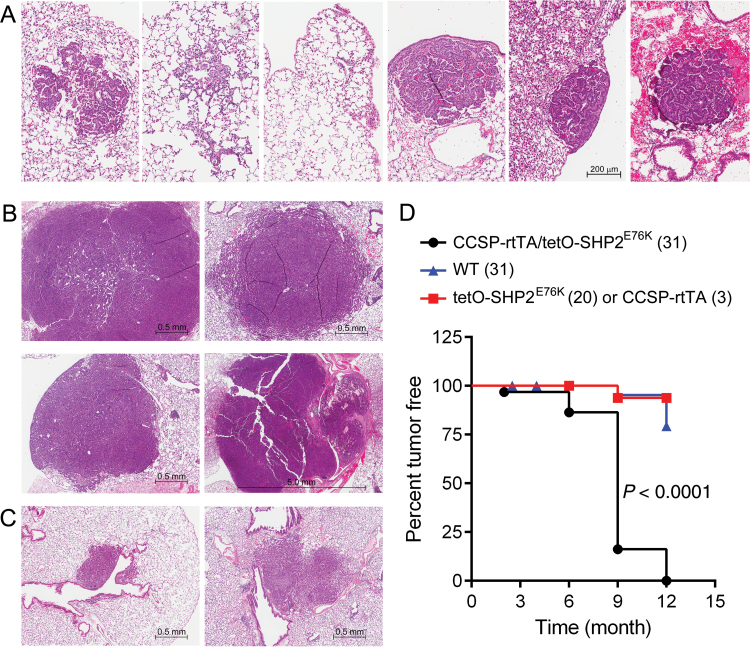
Lung cancer is a major disease carrying heterogeneous molecular lesions and many of them remain to be analyzed functionally in vivo. Gain-of-function (GOF) SHP2 (PTPN11) mutations have been found in various types of human cancer, including lung cancer. However, the role of activating SHP2 mutants in lung cancer has not been established. We generated transgenic mice containing a doxycycline (Dox)-inducible activating SHP2 mutant (tetO-SHP2(E76K)) and analyzed the role of SHP2(E76K) in lung tumorigenesis in the Clara cell secretory protein (CCSP)-reverse tetracycline transactivator (rtTA)/tetO-SHP2(E76K) bitransgenic mice. SHP2(E76K) activated Erk1/Erk2 (Erk1/2) and Src, and upregulated c-Myc and Mdm2 in the lungs of bitransgenic mice. Atypical adenomatous hyperplasia and small adenomas were observed in CCSP-rtTA/tetO-SHP2(E76K) bitransgenic mice induced with Dox for 2-6 months and progressed to larger adenoma and adenocarcinoma by 9 months. Dox withdrawal from bitransgenic mice bearing magnetic resonance imaging-detectable lung tumors resulted in tumor regression. These results show that the activating SHP2 mutant promotes lung tumorigenesis and that the SHP2 mutant is required for tumor maintenance in this mouse model of non-small cell lung cancer. SHP2(E76K) was associated with Gab1 in the lung of transgenic mice. Elevated pGab1 was observed in the lung of Dox-induced CCSP-rtTA/tetO-SHP2(E76K) mice and in cell lines expressing SHP2(E76K), indicating that the activating SHP2 mutant autoregulates tyrosine phosphorylation of its own docking protein. Gab1 tyrosine phosphorylation is sensitive to inhibition by the Src inhibitor dasatinib in GOF SHP2-mutant-expressing cells, suggesting that Src family kinases are involved in SHP2 mutant-induced Gab1 tyrosine phosphorylation.


Figures

Fig. 1.
Fig. 1.
The tetO-SHP2E76K transgenic construct. (A) L3/L2-tetO transgenic vector. 3 and 2 indicate L3 and L2 loxP sequences. cHS4 represents chicken β-globin insulator sequence. (B) The tetO-SHP2E76K transgene. Complementary DNA encoding human SHP2E76K with a C-terminal Flag-tag (29) was inserted into the EcoRV site of the L3/L2-tetO vector. The tetO-SHP2E76K transgene can be induced in the mouse lung type II epithelial cells by in CCSP-rtTA/tetO-SHP2E76K bitransgenic mice by Dox. Dash box, Flag-tag coding sequence.
Fig. 2.
Fig. 2.
SHP2E76K expression and signaling in transgenic mice. (A) Upper panels: RT–PCR assessment of SHP2E76K mRNA expression in various tissues of tetO-SHP2E76K transgenic mice lines 398 and 425. Wt, wild-type mouse lung as a negative control; Lu, lung; Li, liver; Kd, kidney; Co, colon. +, positive control of human SHP2 mRNA from HCC827 cells; −, negative control in which no mRNA was included. Lower panels: tissue lysates were immunoprecipitated with an anti-Flag antibody (M2) and the immunoprecipitates were analyzed by immunoblotting with another anti-Flag antibody (rabbit). +, cell lysate of TF-1/SHP2E76K cells (29); −, no cell lysates. The same data were obtained from repeated experiments. (B) Upper panel: CCSP-rtTA/tetO-SHP2E76K bitransgenic (B) or wild-type (Wt) mice were fed with Dox diet for 1 month. tetO-SHP2E76K mRNA expression in the lung was determined by RT–PCR as in (A). M, DNA molecular weight marker. Lower panel: lung tissues from bitransgenic or wild-type mice as in the upper panel were subjected to immunoprecipitation-immunoblotting analysis of SHP2E76K expression using anti-Flag antibodies. Similar data were obtained from additional experiments. (C) Comparison of signaling proteins in the lungs of transgenic mice. Wild-type (W), monotransgenic (M) or CCSP-rtTA/tetO-SHP2E76K bitransgenic (B) mice were treated with Dox for 1 month. Lung tissue lysates were analyzed by immunoblotting with the indicated antibodies. Similar data were obtained from repeated experiments. (D) Mdm2 quantitative RT–PCR. In each experiment, lung tissues from two animals in each group were assayed in triplicates and the experiment was repeated (a total of four animals in each group). The average Ct values were 27.5 and 25.8, respectively, for samples from the wild-type and Dox-induced CCSP-rtTA/tetO-SHP2E76K mice. Statistically analysis was performed using the non-parametric Mann–Whitney test.
Fig. 3.
Fig. 3.
Histology of lung proliferative lesions and tumor incidence in animals. (A) Proliferative lesions in the lung of CCSP-rtTA/tetO-SHP2E76K bitransgenic mice 6 months after Dox induction. Images (magnification: ×200) of H&E stained sections of lungs from CCSP-rtTA/tetO-SHP2E76K bitransgenic mice at 6 months after Dox induction. Hyperplasia (left 3 panels) and adenoma (right 3 panels) are shown. (B and C) Lung tumors 9 months after Dox treatment. (B) Examples of lung adenoma and adenocarcinoma in CCSP-rtTA/tetO-SHP2E76K bitransgenic mice 9 months after Dox induction (magnification: ×100 or ×40). (C) The only two adenomas found among 13 control monotransgenic (left) and wild-type (right) mice after 9 months Dox treatment (magnification: ×100). (D) Kaplan–Meier tumor-free survival curves of animals. The numbers inside parentheses in the graph legends indicate the total numbers of animals in each group. Statistical comparisons of bitransgenic versus wild-type and bitransgenic versus monotransgenic mice were performed using the Log rank test and both yielded P < 0.0001.
Fig. 4.
Fig. 4.
Lung tumors in CCSP-rtTA/tetO-SHP2E76K mice regress after Dox withdrawal. (A) 3D FSE datasets (TE/TR = 64/1000ms) demonstrating coronal sections of tumor-bearing mice before and 1 month after Dox withdrawal, as indicated. The tumor sizes were 27.2 (mouse #1) and 22.3mm3 (mouse #2) prior to Dox withdrawal. Arrows in panel indicate the positions of tumors or where tumors were detected prior to Dox withdrawal. (B) H&E sections of lung tissue corresponding to where tumors were detected by MRI. Residual atypical adenomatous hyperplasia and scar tissues are indicated by arrows. (C) Lung tissues from Dox withdrawn mice were analyzed by RT–PCR (left) or immunoprecipitation-immunoblotting (right) to verify the absence of SHP2E76K mRNA or protein following deinduction. (D) Immunohistochemical analysis of pErk1/2 in mouse lung tissues. Slides were processed under identical conditions in the same experiment using a Ventana Discovery XT automated system.
Fig. 5.
Fig. 5.
SHP2E76K autoregulates Gab1 tyrosine phosphorylation. (A) Lung tissue from a Dox-induced CCSP-rtTA/tetO-SHP2E76K mouse was immunoprecipitated with an anti-Flag (M2) antibody. Immunoprecipitated proteins were eluted from the Protein-G agarose with a Flag peptide. One-tenth of the eluted immunoprecipitate was used for immunoblotting with an anti-pY antibody. The rest of eluted immunoprecitate was processed for mass spectrometric identification of proteins from corresponding slides of Coomassie blue-stained gel. Major proteins (excluding keratins) identified in each band were searched against PhosphoSitePlus (www.phosphosite.org) database and those that have been reported as tyrosine-phosphorylated proteins are shown. (B) Lung tissue from a Dox-induced CCSP-rtTA/tetO-SHP2E76K mouse was immunoprecipitated with an anti-Gab1 antibody. The immunoprecipitate was analyzed by immunoblotting with antibodies to pGab1 (Y627) and Flag-tag. After removal of antibodies, the membranes were re-probed with antibodies to Gab1 and SHP2. (C) Immunoblot analyses of lung tissue lysates from the wild-type (W), Dox-induced CCSP-rtTA/tetO-SHP2E76K (P), or after Dox withdrawal of CCSP-rtTA/tetO-SHP2E76K mouse with MRI-detected tumor (A). (D) Left panels, Gab1 was immunoprecipitated from cytokine-starved TF-1 cells containing control vector (V), wild-type SHP2 (W) or SHP2E76K (K). The immunoprecipitates were analyzed by immunoblotting with antibodies to pY or SHP2. Right panels, LYN was immunoprecipitated and its tyrosine kinase activity was assayed using a glutathione S-transferase-GAB1 fusion protein (12) as the substrate. (E) H292 cells expressing a control vector (V), wild-type SHP2or SHP2E76K (K) were analyzed by immunoblotting with indicated antibodies. Note that the anti-pSRC antibody cross-reacts with other SFKs. (F) H292/SHP2E76K cells were treated with indicated concentrations of ruxolitinib, dasatinib or erlotinib for 24h. Cell lysates were analyzed for pGAB1 by immunoblotting. (G) H661 cells were treated with dasatinib for 24h. Gab1 was immunoprecipitated from cell lysates and the immunoprecipitates were analyzed by immunoblotting with indicated antibodies (upper panels). Cell lysates were analyzed by immunoblotting as indicated (lower panels). (H) H292/SHP2E76K or H661 cells were transfected with non-targeting (NT), LYN or c-SRC (SRC) siRNAs or left untransfected (N). Cell lysates were prepared and analyzed by immunoblotting with indicated antibodies.

Similar articles

Cited by

References

    1. Tonks N.K. (2006). Protein tyrosine phosphatases: from genes, to function, to disease. Nat. Rev. Mol. Cell Biol., 7, 833–846 - PubMed
    1. Julien S.G., et al. (2011). Inside the human cancer tyrosine phosphatome. Nat. Rev. Cancer, 11, 35–49 - PubMed
    1. Scott L.M., et al. (2010). Targeting protein tyrosine phosphatases for anticancer drug discovery. Curr. Pharm. Des., 16, 1843–1862 - PMC - PubMed
    1. Xu W., et al. (1999). Crystal structures of c-Src reveal features of its autoinhibitory mechanism. Mol. Cell, 3, 629–638 - PubMed
    1. Enders G.H. (2010). Gauchos and ochos: a Wee1-Cdk tango regulating mitotic entry. Cell Div., 5, 12. - PMC - PubMed

Publication types

MeSH terms

Substances