. 2014 Aug;28(8):1627-35.
doi: 10.1038/leu.2014.52. Epub 2014 Jan 31.

Loss of wild-type Jak2 allele enhances myeloid cell expansion and accelerates myelofibrosis in Jak2V617F knock-in mice

Affiliations

Loss of wild-type Jak2 allele enhances myeloid cell expansion and accelerates myelofibrosis in Jak2V617F knock-in mice

H Akada et al. Leukemia. 2014 Aug.

Abstract

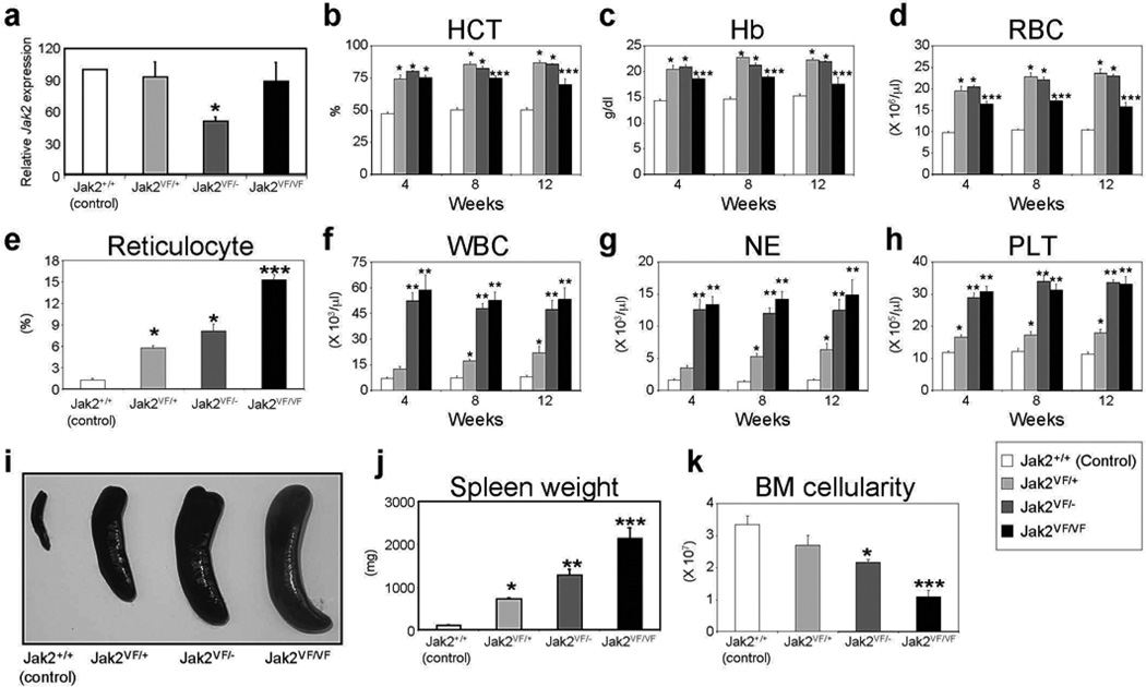
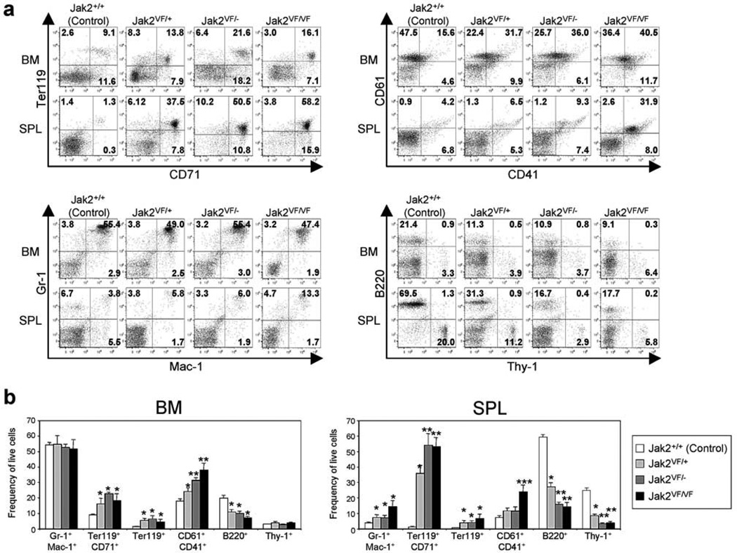
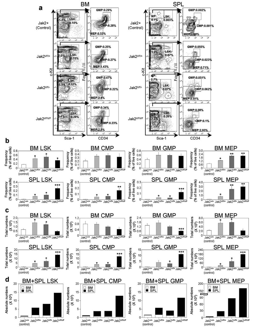
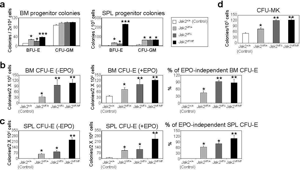
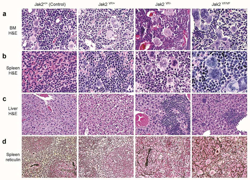
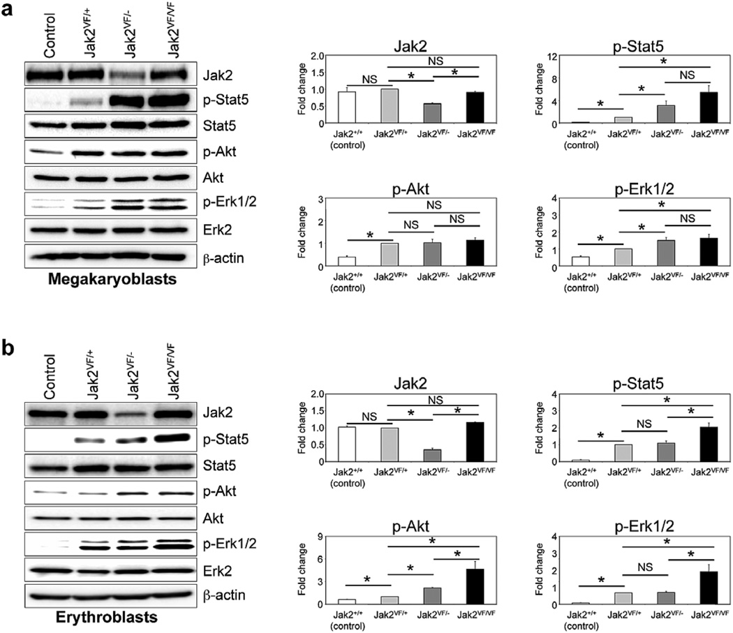
JAK2V617F is the most common mutation found in Philadelphia chromosome-negative myeloproliferative neoplasms (MPNs). Although a majority of MPN patients carry heterozygous JAK2V617F mutation, loss of heterozygosity (LOH) on chromosome 9p (9pLOH) involving the JAK2 locus has been observed in ∼30% of MPN patients. JAK2V617F homozygosity via 9pLOH has been associated with more severe MPN phenotype. However, the contribution of 9pLOH in the pathogenesis of MPNs remains unclear. To investigate the roles of wild-type JAK2 (JAK2 WT) and JAK2V617F alleles in the development of MPNs, we have used conditional Jak2 knock-out and Jak2V617F knock-in mice and generated heterozygous, hemizygous and homozygous Jak2V617F mice. Whereas heterozygous Jak2V617F expression results in a polycythemia vera-like MPN in mice, loss of Jak2 WT allele in hemizygous or homozygous Jak2V617F mice results in markedly increased white blood cells, neutrophils, reticulocytes and platelets in the peripheral blood, and significantly larger spleen size compared with heterozygous Jak2V617F mice. Hemizygous or homozygous Jak2V617F mice also exhibit accelerated myelofibrosis compared with mice expressing heterozygous Jak2V617F. Together, these results suggest that loss of Jak2 WT allele increases the severity of the MPN. Thus, the Jak2 WT allele functions as a negative regulator of MPN induced by Jak2V617F.


Conflict of interest statement

CONFLICT OF INTEREST

The authors declare no conflict of interest.

Figures

Figure 1
Figure 1. Deletion of Jak2 WT allele increases the severity of MPN in Jak2V617F knock-in mice
(a) Quantitative real-time PCR analysis of total Jak2 expression in the BM of control (Jak2+/+), heterozygous (Jak2VF/+), hemizygous (Jak2VF/−) or homozygous (Jak2VF/VF) mice. Peripheral blood hematocrit (b), hemoglobin (c), RBC (d), reticulocyte (e), WBC (f), neutrophil (g) and platelet (h) counts were assessed at 4, 8, and 12 weeks after pI-pC induction in control (Jak2+/+), heterozygous (Jak2VF/+), hemizygous (Jak2VF/−) or homozygous (Jak2VF/VF) mice; (n = 10). Spleen size (i) and weight (j) were significantly increased in mice expressing heterozygous Jak2V617F. Deletion of Jak2 WT allele further enhanced splenomegaly in hemizygous (Jak2VF/−) or homozygous (Jak2VF/VF) mice (n = 10). (k) BM cellularity (total BM cell count in femur and tibia) was significantly reduced in hemizygous and homozygous Jak2V617F mice at 12 weeks after pI-pC induction. Asterisks indicate significant differences (p < 0.05) (* indicates significance when compared with controls; ** indicates significance when compared with controls and heterozygous Jak2VF/+; *** indicates significance when compared with controls, heterozygous Jak2VF/+ and hemizygous Jak2VF/−).
Figure 2
Figure 2. Flow cytometric analysis of BM and spleens from heterozygous, hemizygous and homozygous Jak2V617F knock-in mice
(a) Dot plots demonstrate significant increase in erythroid (Ter119+/CD71+) and megakaryocytic (CD61+/CD41+) precursors in the BM and/or spleens of heterozygous (Jak2VF/+), hemizygous (Jak2VF/−) and homozygous (Jak2VF/VF) mice compared with control (Jak2+/+) animals. (b) Percentages of granulocyte/monocyte (Gr-1+/Mac-1+), erythroid (Ter119+/CD71+) and megakaryocytic (CD61+/CD41+) precursors as well as B-cells (B220+) and T-cells (Thy-1+) are shown in bar graphs as mean ± SEM. Data from five independent experiments are presented. Asterisks indicate significant differences (p < 0.05) (* indicates significance when compared with controls; ** indicates significance when compared with controls and heterozygous Jak2VF/+; *** indicates significance when compared with controls, heterozygous Jak2VF/+ and hemizygous Jak2VF/−). Note that deletion of Jak2 WT allele results in further expansion of megakaryocytic (CD61+/CD41+) and erythroid (Ter119+/CD71+) precursors in the BM and/or spleens of hemizygous and homozygous Jak2V617F mice.
Figure 3
Figure 3. Deletion of Jak2 WT increases megakaryocyte-erythroid progenitor (MEP) compartment in Jak2V617F knock-in mice
(a) Flow cytometric analysis of LSK (LinSca-1+c-Kit+) and subsets of myeloid progenitors including CMP (LinSca-1c-Kit+CD34+FcγRII/IIIlo), GMP (LinSca-1c-Kit+CD34+FcγRII/IIIhigh), and MEP (LinSca-1c-Kit+CD34FcγRII/III) in the BM and spleens from control (Jak2+/+), heterozygous (Jak2VF/+), hemizygous (Jak2VF/−) or homozygous (Jak2VF/VF) Jak2V617F mice. Representative contour plots from five independent experiments are shown. (b) Percentages of LSK, CMP, GMP, and MEP are shown in histograms as mean ± SEM. Data is presented as percentage of live cells. (c) Total numbers of LSK, CMP, GMP and MEP in BM (femur plus tibia) or whole spleens are shown in histograms as mean ± SEM. (d) Absolute numbers (BM plus spleens) of LSK, CMP, GMP and MEP are shown in histograms as mean ± SEM. Asterisks indicate significant differences (p < 0.05) (* indicates significance when compared with controls; ** indicates significance when compared with controls and heterozygous Jak2VF/+; *** indicates significance when compared with controls, heterozygous Jak2VF/+ and hemizygous Jak2VF/−). Note that deletion of Jak2 WT allele increases absolute numbers of LSK and MEP in hemizygous and homozygous Jak2V617F mice.
Figure 4
Figure 4. Deletion of Jak2 WT increases hematopoietic progenitor colony formation in Jak2V617F mice
(a) BM and spleen cells from control (Jak2+/+), heterozygous (Jak2VF/+), hemizygous (Jak2VF/−) or homozygous (Jak2VF/VF) mice (n = 4) were plated in complete methylcellulose medium (Methocult M3434) in presence of cytokine cocktail. BFU-E and CFU-GM colonies were scored on day 7. BM (b) or spleen (c) cells from control, heterozygous, hemizygous and homozygous Jak2V617F mice (n = 4) were plated in methylcellulose media (Methocult M3234) in the absence of cytokine or in the presence of EPO (3 U/ml). CFU-E colonies were scored after 2 days. Percentages of EPO-independent CFU-E colonies in the BM and spleens are shown in the right panels. (d) BM cells were plated in collagen-based (MegaCult-C) media and CFU-Mk colonies were scored 8 days after plating. Asterisks indicate significant differences (p < 0.05) (* indicates significance when compared with controls; ** indicates significance when compared with controls and heterozygous Jak2VF/+; *** indicates significance when compared with controls, heterozygous Jak2VF/+ and hemizygous Jak2VF/−). Notably, deletion of Jak2 WT allele increases erythroid (CFU-E) and megakaryocytic (CFU-Mk) colonies in hemizygous and homozygous Jak2V617F mice.
Figure 5
Figure 5. Histopathologic analysis of heterozygous, hemizygous and homozygous Jak2V617F knock-in mice
(a) Hematoxylin and eosin (H&E) staining of the BM sections (500X) from heterozygous (Jak2VF/+), hemizygous (Jak2VF/−) and homozygous (Jak2VF/VF) mice show trilineage hyperplasia. Jak2VF/− and Jak2VF/VF mice BM exhibit significantly more megakaryocytes with atypical nuclear features compared with heterozygous (Jak2VF/+) mice BM. (b) Spleen sections (H&E staining; 500X) from heterozygous, hemizygous, and homozygous Jak2V617F mice exhibit destruction of normal splenic architecture with attenuated white pulp and markedly expanded red pulp, increased numbers of megakaryocytes, and clusters of immature erythroid and granulocyte precursors. Spleens from hemizygous or homozygous Jak2V617F mice show increased numbers of abnormal megakaryocytes with enlarged nuclei. (c) Liver sections (H&E staining; 200X) from hemizygous, and homozygous Jak2V617F mice display infiltration of myeloid precursors. (d) Reticulin staining of the spleen sections (200X) from hemizygous and homozygous Jak2V617F mice show extensive reticulin fibrosis in both red and white pulp at 12 weeks after pI-pC induction. Heterozygous Jak2V617F mice spleens exhibit very slight myelofibrosis at this stage (12 weeks after pI-pC induction).
Figure 6
Figure 6. Deletion of Jak2 WT enhances constitutive signaling in primary hematopoietic cells expressing Jak2V617F
Primary megakaryoblasts (a) and erythroblasts (b) were derived from the BM of control, heterozygous (Jak2VF/+), hemizygous (Jak2VF/−) and homozygous (Jak2VF/VF) Jak2V617F mice. Cells were serum and factor depleted for 6 hours and lysed in RIPA buffer. Immunoblotting was performed using phospho-specific antibodies against Stat5, Akt and Erk1/2. Membranes were re-probed with respective total antibodies. Total Jak2 protein levels were also determined by immunoblotting using anti-Jak2 antibody. β-actin was used as a loading control. Histograms (in the right panels) demonstrate the fold changes in phosphorylation of Stat5, Akt and Erk1/2 when compared to the phosphorylation levels of those proteins in heterozygous Jak2V617F-expressing megakaryoblasts or erythroblasts in the absence of cytokines. Data from three to four independent experiments are shown in histograms as mean ± SEM. Asterisks indicate significant differences (p < 0.05). Notably, deletion of Jak2 WT allele enhanced constitutive phosphorylation/activation of Stat5, Erk1/2 and/or Akt in primary hematopoietic cells from hemizygous and homozygous Jak2V617F mice.

References

    1. James C, Ugo V, Le Couédic JP, Staerk J, Delhommeau F, Lacout C, et al. A unique clonal JAK2 mutation leading to constitutive signaling causes polycythemia vera. Nature. 2005;434:1144–1148. - PubMed
    1. Levine RL, Wadleigh M, cools J, Ebert BL, Wernig G, Huntly BJ, et al. Activating mutation in the tyrosine kinase JAK2 in polycythemia vera, essential thrombocythemia, and myeloid metaplasia with myelofibrosis. Cancer Cell. 2005;7:387–397. - PubMed
    1. Baxter EJ, Scott LM, Campbell PJ, East C, Fourouclas N, Swanton S, et al. Acquired mutation of the tyrosine kinase JAK2 in human myeloproliferative disorders. Lancet. 2005;365:1054–1061. - PubMed
    1. Kralovics R, Passamonti F, Buser AS, Teo SS, Tiedt R, Passweg JR, et al. A gain-of-function mutation of JAK2 in myeloproliferative disorders. N. Eng. J. Med. 2005;352:1779–1790. - PubMed
    1. Zhao R, Xing S, Li Z, Fu X, Li Q, Krantz SB, et al. Identification of an Acquired JAK2 Mutation in Polycythemia Vera. J. Biol. Chem. 2005;280:22788–22792. - PMC - PubMed

Publication types