Review
. 2014 Jan;26(1):21-37.
doi: 10.1105/tpc.113.119446. Epub 2014 Jan 30.

The UV-B photoreceptor UVR8: from structure to physiology

Affiliations
Review

The UV-B photoreceptor UVR8: from structure to physiology

Gareth I Jenkins. Plant Cell. 2014 Jan.

Abstract

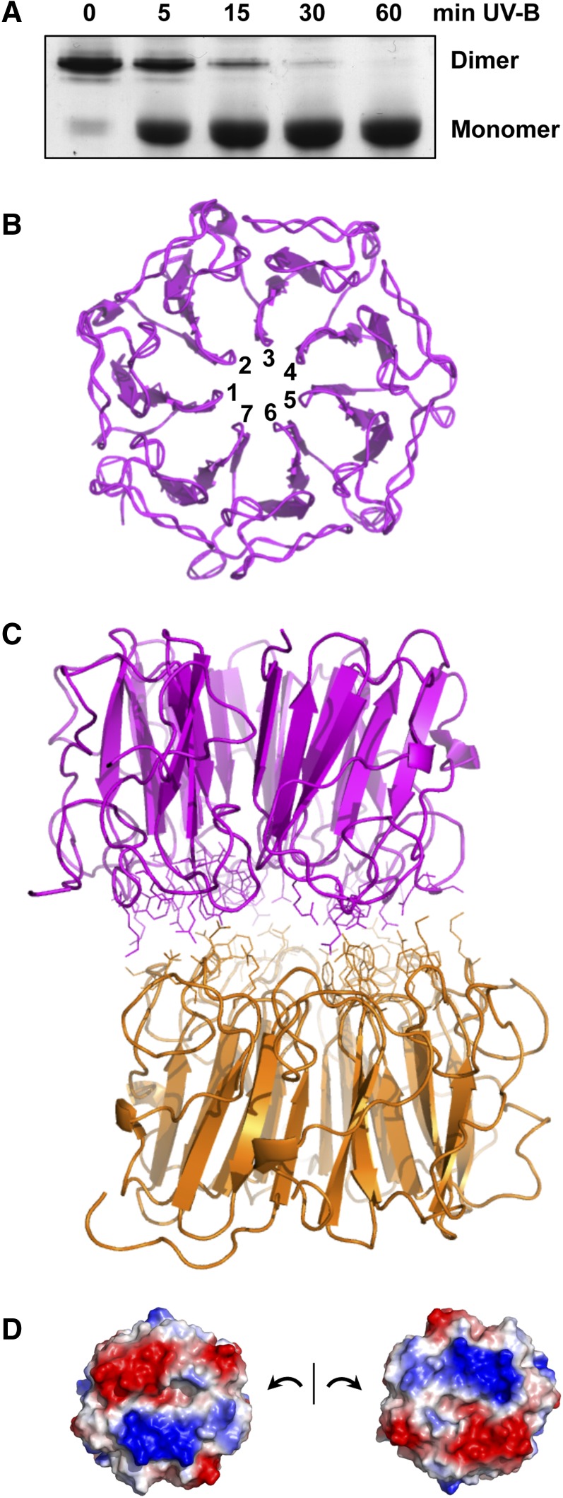
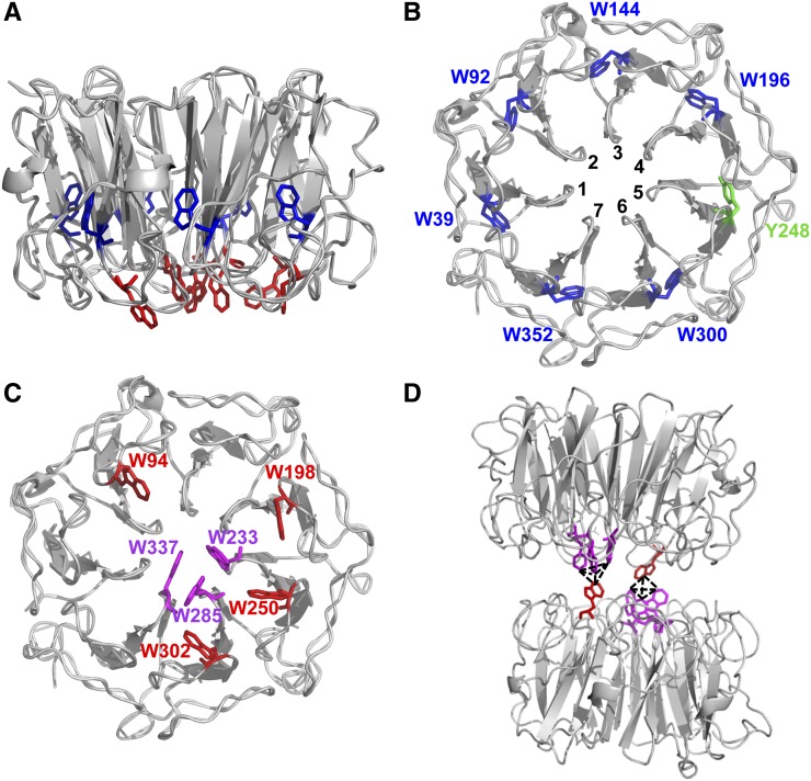
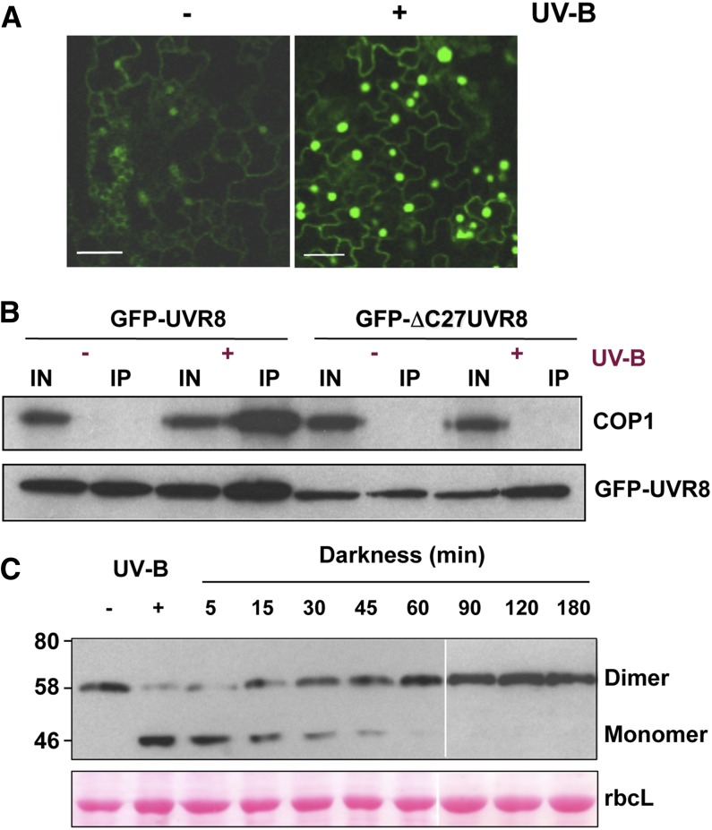
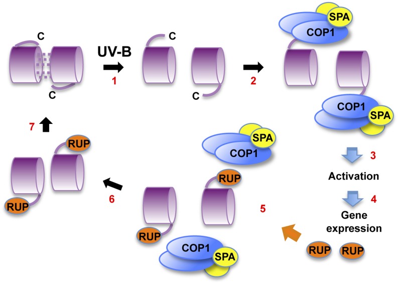
Low doses of UV-B light (280 to 315 nm) elicit photomorphogenic responses in plants that modify biochemical composition, photosynthetic competence, morphogenesis, and defense. UV RESISTANCE LOCUS8 (UVR8) mediates photomorphogenic responses to UV-B by regulating transcription of a set of target genes. UVR8 differs from other known photoreceptors in that it uses specific Trp amino acids instead of a prosthetic chromophore for light absorption during UV-B photoreception. Absorption of UV-B dissociates the UVR8 dimer into monomers, initiating signal transduction through interaction with CONSTITUTIVELY PHOTOMORPHOGENIC1. However, much remains to be learned about the physiological role of UVR8 and its interaction with other signaling pathways, the molecular mechanism of UVR8 photoreception, how the UVR8 protein initiates signaling, how it is regulated, and how UVR8 regulates transcription of its target genes.


Figures

Figure 1.
Figure 1.
UVR8 Dimer Structure and Monomerization. (A) UV-B induces monomerization of UVR8. Coomassie blue–stained SDS-PAGE gel of purified UVR8 exposed for the times shown to 1.5 μmol m−2 s−1 narrowband UV-B (λmax 311 nm). Samples were prepared for electrophoresis without boiling. The UVR8 dimer and monomer are indicated. (B) Seven-bladed β-propeller structure of the UVR8 monomer. The structure is shown for amino acids 14 to 380. (C) Structure of the UVR8 dimer showing residues at the dimer interaction surface. (D) The dimer interaction surfaces of two UVR8 monomers displayed to show patches of complementary electrostatic potential. Basic (blue) and acidic (red) amino acids contribute positive and negative charges, respectively. Images in (B) to (D) were produced using PyMOL. (All panels produced from data presented by Christie et al. [2012].)
Figure 2.
Figure 2.
UVR8 Has Distinct Groups of Trps. (A) The arrangement of all UVR8 Trps (except W400) in the monomer viewed from the side. Trps in the protein core and at the dimer interaction surface are shown in blue and red, respectively. (B) The Trps in the core viewed from the dimer interaction surface. Each Trp is associated with a different propeller blade (numbered). Y248 from blade 5 completes the ring of aromatic residues. (C) The Trps at the dimer interaction surface. The triad Trps are shown in magenta. (D) The UVR8 dimer has two pyramid clusters of excitonically coupled Trps, each consisting of the triad Trps together with W94 on the opposing monomer. The images were produced using PyMOL. ([A] to [C] are reprinted from O’Hara and Jenkins [2012], Figures 1A, 1B, and 1D, respectively; [D] is produced from data presented in Christie et al. [2012].)
Figure 3.
Figure 3.
UVR8 Signaling and Regulation. (A) UV-B–induced nuclear accumulation of UVR8. Confocal image of epidermal cells of Arabidopsis uvr8-1 plants expressing UVR8pro:GFP-UVR8. Plants were grown in 20 μmol m−2 s−1 white light lacking UV-B (−) and exposed to 3 μmol m−2 s−1 broadband UV-B for 4 h (+). Bar = 20 μm. (Modified from Kaiserli and Jenkins [2007], Figure 3A.) (B) Interaction of UVR8 and COP1 in plants. Whole-cell protein extracts were obtained from uvr8-1/UVR8pro:GFP-UVR8 and uvr8-1/UVR8pro:GFP-ΔC27UVR8 plants treated (+) or not (−) with 3 μmol m−2 s−1 narrowband UV-B. Coimmunoprecipitation assays were performed under the same illumination conditions. Input samples (15 μg; IN) and eluates (IP) were fractionated by SDS-PAGE, and protein gel blots were probed with anti-COP1 and anti-GFP antibodies. (Reprinted from Cloix et al. [2012], Figure 3A.) (C) Regeneration of the UVR8 dimer. Immunoblot of whole-cell protein extracts from wild-type Landsberg erecta plants probed with anti-UVR8 antibody. Plants were exposed to 2.5 μmol m−2 s−1 narrowband UV-B for 3 h (+ UV-B) and then transferred to darkness for the indicated time periods before extracts were made. Extract samples were prepared for electrophoresis without boiling prior to SDS-PAGE and immunoblotting. The UVR8 dimer and monomer are indicated. Ponceau staining of Rubisco large subunit (rbcL) is shown as a loading control. (Reprinted from Heilmann and Jenkins [2013], Figure 1A.)
Figure 4.
Figure 4.
Model of UVR8 Action. (1) UV-B photoreception by dimeric UVR8 forms monomers. A proposed conformational change makes the C terminus of the protein available for interaction with COP1. However, the location of the C terminus is not known. (2) COP1, bound to SPA proteins, binds to UVR8 via the C27 region; it is not known whether other regions of UVR8 are involved in the interaction. (3) UVR8 bound to COP1/SPA adopts an active conformation ready to initiate gene expression. (4) UVR8 together with COP1/SPA regulates transcription of target genes, leading to photomorphogenic UV-B responses. (5) Among the genes induced are those encoding the RUP proteins, which negatively regulate UVR8. RUP1 and RUP2 bind to the C27 region of UVR8 and displace COP1. (6) The RUP proteins facilitate reassociation of UVR8 monomers to form the dimer. (7) The dimer is regenerated for photoreception.

References

    1. Ang L.H., Chattopadhyay S., Wei N., Oyama T., Okada K., Batschauer A., Deng X.W. (1998). Molecular interaction between COP1 and HY5 defines a regulatory switch for light control of Arabidopsis development. Mol. Cell 1: 213–222 - PubMed
    1. Ballaré C.L., Barnes P.W., Flint S.D. (1995). Inhibition of hypocotyl elongation by ultraviolet-B radiation in de-etiolating tomato seedlings. 1. The photoreceptor. Physiol. Plant. 93: 584–592
    1. Ballaré C.L., Mazza C.A., Austin A.T., Pierik R. (2012). Canopy light and plant health. Plant Physiol. 160: 145–155 - PMC - PubMed
    1. Belles-Boix E., Babiychuk E., Van Montagu M., Inzé D., Kushnir S. (2000). CEO1, a new protein from Arabidopsis thaliana, protects yeast against oxidative damage. FEBS Lett. 482: 19–24 - PubMed
    1. Boccalandro H.E., Mazza C.A., Mazzella M.A., Casal J.J., Ballaré C.L. (2001). Ultraviolet B radiation enhances a phytochrome-B-mediated photomorphogenic response in Arabidopsis. Plant Physiol. 126: 780–788 - PMC - PubMed

Publication types

MeSH terms

Substances

LinkOut - more resources